1Department of Biology, Damghan Branch, Islamic Azad University, Semnan, Iran
2Department of Biology, Damghan Branch, Islamic Azad University, Semnan, Iran
3Institute of Biochemistry and Biophysics, University of Tehran, Tehran, Iran
4Department of Pathology, Sari Branch, Islamic Azad University, Sari, Iran
5Pharmaceutical Sciences Research Center, Mazandaran University of Medical Sciences, Sari, Iran.
6Department of Toxicology and Pharmacology, Faculty of Pharmacy, Mazandaran University of Medical Sciences, Sari, Iran
Article Publishing History
Received: 01/03/2017
Accepted After Revision: 19/06/2017
Several studies have shown that chronic exposure to arsenic trioxide risk factor for many cancers such as lung, liver, kidney and bladder. Free radicals in various ways such as lipid peroxidation, protein oxidation and DNA damage that leads to many diseases. Chlorogenic acid, an antioxidant plant and prevent many diseases caused by oxidative stress such as cancer. In this study, the protective effect of chlorogenic acid on arsenic trioxide-induced liver toxicity were studied. Biochemical parameters including Alkaline phosphatase (ALP), Alanine aminotransferase (ALT), Aspartate aminotransferase (AST), and Glutathione (GSH) were assessed for liver damage.In compare to positive control group (arsenic trioxide, 10 mg/kg), the serum levels of ALP, ALT, and AST have significantly decreased (p<0.05) and the level of GSH has significantly increased (p<0.05) in the groups administered with chlorogenic acid (10, 50, and 100 mg/kg).The results showed that chlorogenic acid has significant protective effects against the hepatotoxicity caused by arsenic trioxide.
Alanine Aminotransferase, Alkaline Phosphatase, Arsenic Trioxide, Aspartate Aminotransferase, Glutathione
Ghahhari J, Vaezi G, Riazi G, Abbasi A, Modanloo M, Shokrzadeh M. The Protective Effect of Chlorogenic Acid on Arsenic Trioxide Induced Hepatotoxicity in Mice. Biosc.Biotech.Res.Comm. 2017;10(2).
Ghahhari J, Vaezi G, Riazi G, Abbasi A, Modanloo M, Shokrzadeh M. The Protective Effect of Chlorogenic Acid on Arsenic Trioxide Induced Hepatotoxicity in Mice. Biosc.Biotech.Res.Comm. 2017;10(2). Available from: https://bit.ly/2JyHeN7
Introduction
Exposure to arsenic trioxide take place on variety ways, for example, fuel, drinking water, pesticides, air, and food that is a threat to human health (Holt et al., 2009; Mittal et al., 2012). Researches have shown that exposure to arsenic can cause acute and chronic effects on different organs of the body, like cardiovascular system, digestive system, respiratory system, and nervous system(Centeno et al., 2002; Abernathy et al., 2003). Kidney and liver are the most important organs for metals and have the highest level of metallothionein binding to metals (Sarvankumar et al., 2011). Liver is an important site for the metabolism of arsenic trioxide and chronic exposure to arsenic trioxide cause liver disease (Flora et al., 2007). Arsenic toxicity is due to its ability to react with sulfhydryl groups in proteins and enzymes and can be replaced phosphorus in many biochemical reactions (Stevens et al., 2010, Šeruga and Tomac, 2014 and Aoyama and Nakaki, 2015).
Damages caused by arsenic in the liver are determined by changes in liver enzymes, including Alkaline phosphatase (ALP), Alanine aminotransferase (ALT), and Aspartate aminotransferase (AST) (De Azevedo et al., 2008). Free radicals can lead to various injuries at cells, such as DNA damage, inhibition of mitochondrial respiratory chain enzymes, and membrane lipid peroxidation (Umamaheswari and Chatterjee, 2008). The free radicals by creating oxidative stress and toxic oxidative leads to various diseases (Halliwell and Whiteman, 2004). Antioxidants by neutralizing free radicals prevent damages caused by free radicals in the body organs (Gradecka
et al., 2001). Glutathione synthesis of glutamate, cysteine, and glycine has done with the help of two enzymes, including glutamylcysteine synthetase and glutathione synthetase. The liver is an important location for manufacturing and exporting of glutathione (Wu et al., 2004). Glutathione is caused failure of reactive oxygen species such as lipid peroxyl radical, peroxynitrite, hydroxyl radical, and other free radicals (Fang et al., 2002). Glutathione deficiency leads to oxidative stress. Oxidative stress plays an important role in various diseases such as cancer, diabetes, Alzheimer’s disease, kwashiorkor, seizures, liver disease, and Parkinson’s disease (Wu et al., 2004) .
Researches have shown that phenolic compounds as natural antioxidants play an important role in protecting the body’s cells against the toxic oxidative (Bralley et al., 2008). Chlorogenic acid as a polyphenol compound is an ester between caffeic acid and quinic acid, and there is in many foods like coffee and apple. Chlorogenic acid is a strong antioxidant that has pharmacological properties like anti cancer and antibacterial (Um et al., 2006; Das et al., 2012). The present study evaluates the protective effects of chlorogenic acid on the liver toxicity caused by arsenic trioxide in vitro by assessing the biochemical parameters including Alkaline phosphatase (ALP), Alanine aminotransferase (ALT), Aspartate aminotransferase (AST), and Glutathione (GSH).
Material and Methods
42 male mice (27 ± 2 g) procured from the animal house of the Mazandaran University of Medical Sciences, Sari, Iran. They were maintained in a controlled environment (12 h light/dark cycles) and temperature (28 ± 1°C). The mice were fed with drinking water and standard diet.
Chlorogenic acid was obtained from Sigma-Aldrich Company (USA). Arsenic trioxide was purchased from Merck Company (Germany).Mice were divided in 7 groups and 6 mice in each group. In these experiments, the effects of intraperitoneal administration of different doses of chlorogenic acid on the biochemical parameters of the kidney were investigated. The first group was administered with normal saline (0.9%) (10 mg/kg) as control, the second group was administered with arsenic trioxide (10 mg/kg) as positive control, the third group was administered with chlorogenic acid (100 mg/kg) as negative control, and the fourth to the seventh groups were administered with different doses of chlorogenic acid (5, 10, 50, 100 mg/kg), then after 2 hours the fourth to the seventh groups were administered with arsenic trioxide (10 mg/kg) (Sabath and Robles-Osorio,
2012).
Blood samples were collected of heart with the syringe, then transferred to centrifuge tubes, and centrifuged at 4000 rpm for 15 minutes. After centrifuging, the serum was separated and stored in the refrigerator (Tapio and Grosche, 2006). The level of serum Alkaline phosphatase (ALP), Alanine aminotransferase (ALT), and Aspartate aminotransferase (AST) were measured on a chemistry auto analyzer (Tchounwou et al., 2004).Homogenized 0.1 g of liver tissue sample with 1ml assay buffer (EDTA) was taken then, its contents were transferred to centrifuge tubes and 0.5 ml EDTA was added to those. In the next step, 1.5 ml TCA 10% was added to centrifuge tubes. Centrifuge samples for 15 min at 3000 rpm. Remove 1 ml of supernatants and place in new tubes. Then, was added 2.5 ml Tris buffer (0.4 M) and 0.5 DTNB. Absorbance was measured of solutions at 412 nm with spectrophotometry (Saeedi Saravi and Shokrzadeh, 2008; Shokrzadeh et al., 2015). The data were analyzed with SPSS 16 software. Statistical analysis of data was carried out with one way analysis of variance and Tukey test. The differences were considered significant at p < 0.05. Results and Discussion
The serum level of ALP has significantly increased (p<0.05) in the groups administered with of chlorogenic acid (5, 10, 50, and 100 mg/kg) when compared to the control group (normal saline, 10 mg/kg) (Figure 1). The serum level of ALP has significantly decreased (p<0.05) in the groups administered with of chlorogenic acid (10, 50, and 100 mg/kg) when compared to the positive control group (arsenic trioxide, 10 mg/kg) but didn’t show significant difference in dose of 5 mg/kg (Figure 2). The serum level of ALP has significantly increased (p<0.05) in the groups administered with of chlorogenic acid (5, 10, 50, and 100 mg/kg) when compared to the negative control group (chlorogenic acid, 100 mg/kg) (Figure 3).
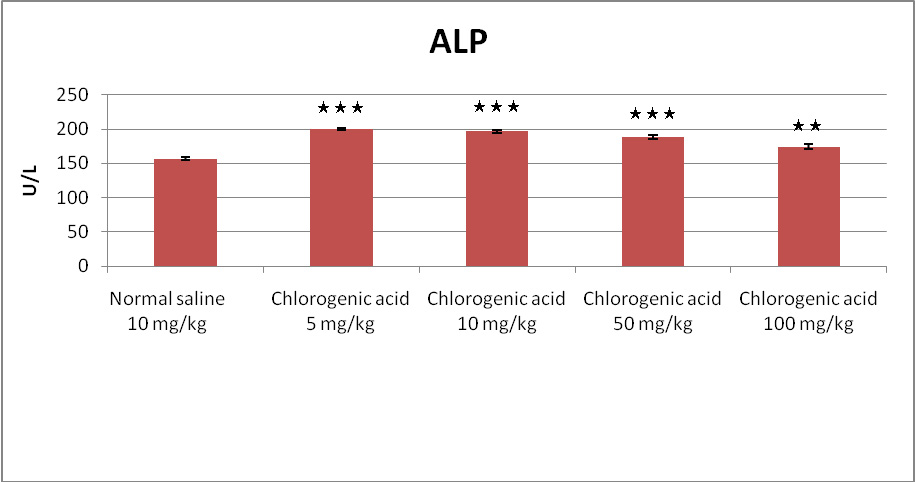 |
Figure 1: The serum level of ALP has significantly increased (p<0.05) in the groups administered with of chlorogenic acid (5, 10, 50, and 100 mg/kg). |
 |
Figure 2: The serum level of ALP has significantly decreased (p<0.05) in the groups administered with of chlorogenic acid (10, 50, and 100 mg/kg). |
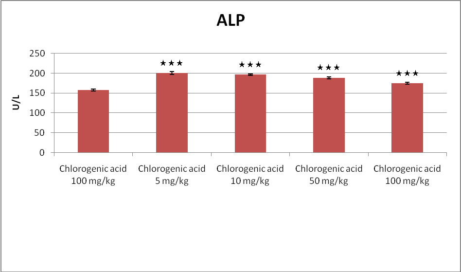 |
Figure 3: The serum level of ALP has significantly increased (p<0.05) in the groups administered with of chlorogenic acid (5, 10, 50, and 100 mg/kg). |
The serum level of ALT has significantly increased (p<0.05) in the groups administered with of chlorogenic acid (5, 10, 50, and 100 mg/kg) when compared to the control group (normal saline, 10 mg/kg) (Figure 4). The serum level of ALT has significantly decreased (p<0.05) in the groups administered with of chlorogenic acid (10, 50, and 100 mg/kg) when compared to the positive control group (arsenic trioxide, 10 mg/kg) but didn’t show significant difference in dose of 5 mg/kg (Figure 5). The serum level of ALT has significantly increased (p<0.05) in the groups administered with of chlorogenic acid (5, 10, 50, and 100 mg/kg) when compared to the negative control group (chlorogenic acid, 100 mg/kg) (Figure 6).
 |
Figure 4: The serum level of ALT has significantly increased (p<0.05) in the groups administered with of chlorogenic acid (5, 10, 50, and 100 mg/kg). |
 |
Figure 5: The serum level of ALT has significantly decreased (p<0.05) in the groups administered with of chlorogenic acid (10, 50, and 100 mg/kg) |
 |
Figure 6: The serum level of ALT has significantly increased (p<0.05) in the groups administered with of chlorogenic acid (5, 10, 50, and 100 mg/kg). |
The serum level of AST has significantly increased (p<0.05) in the groups administered with of chlorogenic acid (5, 10, 50, and 100 mg/kg) when compared to the control group (normal saline, 10 mg/kg) (Figure 7). The serum level of AST has significantly decreased (p<0.05) in the groups administered with of chlorogenic acid (10, 50, and 100 mg/kg) when compared to the positive control group (arsenic trioxide, 10 mg/kg) but didn’t show significant difference in dose of 5 mg/kg (Figure 8). The serum level of AST has significantly increased (p<0.05) in the groups administered with of chlorogenic acid (5, 10, 50, and 100 mg/kg) when compared to the negative control group (chlorogenic acid, 100 mg/kg) (Figure 9).
 |
Figure 7: The serum level of AST has significantly increased (p<0.05) in the groups administered with of chlorogenic acid (5, 10, 50, and 100 mg/kg). |
 |
Figure 8: The serum level of AST has significantly decreased (p<0.05) in the groups administered with of chlorogenic acid (10, 50, and 100 mg/kg). |
 |
Figure 9: The serum level of AST has significantly increased (p<0.05) in the groups administered with of chlorogenic acid (5, 10, 50, and 100 mg/kg). |
The level of GSH has significantly decreased (p<0.05) in the groups administered with of chlorogenic acid (5, 10, 50, and 100 mg/kg) when compared to the control group (normal saline, 10 mg/kg) (Figure 10). The level of GSH has significantly increased (p<0.05) in the groups administered with of chlorogenic acid (10, 50, and 100 mg/kg) when compared to the positive control group (arsenic trioxide, 10 mg/kg) but didn’t show significant difference in dose of 5 mg/kg (Figure 11). The level of GSH has significantly decreased (p<0.05) in the groups administered with of chlorogenic acid (5, 10, 50, and 100 mg/kg) when compared to the negative control group (chlorogenic acid, 100 mg/kg) (Figure 12).
 |
Figure 10: The level of GSH has significantly decreased (p<0.05) in the groups administered with of chlorogenic acid (5, 10, 50, and 100 mg/kg). |
 |
Figure 11: The level of GSH has significantly increased (p<0.05) in the groups administered with of chlorogenic acid (10, 50, and 100 mg/kg). |
 |
Figure 12: The level of GSH has significantly decreased (p<0.05) in the groups administered with of chlorogenic acid (5, 10, 50, and 100 mg/kg). |
Arsenic compounds are one of the pollutants of environment which are a serious threat to human health. Millions of people around the world through drinking water are exposed to arsenic compounds (Sabath and Robles-Osorio, 2012). Exposure to arsenic and its compounds can have dangerous effects on health (Tapio and Grosche, 2006) and can lead to many types of cancers, such as skin, liver, kidney, lung, intestine, and bladder (Tchounwou et al., 2004). Inorganic arsenic compounds as carcinogenic compounds are known. Epidemiological studies have shown that these compounds are related to the types of cancers like liver, kidney, and bladder (Gradecka et al., 2001). Researches have shown that consumption of drinking water containing inorganic arsenic compounds is related to liver diseases such as liver cancer (Islam et al., 2011). In clinical studies of the liver, liver enzymes, including Alkaline phosphatase (ALP), Alanine aminotransferase (ALT), and Aspartate aminotransferase (AST) are evaluated. The enzymes levels of ALT and AST are an indicator to assess the integrity of liver cells and the enzyme level of ALP represents the perfect synthesis of albumin and bile by the liver (Islam et al., 2011). Our research has shown that the serum levels of ALP, ALT, and AST have significantly increased (p<0.05) in all groups administered with of chlorogenic acid when compared to the control and negative control groups. Antioxidants are including two major categories, enzymatic and non enzymatic. Enzymatic antioxidants like catalase, glutathione peroxidase, and superoxide dismutase, which are produced by the body and non enzymatic antioxidants like flavonoids, tannins, and carotenoids that obtained from plants (Lee et al., 2004). Phenolic compounds as antioxidants in plants are caused by the elimination of free radicals. The antioxidant properties of phenolic compounds are related to the characteristic of the redox (oxidation and reduction) that reduce free radicals. Also, they are caused chelating of metals (Hsu, 2006). There is chlorogenic acid as a polyphenolic compound widely in various plants (Šeruga and Tomac, 2014).
Studies, on the health benefits of consuming chlorogenic acid found in foods have shown that this antioxidant to reduce diseases such as cancer, stroke, obesity, diabetes, and Alzheimer’s disease (Zhao et al., 2012). The serum levels of ALP, ALT, and AST have significantly decreased (p<0.05) in the groups administered with of chlorogenic acid (10, 50, and 100 mg/kg) when compared to the positive control group (arsenic trioxide, 10 mg/kg). It sounds that the antioxidant property of chlorogenic acid plays an important role in the protection of cells against free radicals caused by arsenic trioxide. The free radical is an atom or molecule that has an unpaired electron. The cells use of oxygen for produce energy that lead to the production of free radicals such as superoxide anion, hydroxyl, and peroxyl that are known as reactive oxygen species (ROS) and mainly produced by mitochondria. Free radicals can lead to a wide range of toxic oxidative reactions like membrane lipid peroxidation, the failure of enzymes and proteins that could cause cell death (Umamaheswari and Chatterjee, 2008). Free radicals cause oxidative stress that plays an important role in the development of many diseases such as diabetes, cardiovascular, and cancer (Bandyopadhyay et al., 1999). The studies show that many metals such as iron (Fe), lead (Pb), and copper (Cu) with modulating the redox (oxidation and reduction) in cell are causing the level change of thiols like glutathione in the cells (Tchounwou et al., 2002). The glutathione (GSH) as a thiol compound and important in cells have antioxidant role and are caused destroy of free radicals. The glutathione (GSH) is in various body organs such as liver, kidney, brain, pancreas, and heart and highest levels of that found in the liver (Aoyama and Nakaki, 2015).
In this study, the level of GSH has significantly decreased (p<0.05) in the groups administered with of chlorogenic acid (5, 10, 50, and 100 mg/kg) when compared to the control and negative control groups that was specified arsenic trioxide reduced GSH in the liver but the level of GSH has significantly increased (p<0.05) in the groups administered with of chlorogenic acid (10, 50, and 100 mg/kg) when compared to the positive control group that was identified chlorogenic acid as an antioxidant reduces free radicals and increases the glutathione level of the liver. The glutathione (GSH) with formation reversible disulfide bonds between thiols of protein can inhibit the oxidation of proteins during oxidative stress (Giustarini et al., 2004). The glutathione (GSH) as an antioxidant have an important role in balancing of intracellular redox processes (Aoyama and Nakaki, 2015). Overall, our results showed that chlorogenic acid as a powerful and important antioxidant had significant protective effects against the toxicity of arsenic trioxide.
Acknowledgment
We appreciate Of all colleagues who have worked on this project with us include the Laboratory of Toxicology in the Faculty of Pharmacy of Mazandaran University of Medical Sciences and Noor medical laboratory.
References
- Abernathy, C.O., Thomas, D.J., Calderon, R.L., 2003. Health effects and risk assessment of arsenic. The Journal of nutrition 133, 1536S-1538S.
- Aoyama, K., Nakaki, T., 2015. Glutathione in cellular redox homeostasis: association with the excitatory amino acid carrier 1 (EAAC1). Molecules 20, 8742-8758.
- Bandyopadhyay, U., Das, D., Banerjee, R.K., 1999. Reactive oxygen species: oxidative damage and pathogenesis. Current Science-Bangalore- 77, 658-666.
- Bralley, E.E., Greenspan, P., Hargrove, J.L., Wicker, L., Hartle, D.K., 2008. Topical anti-inflammatory activity of Polygonum cuspidatumextract in the TPA model of mouse ear inflammation. Journal of Inflammation 5, 1.
- Centeno, J.A., Mullick, F.G., Martinez, L., Page, N.P., Gibb, H., Longfellow, D., Thompson, C., Ladich, E.R., 2002. Pathology related to chronic arsenic exposure. Environmental Health Perspectives 110, 883.
- Das, N., Paul, S., Chatterjee, D., Banerjee, N., Majumder, N.S., Sarma, N., Sau, T.J., Basu, S., Banerjee, S., Majumder, P., 2012. Arsenic exposure through drinking water increases the risk of liver and cardiovascular diseases in the population of West Bengal, India. BMC Public Health 12, 639.
- De Azevedo, A., Mazzafera, P., Mohamed, R., Melo, S., Kieckbusch, T.G., 2008. Extraction of caffeine, chlorogenic acids and lipids from green coffee beans using supercritical carbon dioxide and co-solvents. Brazilian Journal of Chemical Engineering 25, 543-552.
- Fang, Y.-Z., Yang, S., Wu, G., 2002. Free radicals, antioxidants, and nutrition. Nutrition 18, 872-879.
- Flora, S., Bhadauria, S., Kannan, G., Singh, N., 2007. Arsenic induced oxidative stress and the role of antioxidant supplementation during chelation: a review. Journal of Environmental Biology 28, 333.
- Giustarini, D., Rossi, R., Milzani, A., Colombo, R., Dalle‐Donne, I., 2004. S‐Glutathionylation: from redox regulation of protein functions to human diseases. Journal of cellular and molecular medicine 8, 201-212.
- Gradecka, D., Palus, J., Wasowicz, W., 2001. Selected mechanisms of genotoxic effects of inorganic arsenic compounds. International journal of occupational medicine and environmental health 14, 317-328.
- Halliwell, B., Whiteman, M., 2004. Measuring reactive species and oxidative damage in vivo and in cell culture: how should you do it and what do the results mean? British journal of pharmacology 142, 231-255.
- Holt, E.M., Steffen, L.M., Moran, A., Basu, S., Steinberger, J., Ross, J.A., Hong, C.-P., Sinaiko, A.R., 2009. Fruit and vegetable consumption and its relation to markers of inflammation and oxidative stress in adolescents. Journal of the American Dietetic Association 109, 414-421.
- Hsu, C.-Y., 2006. Antioxidant activity of extract from Polygonum aviculare L. Biological research 39, 281-288.
- Islam, K., Haque, A., Karim, R., Fajol, A., Hossain, E., Salam, K.A., Ali, N., Saud, Z.A., Rahman, M., Rahman, M., 2011. Dose-response relationship between arsenic exposure and the serum enzymes for liver function tests in the individuals exposed to arsenic: a cross sectional study in Bangladesh. Environmental health 10, 64.
- Lee, J., Koo, N., Min, D., 2004. Reactive oxygen species, aging, and antioxidative nutraceuticals. Comprehensive reviews in food science and food safety 3, 21-33.
- Mittal, D.K., Joshi, D., Shukla, S., 2012. Hepatoprotective Role of Herbal Plants–A Review. International Journal of Pharmaceutical Sciences 3, 150-157.
- Sabath, E., Robles-Osorio, M.L., 2012. Renal health and the environment: heavy metal nephrotoxicity. Nefrologia 32, 279-286.
- Saeedi Saravi, S., Shokrzadeh, M., 2008. Histopathological and biochemical disorders following administration of Sambucus ebulus extract on mice and rats and preventive effects of vitamins C and E on renal and hepatic disorders. Phcog Mag 5, 131-135.
- Sarvankumar, G., Lalitha, V., Sengottuvelu, S., SHARIF, S.H., SIVAKUMAR, T., 2011. Nephroprotective activity of Vitex negundoLinn bark against chemical induced toxicity in experimenal Rats. An International Journal of Advances in Pharmaceutical Sciences 2, 462-470.
- Šeruga, M., Tomac, I., 2014. Electrochemical behaviour of some chlorogenic acids and their characterization in coffee by square-wave voltammetry. International journal of electrochemical science 9, 6134-6154.
- Shokrzadeh, M., Chabra, A., Ahmadi, A., Naghshvar, F., Habibi, E., Salehi, F., Assadpour, S., 2015. Hepatoprotective effects ofZataria multiflora ethanolic extract on liver toxicity induced by cyclophosphamide in mice. Drug research 65, 169-175.
- Stevens, J.J., Graham, B., Walker, A.M., Tchounwou, P.B., Rogers, C., 2010. The effects of arsenic trioxide on DNA synthesis and genotoxicity in human colon cancer cells. International journal of environmental research and public health 7, 2018-2032.
- Tapio, S., Grosche, B., 2006. Arsenic in the aetiology of cancer. Mutation Research/Reviews in Mutation Research 612, 215-246.
- Tchounwou, P., Wilson, B., Abdelghani, A., Ishaque, A., Patlolla, A., 2002. Differential cytotoxicity and gene expression in human liver carcinoma (HepG2) cells exposed to arsenic trioxide, and monosodium acid methanearsonate (MSMA). International Journal of Molecular Sciences 3, 1117-1132.
- Tchounwou, P.B., Centeno, J.A., Patlolla, A.K., 2004. Arsenic toxicity, mutagenesis, and carcinogenesis–a health risk assessment and management approach. Molecular and cellular biochemistry 255, 47-55.
- Um, M.-Y., Choi, W.-H., Aan, J.-Y., Kim, S.-R., Ha, T.-Y., 2006. Protective effect of Polygonum multiflorum Thunb on amyloid â-peptide 25-35 induced cognitive deficits in mice. Journal of ethnopharmacology 104, 144-148.
- Umamaheswari, M., Chatterjee, T., 2008. In vitro antioxidant activities of the fractions of Coccinia grandis leaf extract. African Journal of Traditional, Complementary and Alternative Medicines 5, 61-73.
- Wu, G., Fang, Y.-Z., Yang, S., Lupton, J.R., Turner, N.D., 2004. Glutathione metabolism and its implications for health. The Journal of nutrition 134, 489-492.
- Zhao, Y., Wang, J., Ballevre, O., Luo, H., Zhang, W., 2012. Antihypertensive effects and mechanisms of chlorogenic acids. Hypertension Research 35, 370-374.

