Valery P. Kartashev![]()
Russian State Social University, 129226, Moscow, Russia
Corresponding author email: ilmedv1@yandex.ru
Article Publishing History
Received: 15/07/2020
Accepted After Revision: 23/09/2020
The course of early ontogenesis is associated with changes in the morphology and physiology of red blood cells. Largely thanks to these changes, the body adapts to any changes in the environment that occur during the transition from the antenatal period to the postnatal period. Red blood cells at the beginning of ontogenesis always demonstrate greater functional stability, which gradually changes in the future. The red bone marrow and endocrine glands are actively involved in the regulation of erythropoiesis. A special place in this process is occupied by the adrenal cortex. The study confirmed that glucocorticoids in the early postnatal period of rat life are an important factor in the regulation of erythropoiesis involving T-lymphocytes. Erythropoiesis, previously considered at this age to be reactive to various stress factors, should be considered sensitive to their effects. This was demonstrated by increasing the concentration of red blood cells in the blood, increasing the number of red blood cells, discocytes and their average diameter after the administration of hydrocortisone to rats at the age of six days.
Erythrocytes, Rats, Erythropoiesis, Early Ontogenesis, Glucocorticoids, Regulation.
Kartashev V. P. The Effect of Glucocorticoid Hormones on the Morphofunctional State of Red Blood Cells in Early Postnatal Ontogenesis. Biosc.Biotech.Res.Comm. 2020;13(3).
Kartashev V. P. The Effect of Glucocorticoid Hormones on the Morphofunctional State of Red Blood Cells in Early Postnatal Ontogenesis. Biosc.Biotech.Res.Comm. 2020;13(3). Available from: https://bit.ly/2P8YwU3
Copyright © This is an Open Access Article distributed under the Terms of the Creative Commons Attribution License (CC-BY). https://creativecommons.org/licenses/by/4.0/, which permits unrestricted use distribution and reproduction in any medium, provided the original author and sources are credited.
INTRODUCTION
Red blood cells are the largest group of blood cells that realize the process of gas exchange in the body and the rheological properties of blood in vessels of any caliber (Zavalishina, 2018a; Glagoleva and Medvedev, 2018). Postnatal ontogenesis is marked by changes in red blood cells in their morphology and their characteristics (Vorobyeva et al., 2018). Largely thanks to these changes, the body adapts to any changes in the environment, primarily during the transition from the antenatal period to the postnatal period (Medvedev and Kumova, 2007a; Tkacheva and Medvedev, 2020). It is very important that red blood cells, especially at the beginning of ontogenesis, demonstrate greater functional stability, which gradually changes during development (Bikbulatova, 2018a; Vorobyeva and Medvedev, 2020).
It is recognized that red bone marrow, nervous system, endocrine glands, liver, spleen, kidney and organs of the gastrointestinal tract are primarily involved in the regulation of erythropoiesis (Medvedev and Kumova, 2007b).Most mammals are born with a fully formed neuroendocrine system. Further improvement of the function of individual links of this system or their consolidation occurs in the process of individual development of the organism. In particular, it is known that in rats whose intrauterine development proceeded during physiological pregnancy, by the time of transition to definitive nutrition, the hypothalamic-pituitary-corticoid system acquires high functional activity (Henning, 1978). In rats, this occurs at the end of the second week of life (Poland et al,, 1979). Nevertheless, it was found that physical activity throughout the pregnancy of rats leads to a delay in the natural dynamics of the morphofunctional features of red blood cells in postnatal ontogenesis in prenatally stressed offspring (Kartashev et al., 2017), which undoubtedly affects the respiratory function of the body.
As before, many issues of postnatal changes in the functional parameters of red blood cells remain insufficiently studied. As a result of this, to date, the available information on the mechanisms of restructuring the functional state of erythrocytes in postnatal ontogenesis is fragmentary (Karpov et al., 2020). The work on this problem was carried out by non-identical methods on different biological objects, in groups of different ages and incomparable. In this regard, the aim of the work was: to find out the role of corticoids in the formation of morphofunctional features of red blood cells in a growing organism.
MATERIAL AND METHODS
The study was conducted in strict accordance with the ethical principles of the European Convention for the Protection of Vertebrates Used for Experimental and Other Scientific Purposes (adopted in Strasbourg on March 18, 1986 and confirmed in Strasbourg on June 15, 2006).
In the experiment, white Wistar rats were used with a total of 50 animals. To obtain rat pups, females of approximately the same body weight of 180-200 g were selected; they were kept in cages of 8 individuals each. After preliminary getting used to the females, 3-4 males of about the same mass for mating were planted for one day. Fertilized females (this was determined by the presence of sperm in their vaginal smears) were placed in individual cells, kept under unlimited nutrition with sufficient drinking. From the first day after birth, the rat pups were distributed 8 for each lactating female.
On the sixth day of life, rat pups, when the natural level of corticosterone in their blood is still at a critically low level (Kozinets et al., 1977), they received a single injection of hydrocortisone (2.5 mg/100g body weight). This stimulated their body, outstripping the natural increase in blood levels of corticoid concentration, which is observed in rats in postnatal ontogenesis only at the end of the second week of life, reaching the level of adult animals (Henning, 1978). The control group received an injection of an equivalent amount of saline. Then, for 8 days every 24 hours, the rat pups of the control (n = 25) and experimental group (n = 25) evaluated the morphofunctional features of red blood cells.
To study the tested parameters in all cases, blood sampling in rat pups was carried out from the tail vein. The number of red blood cells in their blood was counted in the Goryaev’s cell. The erythrocyte morphology was studied by scanning electron microscopy using a S-405 A HITACHI scanning electron microscope (Japan). To determine the percentage of different forms of red blood cells from each animal, 1-2 blood preparations were prepared and the number of scans was taken that provided at least 200 red blood cells.
On the scans, at least 1000 different forms of red blood cells were counted, followed by a percentage transfer. In accordance with the classification (Kozinets et al., 1977) discocytes were counted, discoid type erythrocytes were determined, erythrocytes with an outgrowth, with a crest, with multiple outgrowths were counted; assessed levels of a non-discoid type – red blood cells in the form of a mulberry, domed, spherical, in the form of a deflated ball, degeneratively altered. The average diameter of red blood cells was determined from the calculation of 100 cells.Quantitative data obtained during the study were processed using G.G. Avtondilova morphometric techniques (Avtondilov, 1990). The reliability of the results was determined by student’s criterion.
RESULTS AND DISCUSSION
The concentration of red blood cells in the blood of rat pups (table 1) practically did not change during the first three days of observation in both groups. On the fourth and fifth days after the exposure, both in the control and in the experiment, the level of erythrocytes increases relative to the initial one, no difference was found between the control and experimental groups. On the sixth day after exposure, the concentration of erythrocytes significantly increases compared with the previous days in both control and hydrocortisone-stimulated animals. However, in the latter, the increase in the number of cells is more pronounced (by 28.9%). In the next day, the increase in the number of red blood cells in the blood of experimental animals continues. On the eighth day of the study, the concentration of red blood cells increases even more in the experimental group and becomes significantly higher than in the control.
Table 1. Dynamics of the number of red blood cells (in 1 mm³) in the blood of rats after a single injection of hydrocortisone at six days of age.
| The age of the animals (in days) | Duration of observation (day) | Control,
n=25, М±m |
An experience, n=25, М±m |
| 6 | Drug administration 0(initially) | 2.78±0.08 | |
| 7 | 1 | 2.70±0.08 | 2.84±0.08 |
| 8 | 2 | 2.82±0.16 | 2.81±0.10 |
| 9 | 3 | 2.92±0.21 | 3.00±0.10 |
| 10 | 4 | **
3.54±0.20 |
**
3.47±0.21 |
| 11 | 5 | *
3.21±0.16 |
*
3.29±0.05 |
| 12 | 6 | **
3.81±0.05 |
**
4.24±0.26 |
| 13 | 7 | **
3.54±0.14 |
**
4.39±0.26 |
| 14 | 8 | **
3.84±0.20 |
**
4.57±0.16 p<0.05 |
Legend: significance of differences relative to the initial value in the group: * – p <0.05, ** – p <0.01; significance of differences between groups – p.
Using scanning electron microscopy of red blood cells (Figure 1), it was found that the level of discocytes in the control group of animals increased on the eighth day of the experiment, while under the influence of exogenous hydrocortisone, quantitative growth of discocytes took place already on the sixth day.
An increase in the level of erythrocyte-discocytes after the administration of hydrocortisone in the experimental group was accompanied by a decrease in the discoid and non-discoid types of red blood cells. Against the background of a general decrease in erythrocyte discoid forms on the sixth day, there is a significant decrease in erythrocyte disocytes with a crest and multiple outgrowths (Figure 1). In the group of non-diskoid cell types, by the fourth day of the study, there was a decrease in predominantly dome-shaped red blood cells in the control and in the experiment. On the sixth day in the blood of animals stimulated by hydrocortisone, they became significantly more than in control rat pups.
Figure 1: Changes in the morphology of red blood cells in rat pups after he was injected with hydrocortisone at six days of age. On the abscissa axis, days after exposure; on the ordinate axis, the number of cells in% of the total number taken as 100%.
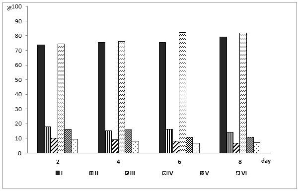
Legend: I – discocytes, II – discoid group of cells, III – non-discoid group of cells in control animals; IY – discocytes, Y – discoid group of cells, YI – non-discoid group of cells in the experiment.
The average erythrocyte diameter (Table 2) did not significantly differ between the observed groups on the second, fourth, and eighth days of the study; on the sixth day only, in experimental animals it was significantly larger compared to the control (p <0.001).
Table 2. Dynamics of the average diameter of red blood cells (in arbitrary units) in rats after the injection of hydrocortisone produced at six days of age
| Observation groups | Duration of observation | |||
| 2 day | 4 day | 6 day | 8 day | |
| Control, n=25, М±m | 5.83±0.06 | 5.73±0.06 | 5.82±0.06 | 5.86±0.05 |
| Experienced, n=25, М±m | 5.74±0.06 | 5.62±0.06 | *6.40±0.10
р<0.001 |
*5.82±0.05 |
Legend: * – significance of differences with the previous observation period (p <0.05), p <0.01 – significance of differences between the experimental and control groups at each observation period.
The results obtained suggest that the introduction of hydrocortisone to six-day-old rats by the end of the experiment increases the concentration of red blood cells, increases the number of red blood cells and their size and reduces the level of discoid and non-discoid red blood cells in animals.The results obtained in the study complement the available data on the structure and functions of rat erythrocytes in postnatal ontogenesis (Zavalishina, 2018b). The data confirm the information that at the beginning of ontogenesis, the number of red blood cells in the blood increases due to the high activity of erythropoiesis, which is clearly regulated by the endocrine system (Medvedev and Gamolina, 2008).
Moreover, an increase in the concentration of red blood cells and the number of discocytes with a large average diameter (macrocytes) in rat pups with age is primarily associated with stimulation of glucocorticoid hormones. In this regard, it can be said that exogenous hydrocortisone at an early age in rats stimulates erythropoiesis, contrary to the opinion that the mechanisms responsible for it in the neonatal period are reactive with respect to specific stimulants erythropoietin (Filimonov and Tabarchuk, 1978) and corticosteroids (Balika and Kartasheva, 1982).
The obtained results to some extent contradict the prevailing opinion that in early postnatal ontogenesis, in particular in the first three weeks of rats (Filimonov and Tabarchuk, 1978) and the first two months in dogs (Gorozhanin, 1978), erythropoiesis is reactive. The authors who adhere to these points of view explain this by the fact that at this age the concentration of erythropoietin is at its maximum and, as a result, erythropoiesis is at its peak. Their experiments with the introduction of plasma animals with a high content of erythropoietin did not change the rate of incorporation of radioactive Fe into red blood cells (Filimonov and Tabarchuk, 1978).
In this work, the action of endogenous erythropoietin after administration of hydrocortisone is excluded, which is observed in adult animals (Malgor et al., 1974; Morschakova et al., 1979; Romashko et al., 1985). Considering that under the conditions of the experiment, an increase in the concentration of erythrocytes after the administration of hydrocortisone occurs only on the sixth to eighth days, it can be assumed that the hormone exerts its effect on the hematopoietic cells not directly, but indirectly with the participation of some additional link (Zavalishina, 2018c).
Disclosure of this missing link in the course of corticosteroid-induced erythropoiesis has been helped by experiments in adult animals. In particular, an increase in the concentration of red blood cells in the experiment described in this article coincides with the timeframes identified earlier (Romashko et al., 1985). It was also previously shown that the effect of corticoids on erythropoiesis is carried out with the participation of the thymus (Moroz et al.,1984; Bespalov et al., 2018a). Graceful experiments (Dygay and Shakhov, 1989; Dygay et al., 1990) demonstrated that, in the chronic immobilization of adult mice, along with an increase in the concentration of glucocorticoids in the blood of animals, there is an increase in T-lymphocyte regulators (expressing lyt-1 + and lyt-2 + antigens on their surface) and their homing in bone marrow tissue with subsequent activation of erythropoiesis, which occurred on the sixth or eighth day, that is, at the same time as in our work with the introduction of hydrocortisone. Prior immobilization, administration of antithymocytic serum, thimectomy, or adrenalectomy inhibited this process. Substitution therapy of adrenalectomized animals restored this process.
It was also noted that an increase in the rate of erythropoiesis in response to the administration of exogenous glucocorticoids would not be observed if anterythropoietin serum was previously introduced (Malgor et al., 1974). This becomes clear when one considers the report of Lipton and Nathan, (1983) that, T-lymphocytes are regulators in bone marrow tissue due to their interaction with monocytes / macrophages with the release of lymphokine or monokine during this process (Bespalov et al, 2018b). Both substances, increasing the sensitivity of early erythroid committed precursors to erythropoietin, stimulate the proliferation and differentiation of committed precursors. It becomes clear that endogenous erythropoietin at the time of action of hydrocortisone in the experiment is the resultant factor in the activation of erythropoiesis.
Given the results, there is reason to adhere to the opinion of O.O. Romashko et al. (1985) on the presence in the organism of mammals of two corticoid-dependent erythropoiesis pathways. One of them is associated with the stimulation of erythropoietin synthesis (Malgor et al, 1974; Morschakova and Pashukov, 1982) and its subsequent action on cells sensitive to it (Medvedev and Kumova, 2007c). Another pathway of erythropoiesis is associated with the activation of its mechanisms sensitive to the action of T-lymphocytes (Bikbulatova, 2018b). This is convincingly demonstrated by Kalinina and Pashukov (1985) in the absence of adrenal glands in animals with high erythropoietic blood activity, where they observed a slight increase in reticulocytes and a lower level of their synthesis in the bone marrow.
It is noteworthy that on the eighth and ninth days of postnatal life, the concentration of red blood cells in humans (Leonova and Rapoport, 1989), productive animals (Vorobyeva and Medvedev, 2018; Tkacheva, 2020), reticulocytes in dogs (Balika and Kartasheva, 1982) and the rate of incorporation of radioactive Fe in rats increase (Filimonov and Tabarchuk, 1978). However, this phenomenon has remained unexplained. Apparently, the activation of erythropoiesis in the second week of fetal life is caused by a high level of hormones of the adrenal cortex in the mother’s blood during birth stress. In this case, corticoids, as is known, freely pass from the bloodstream of the mother to the fetus (Zarrow et al., 1970; Mitskevich, 1978; McEven et al., 1986).
At this point, they include a mechanism for stimulating erythropoiesis with the participation of T-lymphocytes, and the effect of glucocorticoids is detected only after a week, when their basal level is significantly reduced (Henning, 1978; Poland et al., 1979; D’Agostino and Henning, 1982). The physiological significance of the corticoid-dependent activation of erythropoiesis during this period becomes clear, given that the newborn body has to adapt to completely new conditions and type of respiration.
CONCLUSION
Assessing the results obtained during the study taking into account the literature data, it can be concluded that glucocorticoids in the early postnatal period of rat life are an important factor in the regulation of erythropoiesis involving T-lymphocytes. Erythropoiesis, previously considered at this age to be reactive to various stress factors, was found to be sensitive to stimulation. This was demonstrated by an increase in the concentration of red blood cells in the blood, an increase in the number of red blood cells, discocytes and their average diameter after the administration of hydrocortisone to rats at the age of six days.
Ethical Clearance Statement: The Current Research Work Was Ethically Approved by the Institutional Review Board (IRB) of State Social University, 129226, Moscow, Russia.
Conflict of Interest: Author declares no conflicts of interests to disclose.
REFERENCES
Avtondilov, G.G. (1990) Medical morphometry. Leadership. Moscow: Medicine, 384.
Balika, Yu.D. and Kartasheva, V.E. (1982) Reactions of the hematopoietic organs of a newborn to glucocorticoids (experimental study). Obstetrics and gynecology, 7 : 54-55.
Bespalov, D.V. , Medvedev, I.N., Mal, G.S. and Polyakova, O.V. (2018a) Physiological Capabilities Of The Vascular Endothelium With The Developing Arterial Hypertension In People Of Different Ages Who Had Long Had Low Physical Activity. Research Journal of Pharmaceutical, Biological and Chemical Sciences, 9(2) : 972-976.
Bespalov, D.V., Medvedev, I.N., Mal, G.S. and Makurina, O.N. (2018b) Functional activity of the vascular endothelium in patients with initial signs of atherosclerosis against the background of regularly dose-related exercise stress. Research Journal of Pharmaceutical, Biological and Chemical Sciences, 9(2) : 1020-1024.
Bikbulatova, A.A. (2018a) Functional Features Of Microcirculatory Processes In Obese Women Against A Background Of Long Daily Wearing Of Corrective Clothing. Research Journal of Pharmaceutical, Biological and Chemical Sciences, 9(6) : 785-793.
Bikbulatova, A.A. (2018b) Functional Features Of Microcirculatory Processes In Obese Women Against A Background Of Long Daily Wearing Of Corrective Clothing. Research Journal of Pharmaceutical, Biological and Chemical Sciences, 9(6) : 785-793.
D’Agostino, I.B. and Henning, S.J. (1982) Role of thyroxine in coordinate control of corticosterone and CBG in postnatal development. Amer. J. Physiol, 242 : E33-E39.
Dygay, A.M. and Shakhov, V.P. (1989) The role of intercellular interactions in the regulation of hematopoiesis. Tomsk: Tomsk University, 224.
Dygay, A.M., Shakhov, V.P., Kirienkova, E.V., Khlusov, I.A. and Goldberg, I.D. (1990) The role of glucocorticoids in the development of the phenomenon of stimulation of bone marrow hematopoiesis under stress. Biological sciences, 12 : 71-76.
Filimonov, V.I. and Tabarchuk, A.D. (1978) Formation of erythropoiesis regulation mechanisms in postnatal ontogenesis. Regulation mechanisms in the blood system. Part II Krasnoyarsk, 42-43.
Glagoleva, T.I. and Medvedev, I.N. (2018) Physiological Features Of Anti-aggregational Control Of Blood Vessels Over The Shaped Elements Of Blood In Calves At The Onset Of Ontogenesis. Research Journal of Pharmaceutical, Biological and Chemical Sciences, 9(5) : 440-447.
Gorozhanin, L.S. (1978) Ontogenetic development of erythropoiesis regulation mechanisms. Regulation mechanisms in the blood system. Part II. Krasnoyarsk: Krasnoyarsk Medical Institute, 38-39.
Henning, S.J. (1978) Plasma concentrations of total and free conticosterone during developmen in the rat. Am. J. Physiol, 235(5) : E451-E456.
Kalinina, T.T. and Pashchukov, E.N. (1985) On the mechanism of the influence of adrenal hormones on erythropoiesis. II All-Union Congress of Hematologists and Transfusiologists. Abstracts of reports. Moscow, 148.
Karpov, V.Yu., Zavalishina, S.Yu., Komarov, M.N. and Koziakov, R.V. (2020) The Potential of Health Tourism Regarding Stimulation of Functional Capabilities of the Cardiovascular System. Bioscience Biotechnology Research Communications, 13(1) : 156-159. DOI: http://dx.doi.org/10.21786/bbrc/13.1/28
Kartashev, V.P., Shmeleva, S.V., Karpova, N.V. and Dubrovinskaya, E.I. (2017) Effects of physical activity of expectant mothers on morphology and functionality of erythrocytes in postnatal ontogenesis. Teoriya i Praktika Fizicheskoy Kultury, 4 : 61-63.
Kozinets, G.I., Ryapolova, I.V., Shishkanova, Z.G. and Vorobyova, M.G. (1977) Morphological characteristics of the peripheral blood red blood cells of healthy people [scanning electron microscopy]. Hematology problems, 22(7) : 19-21.
Leonova, V.G. and Rapoport, J.Zh. (1989) Quantitative indicators of red blood in children. Novosibirsk: Science, 104.
Lipton, J.M. and Nathan, D.G. (1983) Cell-cell interactions in the regulation of erythropoiesis. Brit. J. Haemat, 53 : 361-367.
Malgor, L., Torales, P. and Klainer. E. (1974) Effects of dexamethasone on bone marrow erythropoiests Hormone Metab. Res, 5 : 269-277.
McEven, B.S., de Kloet, E.R. and Rostene, W. (1986) Adremal steroid receptors and action in the nervos system. Physiol. Review, 66(4) : 1121-1188.
Medvedev, I.N. and Gamolina, O.V. (2008) Lisinopril effects on platelet activity in patients with arterial hypertension and impaired glucose tolerance. Russian Journal of Cardiology, 3 : 45-48.
Medvedev, I.N. and Kumova, T.A. (2007a) Angiotensin II receptor inhibitors: role and place in arterial hypertension and metabolic syndrome treatment. Russian Journal of Cardiology, 5 : 97-99.
Medvedev, I.N. and Kumova, T.A. (2007b) Valsartan effects on platelet activity in patients with arterial hypertension and metabolic syndrome. Russian Journal of Cardiology, 3 : 66-69.
Medvedev, I.N. and Kumova, T.A. (2007c) Angiotensin II receptor inhibitors: role and place in arterial hypertension and metabolic syndrome treatment. Russian Journal of Cardiology, 5 : 97-99.
Mitskevich, M.S. (1978) Hormonal regulation in the ontogenesis of animals. Moscow: Science, 224.
Moroz, B.B., Romashko, O.O. and Adyushkin, A.I. (1984) The effect of dexamethasone on the distribution pattern of colonies produced by CFUc of bone marrow and spleen in thimectomized mice. Hematology and transfusiology, 4 : 32-35.
Morschakova, E.F. and Pashukov, E.N. (1982) Effect of inhibition of protein synthesis on erythropoietin production in the kidneys. In the book: Damage and regulatory processes of the body: Abstracts of the III All-Union Congress of Pathophysiologists. Moscow, 185-186.
Morschakova, E.F., Uzbekova, D.G., Pashukov, E.N., Ivanova, E.F. and Frank, O.D. (1979) The effect of steroid hormones and cyclic nucleotides on erythropoiesis. Molecular mechanisms of erythron adaptation. Ryazan, 77-84.
Poland, R.E., Weinchsel, M.E. and Rubin, R.T. (1979) Postnatal maturation patterns of serum corticosterone and growth hormone in rats: effect of chronic thyroxine administration. Horm. Metabol. Res, 11 : 222-227.
Romashko, O.O., Deshevoy, Yu.B., Lededev, V.G. and Pavlov, A.D. (1985) The effect of repeated administration of dexomethasone on some indicators of erythropoiesis. Hematology and transfusiology, 30(6) : 47-49.
Tkacheva, E.S. (2020) Disaggregated control of blood vessels over platelets in milk piglets 00166. Published online: 28 February 2020.DOI: https://doi.org/10.1051/bioconf/20201700166
Tkacheva, E.S. and Medvedev. I.N. (2020) Functional features of vascular hemostasis in piglets of milk and vegetable nutrition. IOP Conference Series: Earth and Environmental Science. 421 (2020) 022041. doi:10.1088/1755-1315/421/2/022041
Vorobyeva, N.V. and Medvedev, I.N. (2018) Physiological Features Of Platelet Functioning In Calves Of Holstein Breed During The Newborn. Research Journal of Pharmaceutical, Biological and Chemical Sciences, 9(6) : 129-135.
Vorobyeva, N.V. and Medvedev, I.N. (2020) Functional platelet activity in Dutch newborn calves. IOP Conference Series: Earth and Environmental Science. 421 (2020) 022042. doi:10.1088/1755-1315/421/2/022042
Vorobyeva, N.V., Mal, G.S., Zavalishina, S.Yu., Glagoleva, T.I. and Fayzullina, I.I. (2018) Influence Of Physical Exercise On The Activity Of Brain Processes. Research Journal of Pharmaceutical, Biological and Chemical Sciences, 9(6) : 240-244.
Zarrow, M.X, Philpott, J.E. and Denenberg, V.H. (1970) Passage of 140-4-corticosterone from the rat mother to the foetus and neonate. Nature, 226 : 1058-1059.
Zavalishina, S.Yu. (2018a) Functional Antiaggregatory Properties Of Blood Vessels In Calves During Transition From Dairy To Plant Type Of Nutrition. Research Journal of Pharmaceutical, Biological and Chemical Sciences, 9(5) : 1110-1116.
Zavalishina, S.Yu. (2018b) Physiology Of Antiaggregatory Manifestations Of The Vascular Wall In Newborn Calves With Iron Deficiency, Receiving Metabolic Significant Effects. Research Journal of Pharmaceutical, Biological and Chemical Sciences, 9(6) : 1530-1536.
Zavalishina, S.Yu. (2018c) Dynamics Of The Functional State Of Platelet Functions In Newborn Calves Receiving Correction For Dyspepsia. Research Journal of Pharmaceutical, Biological and Chemical Sciences, 9(6) : 1566-1572.

