Diagnostic Science Department, Faculty of Dentistry, King
Abdulaziz University, Jeddah 22252, KSA
Corresponding author email: salhamed@kau.edu.sa
Article Publishing History
Received: 15/10/2021
Accepted After Revision: 20/12/2021
Ectodermal dysplasia (ED) is a rare genetic condition with nearly 200 documented traits. As the name states, ED targets tissues derived from ectoderms, such as hair, skin, nails, sweat glands, and teeth. Other orofacial structures might be affected, such as salivary glands and hard palate. Lack of teeth and diminished facial height can impact negatively on child growth and psychological well-being. Therefore, assessment and an interdisciplinary management plan of orofacial components of ED children should be installed as early as possible. Here we report an early assessment and multi-disciplinary management of ED child’s orofacial structures, which allow restoration of facial height and dental function and saliva reduction by the least invasive restorative approach in the form of the composite build-up of microdont and overdentures. It also highlights the importance of periodic evaluation of growth and treatment plan adjustment as an integral part of the transitional management until the age of a definite dental treatment.
Ectodermal Dysplasia, Salivary Glands, Hyposalivation, Maxillofacial Rehabilitation, Hypodontia
Alhamed S. Noninvasive Multi-Disciplinary Management of Ectodermal Dysplasia With Hyposalivation – A Case Report. Biosc.Biotech.Res.Comm. 2021;14(4).
Alhamed S. Noninvasive Multi-Disciplinary Management of Ectodermal Dysplasia With Hyposalivation – A Case Report. Biosc.Biotech.Res.Comm. 2021;14(4). Available from: <a href=”https://bit.ly/3xyEAQ8“>https://bit.ly/3xyEAQ8</a>
Copyright © This is an Open Access Article distributed under the Terms of the Creative Commons Attribution License (CC-BY) https://creativecommons.org/licenses/by/4.0/, which permits unrestricted use distribution and reproduction in any medium, sources the original author and sources are credited.
INTRODUCTION
Ectodermal dysplasia (ED) consists of an assembly of infrequent congenital disorders with a prevalence of one per 10,000 to 100,000 live births with no known geographical predilection. The condition targets all or some ectodermal-derived tissues, such as hair, skin, nails, sweat glands, and teeth. The most common traits are hypohidrosis and hydrotic, with the main difference being the number and function of sweat glands (Wright et al., 2019, Blattner, 1968).
The faces of these children are often similar due to unique maxillofacial features, including a prominent forehead, flat nasal bridge, thick lips, and periorbital and perioral hyperpigmentation. Oral findings of ED include underdeveloped palate and alveolar ridges, dental anomalies that vary from complete to partial dental agenesis, and teeth malformation, such as microdontia (conical teeth) and enamel hypoplasia (Blattner, 1968, Halai and Stevens, 2015, Wright et al., 2019, Gill et al., 2008).Teeth deficiency and malformation have a direct impact on the child’s growth and general well-being. ED management should commence as early as possible through multi-disciplinary care involving medical and dental teams. Dental rehabilitation for these children is crucial to restoring the required functional, esthetic, and psychosocial aims (Gill et al., 2008, Halai and Stevens, 2015).
Case Report
This is the case of an 8-year-old girl who attended with her parents after a referral for treatment. The child was diagnosed with autosomal recessive hidrotic ED at 3 years old due to delayed teeth eruption. No genetic mapping was done. The main complaint was the appearance of her small spiky front teeth, which result in bullying at school. The patient mentioned that she experiences dryness in her mouth, especially during eating, which improves by drinking sips of water frequently. A detailed dental and medical history was obtained from the child as well as the parents. Family history revealed that HED was affecting family members on her mother’s side.
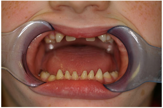
The child presented with some maxillofacial features, such as a prominent forehead, protruding low-set ears, saddle nose deformity, and decreased vertical height with thick everted lips. All primary teeth were present upon intraoral clinical examination, except the upper right and left primary lateral incisors. A dental panoramic tomogram was requested to assess permanent teeth development (Figure 1). Severe hypodontia was diagnosed with 22 missing permanent teeth excluding third molars, with microdontia of the lower anterior teeth.
Skeletal jaw relation assessment showed severe maxillary hypoplasia and reduced lower facial height. We detected a dental class III anterior relationship with severe reverse overjet of 3 mm and full arch crossbite. Orthodontic consultation was recommended to delay orthodontic treatment and reevaluate the case annually to determine the proper approach.Oral medicine consultation was obtained, and the stimulated saliva flow rate was evaluated to be 1 ml per min, which is low. The patient was diagnosed with hyposalivation and present some signs of mucosal dryness such as lobulated tongue and frothy saliva. Artificial saliva gel was prescribed along with instructions to continue water drinking and lubrications using olive oil and petroleum jelly. The periodic revaluation was planned.
Figure 1: Preoperative dental panoramic tomogram showing severe hypodontia.
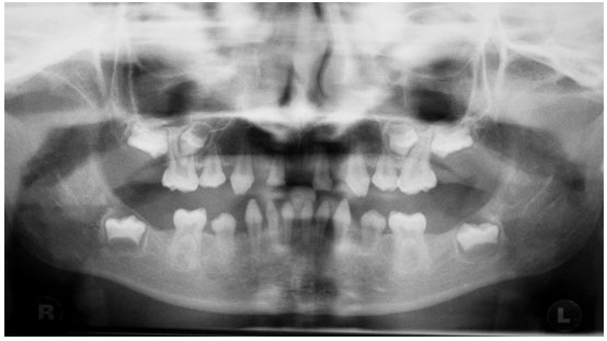
Figure 2: Preoperative clinical pictures.
Dental management was based on noninvasive preservation of the remaining dentition with the aesthetic restorative treatment of teeth morphology and function. Continuous follow-up and adjustment were scheduled until the patient reaches an age at which she can receive definitive treatment. An osseointegrated implant option was not considered for several reasons. First, the patient was anxious, and her parents wanted all the treatment to be atraumatic and conservative. Second, the patient presented with an adequate set of primary dentitions, which allows overdentures as a sufficient alternative plan. Moreover, implant positioning in a growing child has many drawbacks and should be planned only in selective cases.
The treatment plan was divided into preventive, rehabilitation, and maintenance phases. The rehabilitation phase was planned to cosmetically improve the appearance of microdont primary incisors followed by maxillary and mandibular overdenture to increase lower facial height.
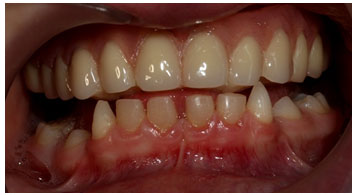
The patient presented for 6 months follow-up visit with good oral hygiene and intact restorations. However, the upper right central incisor showed grade 3 with complete root resorption. For that, an immediate denture was constructed and inserted after extraction. Prosthetic consultation stated that no need for facebow registration at the current stage and anterior teeth should be aligned in edge-to-edge relation (Figure 3,4).
Figure 3: Six months follow-up.
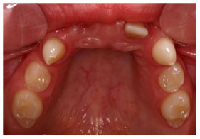
Figure 4: Immediate denture inserted after extraction of URA with an edge to edge occlusion.
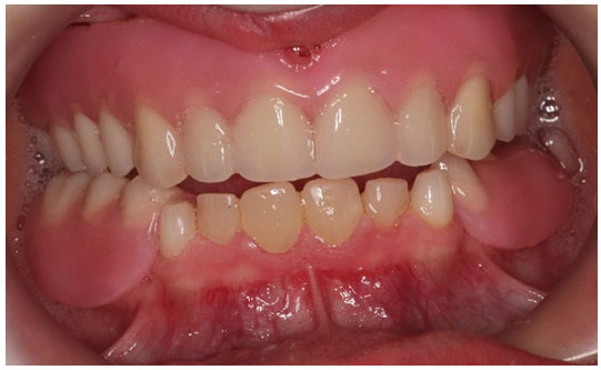
A maintenance phase through periodic recalls was arranged for every 6 months for prevention, evaluation of oral hygiene, teeth development, and eruption. Moreover, this would include evaluation of dentures and planning for adjustment or remaking, when required. The last demonstrated review was 3 years post-treatment (Figure 5).
Figure 5: Three years follow-up with upper and lower overdentures.
DISCUSSION
Hypodontia is the term used to describe the congenital or developmental absence of one or more primary or secondary teeth (excluding third molars) (Gill et al., 2008). In Saudi children, the prevalence of hypodontia was reported to be 2.6%, excluding the third molar (Salama and Abdel-Megid, 1994). Severe hypodontia in children is a very rare condition, most often associated with congenital syndromes, such as Down syndrome or ED (Crawford et al., 1991). Lack of teeth can significantly impact the child’s physical and emotional well-being, especially during adolescence. As in this case, hypodontia could be a reason for bullying, leading to social isolation, loneliness, and underachievement, (Crawford et al., 1991 Bohner et al, 2019).
Prosthetic rehabilitation using an osseointegrated implant in growing children as young as 3 years old was documented in the literature (Bohner et al., 2019, Wang et al., 2016). However, a recent systematic review investigating long-term implant outcomes in children stated that infraocclousion and rotation are common complications in these patients. Moreover, regular adjustments are essential in growing jaws (Bohner et al., 2019). Another systematic review stated that although implants are commonly used in ED children for dental rehabilitation, there is a lack of long-term data on bone augmentation and implant success in the literature (Wang et al., 2016). As osseointegrated implants are considered only in special circumstances, other noninvasive approaches, such as overdentures, are good options to reduce psychological trauma, continuous adjustments, and traumatic treatment (Bohner et al., 2019, Wang et al., 2016).
Hyposalivation is an objective term used to describe an actual reduction in salivary flow (Turner, 2016). There are many reasons for hyposalivation which range from temporary medication Side effects to degenerative exocrine autoimmune conditions such as Sjogren syndrome. As ectodermal dysplasia affects ectoderm-derived tissue, there has been an association with xerostomia and hyposalivation (Lexner et al., 2007, Nordgarden et al., 2003). In addition, qualitative changes have been demonstrated in ED patients such as reduced amylase activity and concentration (Nordgarden et al., 2003).
Management of chronic hyposalivation aims to reduce symptoms and improves patient’s daily activities such as speaking and eating(Lapiedra et al., 2015, Martín et al., 2017). Those patients such as this case learn to continuously lubricate oral mucosa using natural sources such as olive oil and rely on sips of water throughout the day to facilitate oral functions including denture use (Nordgarden et al., 2003, , Ship et al., 2007, Lapiedra et al., 2015, Martín et al., 2017).
CONCLUSION
Instead of invasive procedures with implants, more conservative alternative approaches include using overdentures for dental rehabilitation until completion of growth (Bohner et al., 2019, Wang et al., 2016). The patient was gradually introduced to wearing dentures that satisfactorily restored function, aesthetic, and facial appearance by increasing lower facial height. However, the patient and parents were warned about the problems related to dentures, including limited retention, initial speaking difficulty, dietary limitations, loss of appliance, and the need for adjustments, relining, and replacement.
Data Availability Statement: The database generated and /or analysed during the current study are not publicly available due to privacy, but are available from the corresponding author on reasonable request.
Conflict of Interest: Author declare no conflicts of interests to disclose.
REFERENCES
Blattner, R. J.( 1968) . Hereditary Ectodermal Dysplasia. J Pediatr, 73, 444-7.
Bohner, L., Hanisch, M., Kleinheinz, J. & Jung, S. 2019. Dental Implants In Growing Patients: A Systematic Review. Br J Oral Maxillofac Surg, 57, 397-406.
Crawford, P. J., Aldred, M. J. & Clarke, A. (1991). Clinical And Radiographic Dental Findings In X Linked Hypohidrotic Ectodermal Dysplasia. J Med Genet, 28, 181-5.
Gill, D. S., Jones, S., Hobkirk, J., Bassi, S., Hemmings, K. & Goodman, J. (2008). Counselling Patients With Hypodontia. Dent Update, 35, 344-6, 348-50, 352.
Halai, T. & Stevens, C. (2015). Ectodermal Dysplasia: A Clinical Overview For The Dental Practitioner. Dent Update, 42, 779-80, 783-4, 787-8 Passim.
Lapiedra, R. C., Gómez, G. E., Sánchez, B. P., Pereda, A. A. & Turner, M. D. (2015). The Effect Of A Combination Saliva Substitute For The Management Of Xerostomia And Hyposalivation. J Maxillofac Oral Surg, 14, 653-8.
Lexner, M. O., Bardow, A., Hertz, J. M., Almer, L., Nauntofte, B. & Kreiborg, S. (2007). Whole Saliva In X-Linked Hypohidrotic Ectodermal Dysplasia. Int J Paediatr Dent, 17, 155-62.
Martín, M., Marín, A., López, M., Liñán, O., Alvarenga, F., Büchser, D. & Cerezo, L. (2017). Products Based On Olive Oil, Betaine, And Xylitol In The Post-Radiotherapy Xerostomia. Rep Pract Oncol Radiother, 22, 71-76.
Nordgarden, H., Storhaug, K., Lyngstadaas, S. P. & Jensen, J. L. (2003). Salivary Gland Function In Persons With Ectodermal Dysplasias. Eur J Oral Sci, 111, 371-6.
Salama, F. S. & Abdel-Megid, F. Y. (1994). Hypodontia Of Primary And Permanent Teeth In A Sample Of Saudi Children. Egypt Dent J, 40, 625-32.
Ship, J. A., Mccutcheon, J. A., Spivakovsky, S. & Kerr, A. R. (2007). Safety And Effectiveness Of Topical Dry Mouth Products Containing Olive Oil, Betaine, And Xylitol In Reducing Xerostomia For Polypharmacy-Induced Dry Mouth. J Oral Rehabil, 34, 724-32.
Turner, M. D. (2016). Hyposalivation And Xerostomia: Etiology, Complications, And Medical Management. Dent Clin North Am, 60, 435-43.
Wang, Y., He, J., Decker, A. M., Hu, J. C. & Zou, D. (2016). Clinical Outcomes Of Implant Therapy In Ectodermal Dysplasia Patients: A Systematic Review. Int J Oral Maxillofac Surg, 45, 1035-43.
Wright, J. T., Fete, M., Schneider, H., Zinser, M., Koster, M. I., Clarke, A. J., Hadj-Rabia, S., Tadini, G., Pagnan, N., Visinoni, A. F., Bergendal, B., Abbott, B., Fete, T., Stanford, C., Butcher, C., D’souza, R. N., Sybert, V. P. & Morasso, M. I.( 2019). Ectodermal Dysplasias: Classification And Organization By Phenotype, Genotype And Molecular Pathway. Am J Med Genet A, 179, 442-447.


