Postgraduate Department of Biotechnology Saifia Science College, Bhopal, 462001(M.P)
Corresponding author email: drshariqueali@yahoo.co.in
Article Publishing History
Received: 25/08/2023
Accepted After Revision: 15/12/2023
Increased production of melanin by stimulation of melanocytes leads to various hyperpigmented disorders, and the development of melanolytic agents causing hyperpigmentation is one of the highest priority areas of current research. Natural products with skin-whitening effects are gaining interest among consumers and researchers because they are perceived to be milder, safer, and healthier than synthetic alternatives. The present study was carried out to investigate the effects of crude extract of Aloe vera leaves, on the isolated scale melanocytes of zebrafish.
In this study, the dorso-lateral scales of zebrafish were removed and kept in fish physiological saline with concentrations ranging from 0.02 to 1.52 µM. It was found that the crude extract of Aloe vera leaves induced powerful, dose-dependent, physiologically significant melanin aggregating effects in the isolated scale melanocytes of zebrafish as compared to control untreated embryos. At concentrations ranging from 0.02 to 1.52 µM, the MMSI of Aloe vera leaf extract was found to be 2.5 ± 0.14 to 0.5±0.14 as compared to the control value of 3.58±0.02. These preliminary outcomes clearly reveal that the crude extract of Aloe vera cause dose dependent melanin aggregation and result in skin lightening.
Hyperpigmentation, Melanocytes, Pigmentation, Zebrafish, physiologically significance
Kassab G, Ali S.A. Melanin Inhibitory Effects of Aloe vera Crude Extracts on the Isolated Scale Melanocytes of Zebrafish, Danio rerio. Biosc.Biotech.Res.Comm. 2023;16(4).
Kassab G, Ali S.A. Melanin Inhibitory Effects of Aloe vera Crude Extracts on the Isolated Scale Melanocytes of Zebrafish, Danio rerio. Biosc.Biotech.Res.Comm. 2023;16(4). Available from: <a href=”http://surl.li/pliqz“>http://surl.li/pliqz</a>
INTRODUCTION
Melanin is produced by melanocytes the pigment-producing cells, which serve a variety of vital physiological purposes, such as providing UV protection, preventing photocarcinogenesis, removing reactive oxygen species, and creating vitamin D. The accumulation of melanin-related skin issues such as melasma, post-inflammatory hyperpigmentation, skin cancer, and melanoma poses a major health concern (Agalou et al., 2018). To prevent aberrant melanin, whitening or lightening treatments are used. Dermatological research has been focusing on making new whitening agents from medicinal plants that do not have any adverse side effects in order to treat excessive and abnormal hyperpigmentation of the skin, and screening of plant-origin compounds is a top priority area of research.
Several chemically prepared compounds and molecules are used in cosmetic products for the inhibition of melanin (Kim et al., 2015; Ali and Parveen, 2021). Therefore, pigment cell researchers and dermatologists are now looking for a treatment that is safe, has no or very few side effects, and hardly contaminates the environment (Ali, 2017; Le et al., 2022). As the zebrafish genome has been fully sequenced and has 80% resemblance to the human genome, making it the most widely used model organism globally (Singh et al., 2019). Zebrafish also have melanin pigments on their bodies’ surface, making it possible to see pigmentation without the need for a difficult experimental approach. As a result, the zebrafish is the perfect model to study pigmentation in the context of skin lightening (Jang et al., 2020). Hence, the use of herbs and their ingredients for the treatment of skin hyperpigmentation is gaining much interest as they are found to be safer, healthier, and milder than synthetic products.
Reviewing the literature, it becomes evident that extracts from the leaves of A. vera have been used traditionally as well as medicinally in various ailments such as wound healing, genital herpes, encephalitis, meningitis, eye infections, cold sore, diabetes, and AIDS (Ali et al., 2012; Mahor et al., 2016; Mahor et al., 2019). Many studies report protective effect of Aloe vera and some of its bioactive compounds especially aloin, also called barbaloin is a bitter tasting yellow crystal found in Aloe vera. It is the most important anthraquinone glycoside claimed to be responsible for beneficial effects of Aloe vera (Mahor and Ali 2018, 2019).
The present study has provided vital information on these aspects, and very interesting results have been obtained. As there are no reports available in the literature on the effects of crude extracts of Aloe vera leaves, as skin lightening agents on the isolated scale melanocytes of zebrafish, this study aims to use zebrafish as an in-vitro animal model for the treatment of hyperpigmented disorders.
MATERIAL AND METHODS
The present research work was carried out in the laboratory of the Department of Biotechnology, Saifia Science College, Bhopal, India, and approved by the Institutional Animal Ethics Committee (IAEC) (approval number: SSC/IAEC/2022/02) under the guidelines of the Committee for the Purpose of Control and Supervision of Experiments on Animals (CPCSEA), New Delhi, Government of India.
Collection and identification of plant material and prepare their extraction: Aloe vera leaves were obtained near Bhopal and authenticated from Botanical Society of India Kolkata, India, authentication number- CNH/Tech.II/2022/51. The Aloe vera leaves gel extraction was prepared according to the method of Sánchez-Machado et al., (2017) with slight modification. 250 gm of fresh Aloe vera gel was taken in a Soxhlet apparatus and the extraction process was done for 72 h at 40 °C temperature. The pooled extract was dry in a vacuum evaporator and the resulting dried extract stored at 4 °C. the lyophilized.
Zebrafish maintenance: Zebrafish were obtained from the commercial dealer in Bhopal, (M.P), India and kept in aquaria with provided proper aerating system to maintain the oxygen and water quality required for a healthy environment and then acclimatized for 15 days in an acclimatization tank. The temperature of the tank was maintained at 28±0.5°C, with light conditions of 14:10 hours. pH of tank water was maintained at 7.0±0.5. The zebrafish were fed twice daily with micropellet/ live food (size ±300 mm, Betta fish food and live brine shrimps). Sick and unhealthy fishes were removed from the acclimatization tank and transferred to the quarantine tank for proper care and treatment.
In-vitro fish scale preparation: The zebra fish, (Danio rerio) scales were removed according to the method of Ali et al. (2011). Zebrafish were kept in a dry muslin cloth and selected dorsal lateral area of scales were removed with the help of a scalpel and kept in fish physiological saline containing [130 mM NaCl, 2.7 mM KCl, 5.6 mM D-glucose, 1 mM ethylenediaminetetraacetic acid (EDTA), 5 mM Tris-HCl, in 100 ml of double distilled water pH 7.2] in a small Petri dish and they were equilibrated in the saline medium for 15-20 minutes with frequent stirring.
Dorsal lateral scales of zebrafish containing about 20-50 melanocytes were treated with various concentrations ranging from 0.02 to 1.52 µM crude extract of Aloe vera, untreated scales of zebrafish served as controls under same conditions. The mean melanophore size index (MMSI) was assayed according to the method of Ali (1983) and Bhattacharya et al. (1976), which are modified versions of Hogben and Slome (1931) in which actual diameter (length×breadth) of ten randomly selected melanocytes of zebrafish scale was recorded using Leitz ocular micrometer, calibrated previously with 10×10 magnification. The value thus obtained was then multiplied by the unit of the micrometer, which was 15µ. Thereafter, the arithmetical mean was calculated. This was the mean melanophore size index (MMSI).
Statistical analysis: Statistical data analysis was presented as mean ± standard deviation n=7 represents the number of dose concentrations (treated) used for a particular experiment. Comparisons were made between the treated and control groups using student t-test. All data were analysed using Graphpad Prism 0.5 software p<0.05* indicates a statistically significant difference.
RESULTS AND DISCUSSION
In the present study, depigmentation effect of crude extract of Aloe vera leaves was carried out on the isolated scale melanocytes of zebrafish.To further confirm the effect of crude extract of Aloe vera leaves on the isolated scale melanocytes of zebrafish, two parameters of melanogenesis were considered. First, microscopically observe the aggregation and dispersion pattern of scales melanocytes of zebrafish after treatment with different doses of crude extract of Aloe vera leaves. Second is to measure the mean melanophore size index (MMSI) in the treated isolated scale melanocytes of zebrafish (Danio rerio).
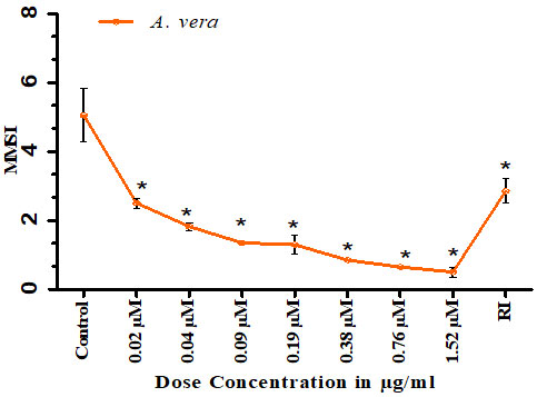
In the present study the crude extract of Aloe vera leaves showed a comparable powerful melanin aggregating effect on the scale melanocytes of zebrafish with concentration ranging from 0.02 to 1.52 µM in which all melanocytes had become perinuclear making the skin pale in colour when compared to untreated control. In response to the maximal concentration of 1.52 µM of the crude extract of Aloe vera leaves, the MMSI of the melanocytes found to be 0.5±0.14 as compared to the control value of 3.58±0.02 in which melanocytes were an intermediate state neither aggregated nor dispersed. After repeated washing and reimmersion (RI) of the Aloe vera extract treated melanocytes in fish physiological saline, it was found that the powerful melanin aggregation effects of the extract completely vanished, as the MMSI became 2.95±0.07 (Fig: 1). It was found that the crude extract of Aloe vera leaves is a powerful melanolytic agent in the scale melanocytes of zebrafish as compared to the control.
Figure 1: Dose-response curve for the melanocytes aggregating effect of crude extract of Aloe vera leaves.
RI signifies the MMSI after the reimmersion of scale melanocytes of zebrafish in physiological saline
after repeated washing. Vertical bars represent the standard error of mean p value <0.05.
The distribution and synthesis of melanin by melanocytes in the epidermis determine skin pigmentation. However, an excess of melanin can cause a number of hyperpigmentary skin diseases that can seriously impair appearance (Mort et al., 2015). Numerous research studies have examined the application of natural products in cosmetics and medicine (Mahor and Ali, 2018). Thus, our aim was to identify new agents from natural sources that can regulate melanogenesis.
As there are no reported inhibitory effects of crude extract of Aloe vera on the skin melanocytes of zebrafish in literature, this study is the first one to elucidate the effect of melanin aggregation by crude extract of Aloe vera, induced skin lightning for creating new depigmentation substances for the treatment of hyperpigmentary disorders and to make the skin a shade lighter.
Our results indicated that, crude extract of Aloe vera leaves exerts significant skin lightening response in dose-dependent manner in the isolated scale melanocytes of zebrafish. It was found that crude extract of Aloe vera leaves aggregated all the isolated scale melanocytes of zebrafish significantly in a dose dependent manner (0.02 to 1.52 µM). At lower concentration, minimal effect of aggregation was perceived under microscope. On increasing concentration, more aggregation effect of melanocytes was observed. Isolated scale melanocytes treated with 0.76 µM of crude extract of Aloe vera showed high aggregation of melanocytes as compared to control. More dense aggregation was observed at 1.52 µM concentration of crude extract of Aloe vera.
At higher concentration melanocytes had become perinuclear making the skin pale in colour. Our findings are substantiated with the findings of Choi et al. (2002) who showed that aloesin from Aloe vera showed significant depigmentation property in human skin exposed to ultraviolet radiation. In our study, scales of zebrafish have been used to observe the aggregation pattern of melanocytes. This method is the preliminary effective method as used in our previous study (Ali and Naaz, 2014; Ali et al., 2012; Ali et al., 2023) where skin melanophores of Bufo melanostictus were used to study the skin pigmentation effect of berberine bioactive compound.
The results indicated that the crude extract of Aloe vera leaves aggregated the melanocytes dispersion effect on the isolated scale melanocytes of zebrafish (Danio rerio). In this study, we reported that Aloe vera induced powerful dose dependent physiologically significance melanin aggregation in the isolated scale melanocytes of zebrafish leading to lightening of the skin. The data of the present study clearly demonstrate that A. vera can have clinical application as nontoxic melanolytic agents for the treatment of hyper pigmentation.
CONCLUSION
It is concluded that the leaf extract of Aloe vera induced powerful, dose-dependent, physiologically significant melanin aggregating effects in the isolated scale melanocytes of zebrafish (Danio rerio). This was confirmed by using control untreated embryos. It was observed that crude extract of Aloe vera leaves significantly aggregates melanin and make the zebrafish skin pale. Our study opens new vistas for the use of Aloe vera regarding its clinical application as a new nontoxic melanolytic agent for the treatment of hyperpigmentation.
ACKNOWLEDGEMENT
The authors are thankful to the Secretary and Principal of Saifia Science College, Bhopal, India for providing the necessary facilities
Consent for publication: Not applicable.
Availability of data and material: All the data generated and analyzed during the study are included in the main manuscript.
Competing interests: The authors declare that they have no competing interests.
Funding: NA
REFERENCES
Agalou, A., Thrapsianiotis, M., Angelis, A., Papakyriakou, A., Skaltsounis, A. L., Aligiannis, N., Beis, D. (2018). Identification of novel melanin synthesis inhibitors from Crataegus pycnoloba using an in vivo zebrafish phenotypic assay. Frontiers In Pharmacology, 9, 1-13.
Ali SA (1983) PhD Thesis Physiology and pharmacology of isolated teleost fish melanophores Bhopal University Bhopal India 1-298
Ali SA and Parveen N. (2021) Alteration in Melanogenesis: Pigmentary Disorders and their Etiopathogenesis. Book Chapter, Pp:40-56 (17), Volume 1. Bentham Science Publishers DOI: 10.2174/9789811491580121010005.
Ali, SA., & Naaz, I. (2014). Comparative light and electron microscopic studies of dorsal skin melanophores of Indian toad, Bufo melanostictus. Journal of Microscopy and Ultrastructure, 2(4), 230-235.
Ali, SA., Galgut, J. M., & Choudhary, R. K. (2012). On the novel action of melanolysis by a leaf extract of Aloe vera and its active ingredient aloin, potent skin depigmenting agents. Planta Medica, 78(08), 767-771.
Ali, SA., Galgut, J. M., & Choudhary, R. K. (2012). On the novel action of melanolysis by a leaf extract of Aloe vera and its active ingredient aloin, potent skin depigmenting agents. Planta medica, 78(08), 767-771.
Ali, SA., Husain, T., Kassab, G., & Khan, D. (2023). Morpho-anatomical analysis of zebrafish scale melanocytes. Journal of Survey in Fisheries Sciences, 987-991.
Ali, SA., Sultan, T., Galgut, J. M., Sharma, R., Meitei, K. V., Ali, A. S. (2011). In vitro responses of fish melanophores to lyophilized extracts of Psoralea corylifolia seeds and pure psoralen. Pharmaceutical Biology, 49(4), 422-427.
Ali, SA., (2017). Recent advances in treatment of skin disorders using herbal products. Journal of skin, 1(1), 6-7.
Bhattacharya SK, Parikh AK, Das PK (1976). Effect of acetylcholine on melanophores of Rana tigrina.Ind J Experimental Biol 32: 1039–1040.
Choi, S., Park, Y. I., Lee, S. K., Kim, J. E., & Chung, M. H. (2002). Aloesin inhibits hyperpigmentation induced by UV radiation. Clinical and experimental dermatology, 27(6), 513-515.
Hogben LT, Slome D (1931). The pigmentary effector system VI. The dual character of endocrine co-ordination in amphibian colour change. Proc R Soc London; 108: 10–53
Jang DK, Jung SH, Jeong JH, Yoo HM, Lee IS, Shin HS (2020). The Antimelanogenic Effect of Inularin Isolated from Flowers of Inula britannica on B16F10 Melanoma Cells and Zebrafish Embryos. J Microbiol Biotechnol. May 28;30(5):749-752. doi: 10.4014/jmb.2003.03025. PMID: 32482941.
Kim, K. N., Yang, H. M., Kang, S. M., Ahn, G., Roh, S. W., Lee, W., … & Jeon, Y. J. (2015). Whitening effect of octaphlorethol A isolated from Ishige foliacea in an in vivo zebrafish model. Journal of Microbiology and Biotechnology, 25(4), 448-451.
Le, D. D., Lee, Y. E., & Lee, M. (2022). Triterpenoids from the leaves of Osmanthus fragrans var. aurantiacus with their anti-melanogenesis and anti-tyrosinase activities. Natural Product Research, 1-7.
Mahor G and Ali SA. (2016) Recent update on the medicinal properties and use of Aloe vera in the treatment of various ailments. Bioscience Biotechnology Research Communication. 9(2):273-288.
Mahor, G., & Ali, S. A. (2018). Protective effects of Aloe vera extract on aluminium sulphate induced alterations in serum lipid profile of male albino rats, Rattus norvegicus. Bioscience Biotechnology Research Communications, 11(4), 727-733.
Mahor, G., Ali, S. A., & Parveen, N. (2019). Aloin from Aloe vera leaves: A potential natural aluminium detoxificant. Biosci. Biotechnol. Res. Commun, 12, 531-538.
Mort, R. L., Jackson, I. J., and Patton, E. E. (2015). The melanocyte lineage in development and disease. Development 142, 620–632. doi: 10.1242/dev.106567.
Sánchez-Machado, D. I., López-Cervantes, J., Mariscal-Domínguez, M. F., Cruz-Flores, P., Campas-Baypoli, O. N., Cantú-Soto, E. U., & Sanches-Silva, A. (2017). An HPLC procedure for the quantification of aloin in latex and gel from Aloe barbadensis Journal of chromatographic science, 55(3), 251-257.
Singh, K. G., Umme Umaima, S., Jangid, K. (2019). In vivo study of Depigmentation Using Tyrosine Ammonia Lyase from Trigonella Foenum-graecum on Zebrafish Embryos. International Journal of Current Research and Review, 11(08), 13-16.

