1Department of Electronics and Communication Engineering, M.Kumarasamy College of Engineering, Karur, India – 639 113
2Department of Mathematics, Bannari Amman Institute of Technology, Erode, India – 638 401.
3Department of Electronics & Communication Engineering, Dr.N.G.P. Institute of Technology, Coimbatore, India – 641 048
Corresponding author email: sakkthisudhan@gmail.com
Article Publishing History
Received: 11/05/2020
Accepted After Revision: 15/06/2020
The study of the mammogram datasets of computer aided design to deploy it on the social networks for the decision-making retrieval report to medical professional is presented in this paper. The existing radiology diagnosis system to integrate with social network has investigated on it. The CAD prototypes of mammogram images have generated by MATLAB such as data acquisition, pre- processing, segmentation, feature extraction and classification. These prototypes deployed by IoT analytics platform (ThinkSpeak) service. This platform generated the instant visualization of live excitations/information which alters to web service. This IoT platform integrates with MATLAB. Hence, these retrieval reports communicate to medical professionals through Twitter/Twilio. The breast cancer is one of the major health diseases for human society. The prevention for the major stage of cancer from human body and exchange the analytical report on medical professions through the social media as like Twitter/Twilio is delivered into this paper. These clinical trials provide the complete the clinical reports, which includes the statistical distribution of cancerous organ. These analytical reports on cancerous organ image has fixed for 81.25% of threshold value. Because Gaussian rules based Gabor filter adapted for pre-processing. Therefore, the smart phone based digitalized mammogram, which demonstrate the image retrieval, classification of trails and display toolset with covers the basic radiology function of today’s medical requirements. These clinical reports fulfilments of medical professionals to analyse the quick suggestion
Breast Cancer, IoT analytics, Instant Visualization, mammoTWEET
Mohanraj S, Vinothini V. R, Sakthisudhan K. MammoTWEET: A Novel Approach for Digitalized Retrieval Report of Breast Cancer for Medical Professionals. Biosc.Biotech.Res.Comm. 2020;13(2).
Mohanraj S, Vinothini V. R, Sakthisudhan K. MammoTWEET: A Novel Approach for Digitalized Retrieval Report of Breast Cancer for Medical Professionals. Biosc.Biotech.Res.Comm. 2020;13(2). Available from: https://bit.ly/2WRpZNs
Copyright © Mohanraj et al., This is an open access article distributed under the terms of the Creative Commons Attribution License (CC-BY) https://creativecommns.org/licenses/by/4.0/, which permits unrestricted use distribution and reproduction in any medium, provide the original author and source are credited.
INTRODUCTION
Breast cancer is the second most common tumour in world and more prevalent in the women population, not only a women disease its present in men even. Since the origin of disease remains unclear, early detection and diagnosis is the optimal solution to prevent tumour progression and allow a successful medical intervention, protect lives and reduce cost. It is identified by using the x-ray mammography technique with the absence of signs. Tiny cancer cells are identified without any symptoms before undergoing x-ray (Brzakovic et al., 1990 Nassar et al., 2017, Sivaranjani 2019).
Early stage cancer is identified in mammography. In the early stage, pre-diagnosing tools are mandatory to identify the cancer cells. The mammography marks abnormal growth of cells exactly from the beginning point of cancer. Even tiny portion of abnormal tumours are also detected in this methodology. Breast cancer can occur in both genders, it can affect any parts in the body. Mainly it affects women, and severe stages it leads to death. Early detection of breast cancer increases the survival rate whereas late diagnosis results in patient to a critical stage and later to death. Digital Mammograms are the images of breast, which are used to find potential signs of breast cancer like tumours and abnormal changes in the skin (Siewertsz et al., 2016, Nassar et al., 2017, Sivaranjani 2019).
Based on the American Cancer Society (ACS) report, more number of women may affect breast cancer, considered the largest disease and it leads to death USA. Suffering from breast cancer usually increases with age. The cancer cells are initially found with X-ray Mammography and this technique is accepted as the suitable imaging technique to detect breast cancer. But the methodology is not producing better results for the patient with dense breasts and also it interpret patients to ionizing radiation. ACS recommended that a person with the family background or finding the symptoms related to breast cancer need to be checked every year, or at the age about 40, or every 10 years from they affected (Boyd et al., 2007). When viewing high risk of women, the overall detection rate of mammography is 36% and combined mammography and MRI detection rates 92.7% (Carkaci et al., 2011 Pradeep and Ribana 2018 Ribana and Pradeep 2018).
Automatic ultrasound imaging allows you to play image quality and reliability, and also eliminates user variability(Cheng et al., 2010). But it has some restrictions to use due to image resolution of scanners is limited (Kelly et al., 2010). Moreover, general practitioner should familiarize themselves with the interpretation of data sets. MRI process uses a larger range of magnets (3 to 5 Tesla) placed along with RF coils (Morris, 2003). MTI produces the 3D picture and locates the cancer cells. This method is too expensive because the Gadolinium is injected in the veins. So the person suffered from kidney disease should not undergo MRI scanning (Morris, 2003). Also, the person placed with Pacemaker or underwent any metal implant surgery are not recommended to take MRI (Morris, 2001).
MATERIAL AND METHODS
The worldwide researcher had authorized the Magnetic Resonance Imaging (MRI) for breast cancer diagnosis application. It is one of the drawback, which follows difficult to indiscriminate the normal cells from the cancerous organ (Nassar et al., 2017). Moreover, the existing methods classified the different kinds of analytical and numerical methods. The proposed methodology is implemented by following major four steps such as, Image Acquisition with display module, image transfer/storage, software development module and report retrieval module. The clinical images of mammogram collected from MIAS (Mammogram Image Analysis Database). These data sets translated into the matrix using 32 bit pixel depths such as 512 grey scale images. These image rescaled to smart phones. These data set can be varied from the colour brightness, quality of smart phone. These personal assistant devices connected with web servers via Internetworking Protocol (IP).
The PACS provide the image transferred to mobile phones over Transmission Control Protocol/Internetworking Protocols (TCP/IP). The proposed application implemented to DICOM receiver over HTTP (Hyper Text Transfer Protocol). Therefore, the web browser read the image of BMP format. It can be stored and delivered based on usage of network services such as, HSDPA (High Speed Downloading Packet Access), EDGE, WiFi (Wireless Fidelity), Bluetooth, GPRS (General Packet Radio access Service), or any other external storage devices such as, removable memory card, pen drive and HDD (Hard derive disk).
The software development module consists of image processing and tumour detection with their classification units. The BMP image is stored in the Android operating system from mobile browser. The DICOM receiver offers the read and write operations for image files. It also capable of offered with web browsers for dynamic tasks such as, rescaling of images, panning image, User Interface (UI), Scalable Vector Graphics (SVG), HTML and interacting huge web pages and their applications. The tumour detection and their classification implemented by the CAD system. At instant, radiologist reviewed the clinical trials and their final report interconnected with medical professional via smart phones with encrypted key. They may be investigated the further medical suggestion. Therefore, the feedback reports interacted with patient through their own social media’s accounts. The proposed system is able extended for continuous monitoring to patient, readiness for further medical processing and make awareness in risky factor of breast cancer.
Figure 1: Functional flow
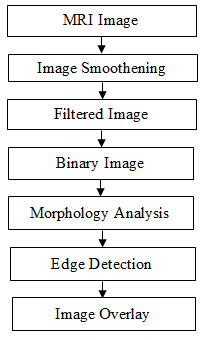
Image processing is a tool for extracting information from a captured digitized image with some mathematical operations on it. Usually image is a two dimensional signal, while performing mathematical operations the useful information can be retrieved. To enhance the quality of MRI images proper denoising filter is to be used. Linear and nonlinear filtering techniques are commonly used on MRI image to increase the quality (Pradeep and Ribana 2018 Ribana and Pradeep 2018).
The Gaussian rule based Gabor filter is used to remove the noises. Little smoothing is done when the larger variance and for smaller variance smoothing is done with preserving edges. Contrast Limited Adaptive Histogram Equalization (CLAHE) algorithm is used to enhance the contrast in medical images. The algorithm is applied on smaller data region of images to improve contrast. The bilinear interpolation is incorporated to connect the neighbouring pixels (Ribana and Pradeep 2018). Sobel edge detection method is used for identifying the edges of tumours. Finally the tumours are detected with the help of Matlab, the identified cancer cells are tweeted to the doctors and care takers immediately to know the severity of the tumour. The flow diagram is depicted in figure 1.
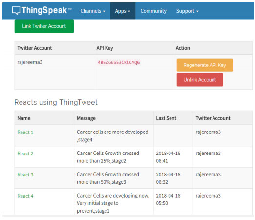
Thing Speak is mainly developed to visualize the data from IoT platform. It is also more helpful to pre-processing the data and analytics (Sivaranjani 2019). It a cloud based platform build on Ruby and helps the researchers and prototyping hobbyist to build their internet of things applications with wired or wireless connectivity. It collects the data from third-party resources and visualize instantly. The ThingSpeak helps the designers to run their applications from the cloud on time bases and it also update the received the information from cloud to Twitter or Twilio. Once the cancer cells are detected via the Matlab, the Thingspeak automatically tweet the information to the doctors and care takers.
RESULTS AND DISCUSSION
Figure 2: The edge detected image and percentage of cancer cells occupied in the cancerous organ
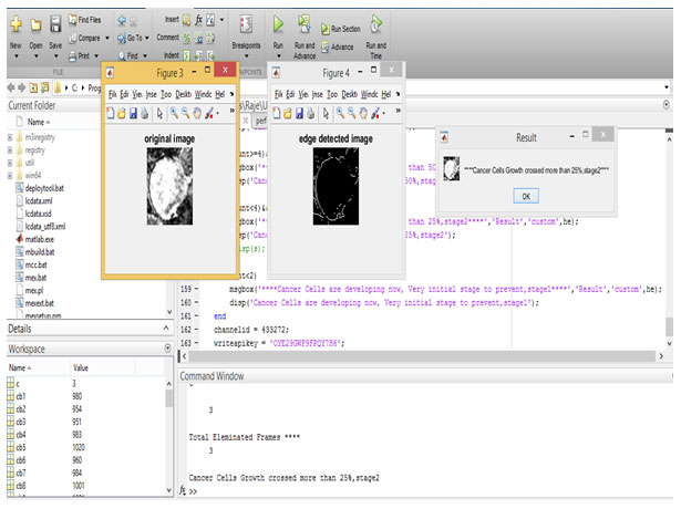
Figure 3: The numerical report simulates to Tweeter social media

Figure 4: Numerical analysed report from Thingspeak with Twitter/Twilio for percentage of the cancer cells with respect to different stages of the cancer
In this proposed system is a mobile communication interacted with web application. Hence, it called as MammoTweet, which uses in the smart personal assistant device for radiologist, clinician and surgeon. It considers the less time computation, technical specification and ability of clinical reports. The MIAS datasets implemented with smart phones and Tweetspeak Apps. These clinical trials based on carcinogen death rate. Hence, it is considered as real time clinical trials. It is to prevent the cancer at early stage. It is to reduce the expenditure of clinical trials since, it provides depth analytical survey about the every trails. Moreover, it is preventing survive of 90% patient life time. Hence, it connected to eHealth care profession.
CONCLUSION
The breast cancer is one of the major health diseases for human society. The prevention for the major stage of cancer from human body and exchange the analytical report on medical professions through the social media as like Twitter/Twilio is delivered into this paper. These clinical trials provide the complete the clinical reports, which includes the statistical distribution of cancerous organ. These analytical reports on cancerous organ image has fixed for 81.25% of threshold value. Because Gaussian rules based Gabor filter adapted for pre-processing. Therefore, the smart phone based digitalized mammogram, which demonstrate the image retrieval, classification of trails and display toolset with covers the basic radiology function of today’s medical requirements. These clinical reports fulfilments of medical professionals to analyse the quick suggestion.
References
Boyd, N.F Helen Guo, Lisa J. Martin (2007) Mammographic density and the risk and detection of breast cancer, New England Journal of Medicine, 356(3), pp. 227-236.
Brzakovic, D. Luo, X. M. and Brzakovic, P.(1990) An approach to automated detection of tumors in MRI’ IEEE Transactions on Medical Imaging, 9, pp. 233-241.
Carkaci, S. Santiago, L. Adrada, B.E. Whitman, G.J. (2011) Screening for breast cancer with sonography Seminars in Roentgenology, 46(4), pp. 285-291.
Cheng, H.D. Shan, J. Ju, W. Guo, Y. Zhang, L. (2010) Automated breast cancer detection and classification using ultrasound images: A survey Journal of Pattern Recognition, 43(1), pp. 299-317.
Kelly, K.M. Dean, J. Comulada, W.S. Lee, S.J. (2010) Breast cancer detection using automated whole breast ultrasound and mammography in radiographically dense breasts’, European Journal of Radiology, 20(3), pp. 734-742.
Morris, E.A. (2001) Review of breast MRI: Indications and limitations Seminars in Roentgenology Journal, 36(3), pp. 226-237.
Morris, E.A. (2003) Screening for breast cancer with MRI Journal of Seminars in Ultrasound, CT, and MRI, 24(1), pp. 45-54.
Nassar, F.J. Nasr, R. Talhouk, R. Micrornas, (2017) As biomarkers for early breast cancer diagnosis, prognosis and therapy prediction Pharmacology & Therapeutics, 172, pp. 34–49.
Pradeep, S. Ribana, K. (2018) Wavelet based medical image denoising technique based on neural network Bioscience Biotechnology Research Communications, 11(2), pp. 82-87.
Ribana, K. Pradeep, S. (2018) Contrast enhancement for magnetic resonance imaging Bioscience Biotechnology Research Communications, 11(2), pp. 1-4.
Siewertsz, V. Reesema, L.L. Lee, M.P. Zheleva, V. (2016) RAS pathway biomarkers for breast cancer prognosis Clinical laboratory international, 40, pp. 18–23.
Sivaranjani, S. (2019) Priority Aware Medical EEG Data Transmission Using Cognitive Radio Network International Journal of Control and Automation, 12(6), pp. 364-370.

