Department of Restorative Dentistry, College of Dentistry King Saud University, Riyadh, Saudi Arabia. 11545.
Corresponding author email: kalahdal@ksu.edu.sa
Article Publishing History
Received: 11/02/2020
Accepted After Revision: 25/04/2020
The present study was designed to evaluate shear bond strength of dentin when bonded with self- etch and self- adhesive resin cements with ErCrYSGG laser (ErCrY) and Er-YAG (ErY) conditioning. Ninety extracted molar teeth were mounted and allocated into nine groups (n = 10) according to the dentine surface conditioning and type of cement. Three type of cement, Panavia, Rely-X and Maxcem were used in the study for comparison among the laser conditioning SBS values. All specimens were tested for shear bond strength using universal testing machine. Ten samples from each group were assessed for modes of failure. Data were assessed using analysis of variance and Tukey multiple comparisons test. The highest mean shear bond strength was observed in conventional treatment with Panavia cement application (21.51 (± 2.13) MPa) whereas the least mean shear bond strength was measured in Er-YAG conditioning along with Maxcem cement application (14.89 (± 3.48) MPa). The hypothesis was partly accepted as the influence of conditioning methods among different cements was comparable, except Panavia cement (p >0.05). Moreover, the SBS values were significantly influenced by the type of resin cement rather than the type of laser used in dentinal conditioning (P < 0.001). Among all groups the most common type of observed failure was adhesive. The study was revealed that the type of Self-etch and self-adhesive cements exhibited significant influence on their bond strength to laser treated (ErCrYSGG and Er-YAG) dentin compared to the type of surface conditioning.
Bond strength; ErCrYSGG ; ErYAG; Self Etch; Self Adhesives; Resin Cement.
Al-Ahdal K. Influence of Er Cr Ysgg, Er Yag and Conventional Treatment on the Shear Bond Strength of Self Etch and Self-Adhesive Resin Cements. Biosc.Biotech.Res.Comm. 2020;13(1).
Al-Ahdal K. Influence of Er Cr Ysgg, Er Yag and Conventional Treatment on the Shear Bond Strength of Self Etch and Self-Adhesive Resin Cements. Biosc.Biotech.Res.Comm. 2020;13(1). Available from: https://bit.ly/2RbE8n2
Copyright © Al-Ahdal, This is an open access article distributed under the terms of the Creative Commons Attribution License (CC-BY) https://creativecommons.org/licenses/by/4.0/, which permits unrestricted use distribution and reproduction in any medium, provide the original author and source are credited.
INTRODUCTION
In the field of restorative dentistry, steps have been taken to simplify the use of adhesive procedures. Initially, a conventional adhesive restoration technique, a standard procedure was employed; however, due to an increased risk of excess demineralisation and salivary contamination these procedures evolved over time. Recently, self- etch, self-adhesive resin cements are introduced for the purpose of easy handling and quick bond formation (Durski et al., 2016). Using the multipurpose system presents with an opportunity for a positive bond formation in a restorative retention. Several studies demonstrated the dentin adhesive bond formation to be considered as a major factor influencing the success of restorative retention (Esteves-Oliveira et al., 2007, Gulec et al., 2018). The self -etch self- adhesive cement exhibited reduction in the need for pre-treatment of dentinal surface, minimises the procedure time and technique sensitivity; however, its viscosity limits decalcification and deep penetration into dentin, which result in compromised bond formation (Ferreira-Filho et al., 2018).
With the advancement in technology and change in the perceptions, minimal invasive treatments are implemented in current practices. Recently, the use laser technology has been introduced to enhance the dentin surface adhesion to the resin cement (Gulec et al., 2018). A contemporary method to enhance bond strength is the use of high intensity lasers. Erbium: yttrium-aluminum-garnet (ErYAG) and erbium, chromium: yttrium-scandium-gallium-garnet (Er, Cr: YSGG) belonging to the Erbium family have an ability to ablate the hard tissues without thermal damage, reduces dentinal hypersensitivity and exhibit bactericidal property (Bandéca et al., 2012, Acar et al., 2014). The use of these lasers increases the resistance of the dentine surface against a caries progression developing under the restoration (Harorli et al., 2015, Giray et al., 2014). The dentinal conditioning has exhibited a positive bond formation at the dentinal interface with the resin cement. The phototherapy causes the hydroxyapatite crystal as well as water to absorb wavelength energy of the laser (Er:YAG, λ= 2.94 m; Er,Cr:YSGG, λ=2.87 m) resulting in the dentin ablation and water evaporation forming imbricate irregularities on the dentin surface. The topographical change along with clearance of the smear layer initiate favourable condition for adhesive bond between resin and dentin (Esteves-Oliveira et al., 2007, Lukac et al., 2016). Moreover, phototherapy conditioned dentinal surface led to open dentinal tubules and peritubular dentin rather than intertubular dentin (Gulec et al., 2018).
Previous studies suggest that the laser irradiation modifies the dentin surface cultivating susceptible conditions for effective adhesive bond between resin and dentine (Ramos et al., 2015, Aras et al., 2016). In addition laser parameters including, wavelength, pulse-energy and power are a critical factor, in dentin conditioning outcomes for adhesive bonding to resins (Ramalho et al., 2015, Ferreira-Filho et al., 2018, Lopes et al., 2015). Esteves-Oliveira et al proclaimed that the use of self-etch adhesives is influenced by the type of erbium laser as the shear bond strength of resin to dentine was greater in the Er: YAG-laser conditioned surface compared to the Er, Cr:YSGG-laser irradiated, (Esteves-Oliveira et al., 2007, David and Gupta, 2015).
This mounting interest in laser therapy encourages further investigation in laser conditioning using different type of self- etch and self -adhesive resin cements. It is hypothesized that use of Er, Cr:YSGG and Er: YAG laser for dentin conditioning presents with comparable self-etch, self- adhesive resin cement bond strength outcomes to conventional conditioning techniques. Therefore, the present study was designed to evaluate shear bond strength of dentin when bonded with self- etch and self- adhesive resin cements with ErCrYSGG laser (ErCrY) and Er-YAG (ErY) conditioning.
MATERIAL AND METHODS
The project was approved by the institutional research review board. The study compared the shear bond strength of dentin when bonded with self- etch and self- adhesive resin cements with ErCrYSGG laser (ErCrY) and Er-YAG (ErY) conditioning. A total of 90 extracted molars, with no caries, restoration or fracture were selected and stored in thymol solution (0.01%) for 1 week. Each tooth was cleansed using the chlorohexidine to remove the debris, calculus and plaque before sample preparation. The samples were shifted temporarily into a jar of distilled water at 4°C.
All the specimen roots were sectioned using a diamond saw (Leitz 1600, Wetzlar, Germany) at 2mm below the cemento-enamel junction. The teeth were then mounted in acrylic resin (Meliodent; Kulzer, Hanau, Germany) and the occlusal enamel was removed to expose the mid coronal dentine.
Dentine surface preparation: Each specimen was cut by the diamond saw (Leitz 1600, Wetzlar, Germany) to prepare an area of 4 mm in the mesiodistal plane followed by 400 – 600 grit carbide paper (Buehler) polish under a water coolant spray. Using the stereomicroscope the surface was closely examined for any enamel residue or pulpal tissue exposure. The total 90 extracted tooth was now divided into three categories: control (no laser treatment – standard bur cut with cylinder diamond burs with medium-sized particles), ErCrYSGG (ErCRY) laser and ErYAG (ErY) laser application.
Composite disc preparation: A putty mould (Easy Composites’ Uni-Mould system, UK) combined with a wax disc was prepared with dimension of 2 mm diameter and 3mm height. Resin composite disc was prepared with a similar dimension of 2mm x 3 mm using technique of placing the putty mold on a glass slide and packing the material (MultiCore Flow bulk-fill composite Ivoclar-Vivadent, Schaan, Liechtenstein) in a mould. The excess was removed using the hand instrument and the disc was photo polymerised using the light source (Bluephase G2; Ivoclar-Vivadent) for 40 sec from top to bottom through another glass placed on the mould. The composite discs were finished followed by polishing and measured for required dimensions.
Sample preparation: The dentinal surface in the all specimens were polished with a 600 grit silicon carbide waterproof abrasive paper before dentinal conditioning, creating an area of 4 mm in diameter under a water coolant spray. The samples were divided randomly into three groups, conventional, ErCrYSGG and ErYAG laser (n=30). Following the distribution experimental groups abide by the subsequent conditioning protocol:
Group 1: Conventional treatment:Specimens were conditioned using a diamond bur with medium sized particles to flatten the dentine followed by polishing. No laser treatment was applied on the dentine.
Group 2: ErCr laser: Dentinal surface was conditioned by Er,Cr:YSGG (Waterlase C100; BioLase Tech, Inc., CA) laser power 4.5 W, wavelength of 2780 nm and frequency 20 Hz in a noncontact mode from a distance of 2 mm using tip (MZ = 8, 6 mm) for a interval of 60 sec.
Group 3: ErY laser: Dentinal surface was conditioned by the ErYAG laser (Kavo Key Laser 3, Kavo Dental GmbH & Co. KG,). The laser frequency 6 Hz, with a working distance of 20 mm, wavelength 2940 nm and directing 300 mJ per pulse.
After the tooth dentin conditioning, each group consisting of 30 specimens was distributed further into three sub groups based on the type of cement applied.
Group A: Panavia 2.0
Group B: RelyX Unicem
Group C: Maxcem
Total of 9 groups were created as a result as shown below:
Gp A1- Control Gp A2- ErCr laser Gp A3- ErY laser
Gp B1-Control Gp B2- ErCr laser Gp B3-ErY laser
Gp C1-Control Gp C2- ErCr laser Gp C3- ErY laser
Each dentinal surface was smeared with a particular cement according to manufacturer’s instructions and composite discs were cemented under a constant load of 15kg for 30 secs. The excess cement was removed using a micro brush. The cement layer was polymerized from all sides for 20 seconds (80 seconds in total). After completion of the process, tooth specimens underwent thermocycling for 30,000 cycles, under temperature of 5 to 55 ºC with a dwell time of 30 sec in (Thermocycler (GMBH, Miebacher Strabe, Germany) and then stored in humid conditions (Incubator, Memmert Universal Oven, Germany) at 37 ºC. The shear bond strength (SBS) was evaluated using universal testing machine (Instron 8500 Plus, Canton) through the controlled application of force at a cross-head speed of 0.5mm/min, resulting in fracture at the cement interface. The chisel-shaped probe was applied parallel to the interface on the composite disc until fracture. Shear bond strength was expressed in Megapascals (MPa). The fractured surface (bonding interface) was evaluated through the stereomicroscope and classified according to the type of failure; adhesive (the interface between dentine and cement), cohesive (within the material) and admixed (cement partly remains on dentine interference).
The normality of data was assessed using Kolmogorov-Smirnov test. Mean and standard deviations (SD) of the observed data were assessed using descriptive statistics. Comparison of means and SD was performed with ANOVA and Multiple comparisons tests (Tukey-Kramer). Statistical software for social sciences (SPSS 20.0 version) was employed and p <0.05 was considered statistically significant.
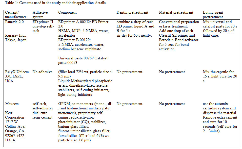
Table 1: Cements used in the study and their application details
RESULTS AND DISCUSSION
The Kolmogorov-Smirnov test displayed the normality of the data to be distributed evenly. The study groups without laser conditioning are designated as A1, B1, and C1 for the Panavia, RelyX Unicem and Maxcem cement, respectively. The laser conditioning, ErCrY laser and ErYAG laser study groups were classified as A2, B2, C2 and A3, B3, C3 for the three types of cement (Panavia, RelyX Unicem and Maxcem), respectively. The highest mean shear bond strength was observed in conventional treatment with Panavia cement application (21.51 (± 2.13)) whereas the least mean shear bond strength was measured in Er-YAG conditioning along with Maxcem cement application (14.89 ( ± 3.48)). Table 2 presents the mean and standard deviation of shear bond strength measured in each study group. Analysis of variance
(ANOVA) was performed among the study groups exhibiting statistical differences in the calculated mean SBS value (p-value <0.05) (table 2).
Table 2: Mean and standard deviation with three different types of cement-based on three different dentine conditioning methods.
Note: Dissimilar superscript small alphabets in same row denote significant difference in dentinal conditioning (p<0.05). Dissimilar superscript capital alphabets in same column denote significant difference in a different types of cement (p<0.05). * denotes a significant
Table 2: Mean and standard deviation with three different types of cement-based on three different dentine conditioning methods.
| Cement Type | No laser
(Control) |
Er-Cr-YSGG | Er-YAG | ANOVA
P value |
|
| Panavia | 21.51 (2.13)a A* | 19.16 (1.70)ab A | 18.43 (1.36)b A | <0.001 | |
| Rely-X | 18.24 (3.66)a AB | 17.39 (2.11)a AB* | 17.54 (3.61)a AB* | ||
| Maxcem | 16.40 (1.89)a B | 15.71 (2.26)a B | 14.89 (3.48)a B |
(ANOVA) was performed among the study groups exhibiting statistical differences in the calculated mean SBS value (p-value <0.05) (table 2).
Note: Dissimilar superscript small alphabets in same row denote significant difference in dentinal conditioning (p<0.05). Dissimilar superscript capital alphabets in same column denote significant difference in a different types of cement (p<0.05). * denotes a significant difference between the two different study groups.
Two – way ANOVA and tukey HSD Post-hoc test revealed that the SBS strength is significantly influenced by the type of resin cement rather than a type of laser used in dentinal conditioning (P < 0.001)(table 2). Except for panavia, which successfully presented with a significant difference between the control group and Er-YAG conditioning (p<0.036); however, similar results were appreciated with the ERYSGG group. As for the Rely X and Maxcem, the results were comparable, each group showing no evident significance (p >0.05).
A comparison between the three types of cement exhibited a significant difference between Panavia and Maxcem in each type of conditioning. Nevertheless, Rely X presented with comparable results with no significance in the study groups (p >0.05). However, tukey HSD Post-hoc test demonstrated that study groups under different circumstances also displayed a significant difference in the mean value (p<0.05) that includes an evident comparable result between panavia control to Rely X Er- YAG and Rely X Er- YSGG.
The failure modes assessment pointed out that Adhesive failure was more evident among the study group compared to cohesive and admixed failure. 100% adhesive failure was observed in the Rely-Er-Cr-YSG, Max-Control and Max-Er-Cr-YSGG groups. Cohesive failure was appreciated only in panavia control group (n=1). Admixed failure ranged from 20 – 40 % mainly among four study groups; Pan control, Pan-Er-YAG, Rely-Er-YAG and Max-Er-YAG. The analysis of the failure mode indicated Er-Cr-YSGG and Er-YAG conditioned exhibited adhesive failures compared to conventional treatment. However, all the cement study groups equally demonstrated adhesive failure except panavia control, which exhibited equal failure of adhesive and admix failure (4 specimens each) and 1 failure of cohesive type (table 3).
Table 3: failure mode between study groups
| Study Groups | Adhesive | Cohesive | Admixed |
| Pan-Control | 40 | 10 | 40 |
| Pan-Er-Cr-YSGG | 70 | 0 | 30 |
| Pan-Er-YAG | 80 | 0 | 20 |
| Rely-Control | 60 | 0 | 40 |
| Rely-Er-Cr-YSGG | 100 | 0 | 0 |
| Rely-Er-YAG | 80 | 0 | 20 |
| Max-Control | 100 | 0 | 0 |
| Max-Er-Cr-YSGG | 100 | 0 | 0 |
| Max-Er-YAG | 80 | 0 | 20 |
The present study was based on the hypothesis, that use of Er, Cr:YSGG (30 Hz 4.5 W) and Er: YAG (6 Hz 2 W) laser for dentin conditioning will show comparable self-etch, self -adhesive resin cement bond strength outcomes to conventional conditioning techniques. The influence of conditioning methods among different cements was comparable, except Panavia cement. The study’s results revealed the formation of a strong shear bond strength depends upon the type of the cement used rather than the type of dentin conditioning. Therefore, the hypothesis was partly accepted in addition to a multitude of explanations can be provided in this regards including each step performed in the study for establishing a certain standard and shear bond failure in the specimens.
The use of universal testing machine to measure the shear bond strength, sets a peculiar standardization, consistency and homogeneity in the variable outcome (Jayasheel et al., 2017). To age the bonded specimen and simulate the oral functions, the specimen were placed in a thermocycler after cement application. As stated by Brunzel et al (2010) thermocycling creates favorable condition at the dentine bonding surface that instigates artificial ageing of the bonding system.The present study aimed to compare the effectiveness of dentinal conditioning between the conventional technique and two different types of lasers, ErCrYSGG and ErYAG. It is proposed that laser conditioning causes the water evaporation from the surface resulting in microburst activity with increased surface tension for bonding (Cassimiro-Silva et al., 2016, Gulec et al., 2018). The low organic content in peritubular dentin is easily opened up by the laser; thus, dentinal tubules produce a cuff like appearance around it (Samad-Zadeh et al., 2011).
In the present study, use of laser showed comparable outcomes of SBS to conventional conditioning technique. It is narrated that the laser conditioning facilitates the removal of the smear layer, which allows the formation of retention tags to improve the shear bond strength (Gulec et al., 2018). In addition, it is suggested that using conventional bur conditioning leads to the formation of a smear layer that limits the retention tags formation compared to laser conditioning, which ablates the interprismatic dentin and removes smear layer, permitting resin tags formation(Naranjo et al., 2015, Cassimiro-Silva et al., 2016).
Kiomarsi et al (2018), presented higher shear bond strength with Er YAG laser-treated dentin compared to the control group. The possible explanation for high SBS was the micro ablation of the dentine surface without any thermal damage to hard tissue and pulp; in addition, to antimicrobial activity (Kiomarsi et al., 2018). However, in the present study, only Panavia demonstrated similar results of high SBS in ErYAG treated dentine compared to the control group; however, other cement did not exhibit an evident difference. Appreciating the descriptive statistics, there was a slight difference between the mean values of the study groups; nevertheless, multiple comparison test proved no significant difference between conditioning treatment in Relay -X and Maxcem cement study groups. Therefore, the viscous nature of the cement can be deduced as a limiting factor for strong SBS observed. Furthermore, the slight difference in the mean value of the Er YAG and ErYSGG study groups indicates structural differences in ablated dentin after laser conditioning. As per Kiomarsi et al (2018), ErCrYSGG creates a scaled surface showing a thermal damaged surface because its wavelength is easily absorbed in the tissue leading to a rise in temperature. In contrast, Er YAG creates a surface with close resemblance to the conventional acid etched surface (Harorli et al., 2015).
In the current study, the effect of dentinal conditioning using three different types of cement; Panavia, Rely x and Maxcem was evaluated. The results displayed a significant difference between the SBS among Panavia and Maxcem specimens. However, SBS among the specimens of Rely-X and other cement groups was comparable. Naranjo, Ali, and Belles (2015) presented in their study no significance in the shear bond strength in self- etch self- adhesive resin cements. These authors recommended that conventional methods and total etch methods are more preferred compared to self- etch and self- adhesive cement. Furthermore, in the present study, no significant relation with Rely X was evident because of its low pH and low surface interaction, which leads to limited resin tags formation (Zidan et al., 2015). Nevertheless, statistical test revealed that if dentinal conditioning was varied in addition to change in cement preference, a significant difference was observed among the Panavia controls and Rely X.
Many studies’ have assessed the tensile bond strength that measures the adhesive nature of the luting cement (Brunzel et al., 2010, Souza et al., 2016, Sekhri et al., 2016). This study, on the contrary, evaluated shear bond strength; the analysis of shear bond indicates the strength of the adhesive bond in an altered configuration (Brunzel et al., 2010). Few authors have reported that cohesive failures are observed in cases of the shear bond test; however, the present study displayed adhesive failure in the majority of specimens (Brunzel et al., 2010, Souza et al., 2016). Despite the effort for dentin conditioning to make the surface more retentive, the self -etch and self – adhesive cements has a limited capacity to demineralized dentine. In addition, they displayed limited inflow of viscous cement leaving a thick area of collagen mesh resulting in 100% adhesive failure (Weiser and Behr, 2015, Aguiar et al., 2014). It is pertinent to mention that there was no evident difference between the three dentin conditioning groups suggesting ablated surface may not influence the interfacial bond between dentine and cement.
The outcomes of the study should be viewed in light of the possible limitations. The study observed an in-vitro design, with extracted teeth; in contrast, in-vivo, dentin shows fluid movement in and out of the tubules possibly challenging the adhesive bond. In addition, the variation in the morphology and composition of dentine along with directions and dimensions of dentinal tubules vary among the teeth employed in the in-vitro design. Despite these limitations, results produced were comparable to previous in vitro studies (Jayasheel et al., 2017, Cassimiro-Silva et al., 2016). However, in vivo studies under clinical conditions are essential to validate these outcomes. As conventional etch and rinse methods has shown a better dentine adhesion compared to the self- etch and self- adhesive cements (Kiomarsi et al., 2018). Therefore, to clinical trials assessing self-etch and total etch adhesives with laser treatments are recommended.
CONCLUSION
The adhesive bond strength of self-etch and self-adhesive resin cements was not influenced by the use of ErCrYSGG and Er-YAG laser for dentin conditioning, except Panavia. The type of Self-etch and self-adhesive cements exhibited significant influence on their bond strength to laser treated (ErCrYSGG and Er-YAG) dentin.
Conflict of interest statement: No conflict of interest
REFERENCES
Acar, O., Tuncer, D., Yuzugullu, B. & Celik, C. (2014). The Effect of Dentin Desensitizers and Nd: Yag Laser Pre-Treatment On Microtensile Bond Strength Of Self-Adhesive Resin Cement To Dentin. The Journal of Advanced Prosthodontics, 6(2):88-95.
Aguiar, T. R., André, C. B., Correr-Sobrinho, L., Arrais, C. A., Ambrosano, G. M. & Giannini, M. (2014). Effect of Storage Times and Mechanical Load Cycling On Dentin Bond Strength of Conventional and Self-Adhesive Resin Luting Cements. The Journal of Prosthetic Dentistry, 111(5):404-10.
Aras, W. M., Barroso, J. S., Blay, A., Rodrigues, J. A. & Cassoni, A. (2016). Er, Cr: Ysgg Laser Irradiation Influence on Y-Tzp Bond Strength to Resin Cement. Ceramics International, 42(12):13790-5.
Bandéca, M. C., Pinto, S. C. S., Calixto, L. R., Saad, J. R. C., Barros, É. L. D. & Shelb, A. (2012). Influence of Er, Cr: Ysgg Laser on Bond Strength of Self-Adhesive Resin Cement. Materials Research, 15(4):491-4.
Brunzel, S., Yang, B., Wolfart, S. & Kern, M. (2010). Tensile Bond Strength of a So-Called Self-Adhesive Luting Resin Cement to Dentin. Journal of Adhesive Dentistry, 12(2):143.
Cassimiro-Silva, P.F., Zezell, D.M., de Melo Monteiro, G.Q., Benetti, C., de Paula Eduardo, C. and Gomes, A.S.L. (2016, February). Evaluation of microshear bond strength of resin composites to enamel of dental adhesive systems associated with Er, Cr: YSGG laser. In Lasers in Dentistry XXII (Vol. 9692, p. 96920N). International Society for Optics and Photonics.
David, C. M. & Gupta, P. (2015). Lasers in Dentistry: A Review. International Journal of Advanced Health Sciences, 2(8):7-13
Durski, M., Metz, M. J., Thompson, J., Mascarenhas, A., Crim, G., Vieira, S. & Mazur, R. (2016). Push-Out Bond Strength Evaluation Of Glass Fiber Posts With Different Resin Cements And Application Techniques. Operative Dentistry, 41(1):103-10.
Esteves-Oliveira, M., Zezell, D. M., Apel, C., Turbino, M. L., Aranha, A. C. C., Eduardo, C. D. P. & Gutknecht, N. (2007). Bond Strength Of Self-Etching Primer To Bur Cut, Er, Cr: Ysgg, And Er: Yag Lased Dental Surfaces. Photomedicine and Laser Surgery, 25(5):373-80.
Ferreira-Filho, R., Ely, C., Amaral, R., Rodrigues, J., Roulet, J., Cassoni, A. & Reis, A. (2018). Effect Of Different Adhesive Systems Used For Immediate Dentin Sealing On Bond Strength Of A Self-Adhesive Resin Cement To Dentin. Operative Dentistry, 43(4):391-7.
Giray, F. E., Duzdar, L., Oksuz, M. & Tanboga, I. (2014). Evaluation of the Bond Strength Of Resin Cements Used To Lute Ceramics On Laser-Etched Dentin. Photomedicine and Laser Surgery, 32(7):413-21.
Gulec, L., Koshi, F., Karakaya, İ., Yanardag, E. C. & Ulusoy, N. (2018). Micro-Shear Bond Strength of Resin Cements To Er, Cr: Ysgg Laser and/or Acid Etched Enamel. Laser Physics, 28(10):105601
Harorli, O., Barutcugil, C., Kirmali, O. & Kapdan, A. (2015). Shear Bond Strength Of A Self‑Etched Resin Cement To An Indirect Composite: Effect Of Different Surface Treatments. Nigerian Journal of Clinical Practice, 18(3):405-10.
Jayasheel, A., Niranjan, N., Pamidi, H. & Suryakanth, M. B. (2017). Comparative Evaluation of Shear Bond Strength of Universal Dental Adhesives-An In Vitro Study. Journal of Clinical and Experimental Dentistry, 9(7):e892
Kiomarsi, N., Arjmand, Y., Fard, M. J. K. & Chiniforush, N. (2018). Effects of Erbium Family Laser on Shear Bond Strength of Composite to Dentin after Internal Bleaching. Journal of Lasers in Medical Sciences, 9(1):58.
Lopes, R. M., Trevelin, L. T., Da Cunha, S. R. B., De Oliveira, R. F., De Andrade Salgado, D. M. R., De Freitas, P. M., De Paula Eduardo, C. & Aranha, A. C. C. (2015). Dental Adhesion to Erbium-Lased Tooth Structure: A Review of the Literature. Photomedicine and Laser Surgery, (8):393-403.
Lukac, N., Suhovršnik, T., Lukač, M. & Jezeršek, M. (2016). Ablation Characteristics of Quantum Square Pulse Mode Dental Erbium Laser. Journal of Biomedical Optics, 21(1):015012.
Naranjo, J., Ali, M. & Belles, D. (2015). Comparison Of Shear Bond Strength Of Self-Etch And Self-Adhesive Cements Bonded To Lithium Disilicate, Enamel And Dentin. Texas Dental Journal, 132(11):914-21.
Ramalho, K. M., Hsu, C.-Y. S., De Freitas, P. M., Aranha, A. C. C., Esteves-Oliveira, M., Rocha, R. G. & De Paula Eduardo, C. (2015). Erbium Lasers for the Prevention of Enamel and Dentin Demineralization: A Literature Review. Photomedicine and Laser Surgery, 33(6):301-19.
Ramos, T. M., Ramos-Oliveira, T. M., De Freitas, P. M., Azambuja, N., Esteves-Oliveira, M., Gutknecht, N. & De Paula Eduardo, C. (2015). Effects of Er: Yag And Er, Cr: Ysgg Laser Irradiation On The Adhesion To Eroded Dentin. Lasers in Medical Science, 30(1):17-26.
Samad-Zadeh, A., Harsono, M., Belikov, A., Shatilova, K. V., Skripnik, A., Stark, P., Egles, C. & Kugel, G. (2011). The Influence of Laser-Textured Dentinal Surface on Bond Strength. Dental Materials, 27(10):1038-44.
Sekhri, S., Mittal, S. & Garg, S. (2016). Tensile Bond Strength Of Self Adhesive Resin Cement After Various Surface Treatment Of Enamel. Journal of Clinical and Diagnostic Research: Jcdr, 10(1):ZC01.
Souza, E. M., De Munck, J., Pongprueksa, P., Van Ende, A. & Van Meerbeek, B. (2016). Correlative Analysis of Cement–Dentin Interfaces Using an Interfacial Fracture Toughness and Micro-Tensile Bond Strength Approach. Dental Materials, 32(12):1575-85.
Weiser, F. & Behr, M. (2015). Self‐Adhesive Resin Cements: A Clinical Review. Journal of Prosthodontics, 24(2):100-8.
Zidan, S., Peeran, S. W. & Ramalingam, K. (2015). Rely-X Unicem Self-Adhesive Universal Resin Cement. Dentistry and Medical Research, 3(1):3.

