1Faculty of Pharmacy, International Campus, Tehran University of Medical Sciences, Tehran, Iran
2Natioanl Institute for Genetic Engineering and Biotechnology (NIGEB)
3Department of Laboratory Animal Science, Pasteur Institute of Iran, Karaj, Iran
4Department of Medicinal Chemistry, Faculty of Pharmacy, Tehran University of Medical Sciences, Tehran, Iran
5Department of Radiopharmacy, Faculty of Pharmacy, Tehran University of Medical Sciences, Tehran, Iran
Corresponding author Email: shafieeardestani@gmail.com
Article Publishing History
Received: 10/07/2017
Accepted After Revision: 28/09/2017
Resulting from many efforts in opportune recognizing and correct treating briefly molecular imaging and therapy, some methods or molecules have been developed by now to overcome any unwanted defaults in imaging and therapy, specifically nanoparticles. Generally, synthesis new compounds, for example Zinc Oxide-Iron nano-complex consisting non-toxic paramagnetic ion [Fe3+] and its cellular uptake vehicle [zinc oxide] as a lowering risk of toxicity and increase in cellular uptake liability, could be useful and noticeable in molecular imaging purposes. The isolated nano-contrast was structurally analyzed by variety of techniques such as EDAX, AFM, Zeta and size measuring, SEM, FTIR and UV spectrums. Finally for monitoring the nano-complex toxicity and cellular uptake on human embryonic kidney cells named as HEK 293 was assessed respectively. The analytical result showed a very good promising size at nanoscale and zeta potential as well as iron content for the suggested ferric containing nano-complex as well as paramagnetic properties. Also outcome of cell study were resulted in no significant cellular toxicity comparing to 62% toxicity of control drug Magnevist and higher cellular uptake of 56% comparing to 9% for that of Magnevist as well. In summary, it seems that the proposed ZnO-Fe nano-complex may be useful as a novel low risk contrast agent to increase resolution in molecular Imaging like MRI and improve the current situation with the minimum cost.
Synthesis, Contrast Agent, Molecular Imaging, Zno-Fe3+ Nanocomplex, Human Embryonic Kidney Cell
Roshani K, Etemadzade M, Farhoudi R, Ebrahimi S. E. S, Hamedani M. P, Assadi A, Ardestani M. S. Fe3+-EDTA-Zinc Oxide Nano-Diagnostics: Synthesis and in Vitro Cellular Evaluation. Biosc.Biotech.Res.Comm. 2017;10(3).
Roshani K, Etemadzade M, Farhoudi R, Ebrahimi S. E. S, Hamedani M. P, Assadi A, Ardestani M. S. Fe3+-EDTA-Zinc Oxide Nano-Diagnostics: Synthesis and in Vitro Cellular Evaluation. Biosc.Biotech.Res.Comm. 2017;10(3). Available from: https://bit.ly/2Y2G1nn
Introduction
As per numerous endeavors in perfect perceiving and right treating [early conclusion and treatment], expansive quantities of creating strategies have been utilized as of recently. One of the imperative one in this field is sub-atomic imaging, an advanced innovation which gives legitimate component to the early recognition and portrayal of the ailments, checking of natural process in body, assessment of treatment and observing reaction, almost assessing drug pharmacokinetics. Atomic imaging as shows is the molecularly focused on, constant, and noninvasive imaging of wonders and procedures at cell and subcellular levels, (Meade et al. 2009). For upgraded determination execution imaging specialist as little particles, built protein nanoparticles has been performed, (James et al. 2012 Rameshwar et al. 2015 Langer et al. 2015).
At present there are a few imaging modalities usually utilized for atomic imaging like Magnetic Resonance which benefits balance operators with paramagnetic owing to properties and the others incorporate positron emission tomography (PET), single-photon emission computed tomography (SPECT), computerized tomography (CT), optical imaging [fluorescence and bioluminescence], photo acoustic imaging. Contrasted with other imaging modalities, the principle points of interest of attractive reverberation imaging is its great determination which can enhance by upgrading contrast specialist. The first kind of clinically applied radio-opaque usually known as contrast agents were salts of chemically designed complexes by paramagnetic diagnostics, such as Ferric (Fe III), gadolinium (III) and manganese (II) which their mechanisms show that such complexes of mentionedparamagnetic radio-metalsdecrease the longitudinal (T1) and transverse (T2) relaxation parameters of surrounded water molecules, (Lauffer et al.1987), (Toth et al. 2002) (Schwert et al. 2002).
Indeed it is notifying about paramagnetic compounds as chemically designed agents ready to go about as differentiation operators in the locales where they disperse in body and improve differentiate amid imaging. Among the difference operator the greater part of them are low sub-atomic weight metal edifices. Also the utilization of paramagnetic metal for this propose, nanomedicine formulation by employing different types of nanoparticles such as polymeric or metal based such as zinc oxide nanoparticles have been vastly used for imaging applications beside for therapeutic goals. Little size of nanoparticles encourage the auspicious identification of little different changes furthermore give a high surface range to stacking different atoms. Attractive nanoparticles have additionally been utilized to convey medications to a sick range, (Barakat, 2009).
It is another test utilizing nanoparticle as a part of both indicative and restorative objectives. In such framework the name as theranostic has been more recognizable in scientist’s mentality, nanoparticles assume an essential part as growing high flag force and limit with regards to various application, (Jain et al. 2008). The fundamental advantages of utilizing nanoparticle as a part of symptomatic and imaging are low lethality, site-particular collection and hours of course time next to their wellbeing and biocompatibility, (Jesse et al. 2011). In spite of broadly utilization of difference operator up to now, many looks into are progressing due symptoms and lethality of complexity specialist especially in crucial organs, for example, kidney and here and there low determination that are the primary issues as dependably uncommonly in the individuals who utilize this strategy to catch up the treatment procedure forever. Even so, synthesis new compounds with all the ideal index have still been restricted and have no complete in vitro or in vivo data. Herein, we synthesis a new Compound including Zink Oxide nanoparticle in chelation with Fe3+-EDTA in optimal condition with the aim of creating low risk contrast agent and improving the current situation along with minimum cost. Briefly in current experimental observations, stable complex of iron with a chelator was formed and then such complex loaded into zinc oxide nanoparticle for increase in cellular uptake liability of tumors.
Material and Methods
Zincsulfate hepta hydrate [ZnSO4.7H2O, Merck], Sodium Hydroxide [Merck], Deionized Water, EthyleneDiamin Tetra Acetic acid [99.5 %, Merck], Ferric-Cholorid hexa hydrate [FeCl2 .6H2O, Merck] were used for the synthesis of the materials. In the following, scanning electron microscope [SEM] with EDAX analysis was provided by Phenom-Prox model made in Holland Atomic-force microscopy[AFM] image was obtained by CP-RESEARCH[CP-R] model, VEECO Manufacturer Company made in America. Zeta potential and size on nanoparticle in water as a solvent and ultimate nanocomplex in DMSO as a solvent were performed by Malvern Nano-Zetasizer from Pasteur Institute in Tehran. Fourier Transform Infrared spectroscopy [FTIR] result was measured by Perkin Elmer, Spectrum Two FT-IR and at the end ultraviolet-visible spectrum was measured on VARIAN, CARY 100 Bio UV-VIS. Cells and related mediums were provided from Pasteur Institute of Iran.
Synthesis Of Zinc Oxide Nanoparticle
In order to synthesize ZnO nanoparticle water based solution [0.2 M] of zinc nitrate as well as a prepared [0.4 M] of NaOH was prepared by using deionized water, respectively. The accessible NaOH solution was dropped into zinc nitrate medium at room temperature[rt] with accurate stirring, which advanced in the accumulation of abeyance with a white color. Resulting white finished compound was elaborately centrifuged at 5000 rpm for at least 20 min and then washed three times with eluting solution of distilled water as well as washing with absolute alcohol finally. The acquired artefact was finally calcified at 500 °C in air atmosphere for 3 hrs to achieve zinc oxide NP respectively.
Synthesis Fe3+-Edta Complex
1 mol EDTA equivalent 0.25g [M=292.2 g/mol] was mixed with 20ml distilled water and after 5 minutes of stirring, 1 mol Ferric Choloride hexa hydrate equivalent 0.27g [M=270 g/mol] was added to the container. The mixture was heated to 90 0C for 10 minutes with stirring. Then after wrapping the foil placed 7-10 days without moving in laboratory condition to form the intended complex. Eventually purified by sephadex G25 column or Dialysis bags [cut off 1000Da] as well.
Synthesis Zno-Fe-Edta Nanocomplex
To synthesis the ultimate complex, half of the amount of Fe-EDTA complex was added to the double amount of Zinc Oxide nanoparticle with 5-7ml distilled water and were stirred for 20 minutes to obtaine light brick color transparent solution and placed 7-10 days without moving in 250C, covering with aluminum foil. Henceforth purification by sephadex G25 was performed as previous step.
In order to confirm the final structure, nanocomplex was structurally analyzed by variety of techniques. To characterize size, morphology, elements Scanning Electron Microscopy[SEM] withEDAX analysis was used, andcalculation of the roughness parameters measured by means of the Atomic Force Microscopy[AFM], Zeta and size measuring ,also Fourier transform infrared spectroscopy[FTIR] is an established tool for the structural characterization of the new complex and in additionUltraviolet[UV] spectrum was used to identify the active site in nanocomplex, measuring zeta potential and size of nanoparticle was done eventually.
In Vitro Cellular Toxicity [XTT]
In order to find and assesse the possible toxic parameters of synthesized zinc oxide nanoparticles containing trivalent iron-EDTA, XTT method was employed as described in detail in the literature. Furthermore, as stated in literature one of the possible route of discretion of nanoparticle from the body as well as major affected body part of human by the contrast agents is kidney, HEK-293 was elected for XTT experimental analysis and observations based on the effects of different dosages of zinc oxide nanoparticles containing trivalent iron-EDTA, Fe3+-EDTA and Magnevist [as a FDA approved MR contrast agent] as controls. XTT data were performed for 72 h period of time.
In Vitro Cellular Uptake
To evaluate the cancerous cellular permeability of zinc oxide nanoparticles containing trivalent iron-EDTA, MCF-7 was chosen and different dosages of zinc oxide nanoparticles containing trivalent iron-EDTA, Fe3+-EDTA and Magnevist [as a FDA approved MR contrast agent] as controls were assessed at an obvious time of 6 hours after exposure. After a subjected times cells treated by the agents were eluted by cell culture medium and the cells were centrifuged and lysed by acidic medium. The resultant solution was transferred to detect amunts of Gd3+ and Fe3+ by ICP-Mass spectroscopy as well. Intercellular concentration of metals were considered as percentage of cellular uptake.
Results and Discussion
In Figure 1 SEM with EDAX analysis for ZnO nanoparticle is presented, in addition the image of the sample in various dimension, the result of SEM also be displayed as spectrums. The EDAX pattern exhibited different peaks that each of them shows a specific element percentage. The higher peak represents greater concentration. In this way, each element has a special atomic structure containing series of peaks. We have followed the same procedure after preparation of Fe-EDTA complex and ultimate nanocomplex and prominent additional peaks for new elements are obvious whereas those peaks are completely absent in the case of EDAX spectra of the ZnO nanoparticle.
![Figure 1: SEM with EDAX analysis, [A] EDAX of ZnO nanoparticle, [B] EDAX of Fe-EDTA complex, [C] EDAX of ZnO nanoparticle with Fe-EDTA as a nanocomplex.](http://bbrc.in/wp-content/uploads/2020/11/fig1a_opt.jpeg) |
Figure 1: SEM with EDAX analysis, [A] EDAX of ZnO nanoparticle, [B] EDAX of Fe-EDTA complex, [C] EDAX of ZnO nanoparticle with Fe-EDTA as a nanocomplex. |
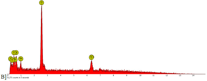 |
Figure 1b |
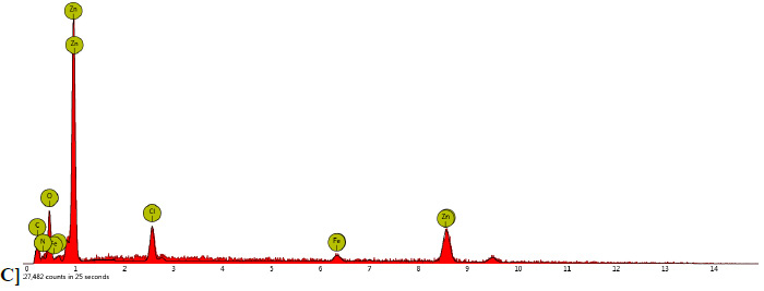 |
Figure 1c |
In Figure 2 AFM topography of the as prepared ZnO nanoparticle and the ultimate nanocomplex are presented. The diagram and two-dimensional images of the initial nanoparticle and conclusive nanocomplex are shown in Figure 2a-b which shows the approximate the height of the sample. Three-dimensional images of ZnO nanoparticle and final complex are displayed in Figure 2c-d that shows peak-valley more concretely. The Fe-EDTA complex is inside ZnO nanoparticle pores randomly resulting in the formation and size of nanocomplex. As the load increases, the values of area roughness, line roughness, and also peak−valley height increases obviously. Thus surface morphology verification of nanoparticle and terminal nanocomplex was determined by AFM technique. The changes are explicated by comparing the graphs. Another physicochemical parameter which has been investigated by Zetasizer is particle size and zeta potential. The average diameter, particle size distribution index, intercept of ZnO nanoparticle and final nanocomplex is shown in Figure 3 and in addition the nanoparticles charge surface before and after reactionsare-7.31mv and -5.63mv respectively.
![Figure 2a: 2D image and diagram of ZnO nanoparticle, [b] 2D image and diagram of ultimate nanocomplex, [c] 3D image of ZnO nanoparticle at the previous zone, [d] 3D image of ultimate nanocomplex at the previous zone.](http://bbrc.in/wp-content/uploads/2020/11/fig2a_opt.jpeg)
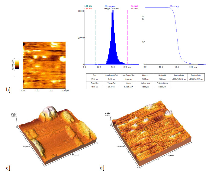 |
Figure 2b |
![Figure 3a: Size of ZnO nanparticle is shown at the left side, and the graph on the right related to final nanocomplex. [b] ZP of ZnO nanoparticle is on the left and the right one is ZP of ultimate nanocomplex.](http://bbrc.in/wp-content/uploads/2020/11/fig3a_opt.jpeg)
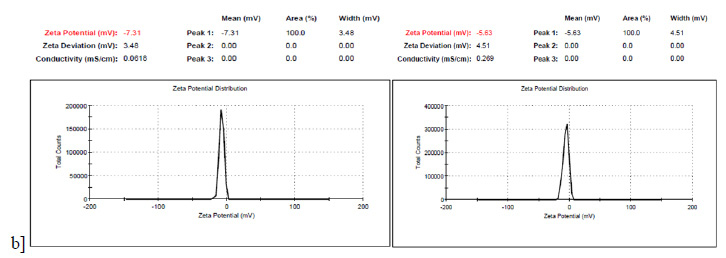 |
Figure 3b |
The FTIR spectrum support the result obtained from SEM, AFM, Zetasizer. After each reaction new peaks appear that proverecent atoms and links as an example a FTIR absorption band is observed at 3436 cm-1 which represents NH link related an amino group of EDTA or the peak in fingerprint zone, about 440 cm-1 reveals presence of halogen of FeCl3 in final nanocomplex and background spectrum of ZnO nanoparticle including a peak at 2926 cm-1 confirms the presence of zinc oxide nanoparticle.Figure.4 give more explanation about spectrums.
![Figure 4a: FTIR spectrums regarding prepared nanoparticle containing Fe-EDTA [A], Zinc Oxide NP [B], Fe-EDTA [C], EDTA [D] as well as FeCl3 [E].](http://bbrc.in/wp-content/uploads/2020/11/fig4a_opt.jpeg) |
Figure 4a: FTIR spectrums regarding prepared nanoparticle containing Fe-EDTA [A], Zinc Oxide NP [B], Fe-EDTA [C], EDTA [D] as well as FeCl3 [E]. |
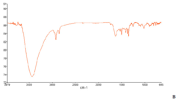 |
Figure 4b |
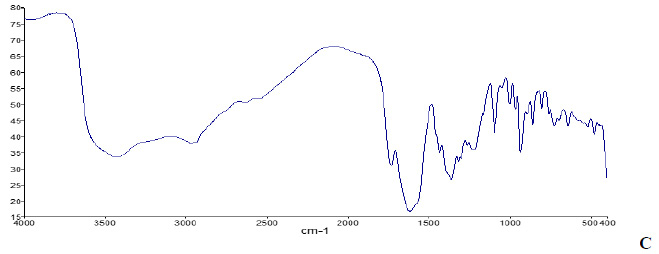 |
Figure 4c |
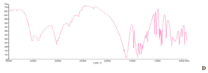 |
Figure 4d |
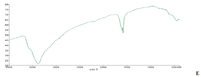 |
Figure 4e |
At the end UV-VIS spectra absorption is recorded between 200-800nm wavelengths. In Figure 5 there is a clear indication of Fe-EDTA complex in the absorption spectrum after loading. The absorption spectrum of the Fe-EDTA complex shows two peaks with absorption between 1.5-2.5 which the first one is related to Carboxylic Acid group of EDTA and the second one demonstrates Ferric Choloride that because of its orbital properties is tended to long wavelengths or visible light. Also the final nanocomplex dissolved in DMSO shows a sharp and narrow peak between 0.2-1nm comparably the previous one.
![Figure 5: U.V spectroscopy data regarding prepared nanoparticle containing Fe-EDTA [A], Fe-EDTA [B], EDTA [C] as well as FeCl3 [D].](http://bbrc.in/wp-content/uploads/2020/11/fig5a_opt.jpeg) |
Figure 5: U.V spectroscopy data regarding prepared nanoparticle containing Fe-EDTA [A], Fe-EDTA [B], EDTA [C] as well as FeCl3 [D]. |
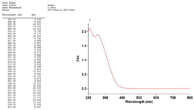 |
Figure 5b |
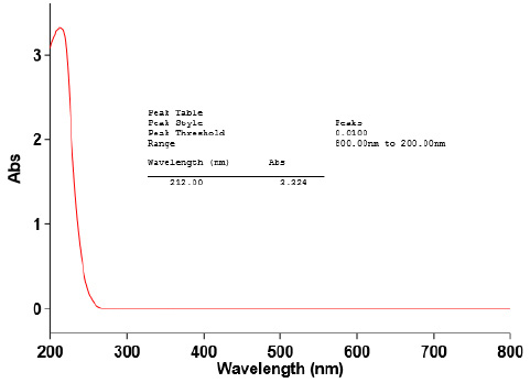 |
Figure 5c |
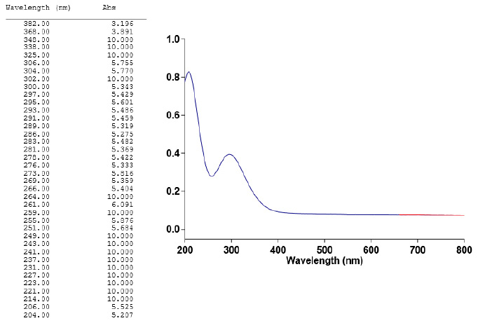 |
Figure 5d |
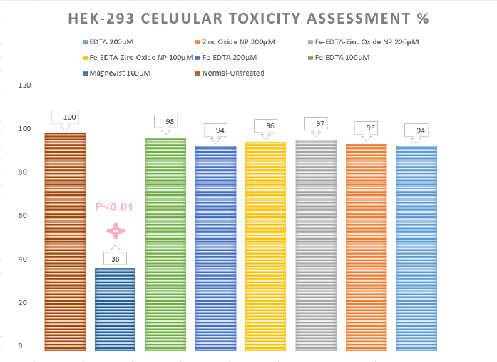
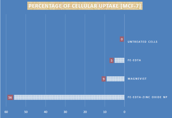
Moreover, as stated in Figure. 6 zinc oxide nanoparticles containing trivalent iron-EDTA, Fe3+-EDTA at all the used dosages did not rise any significant toxic effects on human kidney cells otherwise a significant toxic effects was shown by Magnevist 100 µM [as a FDA approved MR contrast agent] as control caused 62% cellular mortality respectively. Additionally a very good promising cancer cell uptake was recorded for zinc oxide nanoparticles containing trivalent iron-EDTA 56% comparing to 9% for that of Magnevist and 5% for Fe-EDTA as well [more details Figure 7].
As it is clear utilizing contrast specialist as a part of sub-atomic imaging is a discussable subject in atomic prescription, so profited a low dangerous balance operator with ideal impact has dependably been a worry. Too Many diverse diagnosis of some targeted human body sections in molecular imaging methodology including Magnetic Resonance Imaging MRI showed noticeable drawbacks that could be considered as difficulties to remain them also without other confirmation techniques that mostly would be invasive or even defined as impossible, (Rameshwar et al. 2015).
Nephrotoxicity and unfavorably susceptible response of many difference operator are the other test because of their structure and phisico-concoction portrayal. In such cases FDA caution is not immaterial showing the improving shot of Nephrogenic Systemic Fibosis [NSF] and or Nephrogenic Fibrosing Dermopathy [NFD] for the determined patients confounded to have renal disappointment maladies. [Enzo et al. 2010] What’s more moderate discharge and high lethality of numerous macromolecular complex, for example, Gd III, are because of the digestion system which can discharge Gd III particle in body, (Zheng-Rong et al. 2003). Magnetic nano-bio-materials like fe-EDTA-prepared zinc oxide NP nanocomplex using in MRI have been well offered for the next subsequent imaging such viable tumor places (Langer et al. 2015) then again different techniques which require keeps imaging amid treatment. However nano-drugs like nano-complex can defeat natural obstructions which can impact on conclusion and treatment because of passing particles. Then again this nano scale give a fast dissemination amid systemic conveyance furthermore can be cleared up easily (Frank et al. 2008). These qualities gather least danger and time to get the most extreme impact and tissue aggregation. Numerous molecule in view of nanotechnology have been considered to conform the pharmacokinetic properties furthermore upgrade the relaxivity which metal-based nanoparticle can be noted, (Enzo et al. 2010, Na et al. 2008).
In this work we have used FeCl3 chelating with EDTA, a six dentate ligand, and react the resulting complex to ZnO nanoparticle to be placed in its pores and structure properly. Ferric as main recognized paramagnetic ionable to provide adequate magnetic effects on surrounding water atoms and causes to reduce in T2 and or T1 relaxation time quantities. Subsequently this decline makes a brighter picture in light of T1 relaxation time lessening and darker picture consequently diminishing T2 relaxation time, (Brown et al. 2003) (Na et al. 2008). Contrasting with present difference operator, Gd buildings are delegated the specialists that impact on T1 relaxation time and can’t wide water proton resonance and also T2 specialist, for example, paramagnetic and superparamagnetic specialist containing iron. [(Luca et al. 2012))(Na et al. 2008)]. So creating new compound by having both T1 and T2 diminishment can be so useful. Therefor in this paper we synthesized ZnO-Fe nanocomplex besides EDTA as a safe FDA approved and low cost effective chelator in optimum condition.
The result of physico-chemical study showed a good and acceptable data and also low toxicity, at least cost and availability of the material needed are the other positive aspects. The small size of nanocomplex and existing Fe3+ as a paramagnetic agent in ultimate complex displayed a stable and appropriate complex in different features such as size, surface charge, topography, functional groups and fundamental elements. Ease of entry and low risk of cytotoxicity make this nanocomplex as a novel carrier in loading some drugs and having therapeutic effects in addition to diagnostic effects especially in tumor cells with prominent uptake.Consequently, based on these points and outcomes synthesized ZnO-Fe nanocomplex can freely play an important and effective role in the replacement of present contrast agent in MR molecular imaging erelong.
A novel nanocomplex of ZnO-Fe along with EDTA has been prepared by a simple chemical approach as a low toxicMR contrast agent. Thus after chelating iron by EDTA, composed complex reacts with ZnO nanoparticle and a new stable nanocomplex is formed. Small size of ZnO in nano scale helped to uptake more by cells. The presence of Ferric Chlorideas a valuable magnetic agent makes opportune recognizing easier in molecular imaging like Magnetic Resonance Imaging. The physicochemical characteristics were monitored by different analysis resulting in desired achievements that confirm the final nanocomplex without any structural damage. In conclusion, along the development of nanoparticles for targeted diagnosis and therapy for biomedical application, proposed ZnO-Fe nanocomplex may be useful as a novel low risk contrast agent to increase resolution in molecular Imaging like MRI and improve the current situation with the minimum cost. Considering lower normal human cellular toxicity as well as higher and promising cancerous human cellular uptake comparing to Magnevist it could be concluded that such iron containing nanoparticle marked as new target for future in vivo studies as well as clinical practice.
Acknowledgments
The authors would to thank Tehran University of Medical Sciences as well as all laboratories and technicians who provided supported during within the course of the study.
Conflict of Interest
The authors declared no conflict of interest.
References
- Barakat, N. S. (2009). Magnetically modulated nano systems: a unique drug-delivery platform. Nanomedicine, 4[7], 799–812.
- Brown, M. A.; Semelka, R. C. (2003).MRI: Basic Principles and Applications; Wiley-Liss: New York.
- Enzo T, Daniela D C, Alessandra V, Silvio A. (2010).Challenges for Molecular Magnetic Resonance Imaging. Chem. Rev, 110, 3019–3042.
- Frank , Eric P, Linda K. M, Omid C. (2008).Factors Affecting the Clearance and Biodistribution of Polymeric Nanoparticles.Mol. Pharmaceutics,5 [4], pp 505–515
- Jain, P. K.; Huang, X.; El-Sayed, I. H.; El-Sayed, M. A. (2008) .Noble metals on the nanoscale: optical and photothermal properties and some applications in imaging, sensing, biology, and medicine. Acc. Chem. Res, 41[12], 1578–1586.
- James, M. L., and Gambhir, S. S. (2012) . A molecular imaging primer: modalities, imaging agents, and applications. Physiol. Rev. 92, 897−965
- Jesse V. J, Sanjiv S. G. (2011).Molecular Imaging with Theranostic Nanoparticles. Acc. Chem.Res.,44 [10], pp 1050–
1060 - Langer, R. and Weissleder, R. (2015). Nanotechnology. JAMA. 313, 135-136
- Lauffer, R. B. [1987]. Paramagnetic metal complexes as water proton relaxation agents for NMR imaging: theory and design. Chem. Rev. 87, 901−927.
- Luca F, Junqing Z, Changning W, Chunying W, Robert H. M, Yanming W. (2012) Myelin Imaging Compound [MIC] Enhanced Magnetic Resonance Imaging of Myelination. Med. Chem., 55[1], pp 94–105
- Meade, T. J., and Aime, S. (2009). Chemistry of molecular imaging. Acc. Chem. Res. 42, 821−821.
- Na, H. B.; Hyeon, T. (2009). Nanostructured T1 MRI contrast agents. J. Mater. Chem., 19, 6267.
- Na, H. B.; Song, I. C.; Hyeon, T. (2009). Inorganic Nanoparticles for MRI Contrast Agents. Adv. Mater., 21, 2133–2148.
- Rameshwar P, Alexander V. L, Pallavi R. G, Hui D, Jose P.A, Shawn W, Satoshi I, Bindu K, Arthur R, Alexandra C, Janet L. M, Vladimir A. L, Debiao L, Ravi S. P, Keith L. B, Eggehard H, and Julia Y. L. (2015). MRI Virtual Biopsy and Treatment of Brain Metastatic Tumors with Targeted Nanobioconjugates: Nanoclinic in the Brain.ACS Nano,9 [5], pp 5594–5608
- Schwert, D., Davies, J., and Richardson, N. (2002) NonGadolinium-Based MRI Contrast Agents [Krause, W., Ed.] pp 165−199, Springer, Berlin
- Toth, E., Helm, L., and Merbach, A. (2002) Relaxivity of MRI Contrast Agents [Krause, W., Ed.] pp 61−101, Springer,
- Zheng-Rong L, Xinghe W, Dennis L. P, K. Craig G, Henry R. B. (2003). Poly [L-glutamic acid] Gd[III]-DOTA Conjugate with a Degradable Spacer for Magnetic Resonance Imaging. Bioconjugate Chem, 14, 715−719

