1Prosthetic Dental Science Department, College of Dentistry, Jazan University, Jazan 45142, SA
2Department of Microbiology, College of Dentistry, Jazan University, Jazan 45142, SA
3National Center for Disease Prevention and Control, Ministry of Health, Jazan, SA
4Dental Interne, College of Dentistry, Jazan University, Jazan, SA
Corresponding author email: drmoaleem2014@gmail.com
Article Publishing History
Received: 15/01/2020
Accepted After Revision: 19/03/2020
The effects of khat chewing habits on the stainability of feldspathic metal ceramic (MC) materials used in the fabrication of dental restorations remain unexplored. This study investigated the effect of khat extract (KE) on mean color changes (ΔE*) and surface topography among different feldspathic MC materials, namely, VMK VM13, VMK MASTER, and VMK 95). Sixty feldspathic MC specimens were prepared from nickel chromium alloy with the above-mentioned ceramic materials. The ΔE* of the specimens were measured using a spectrophotometer before and after immersion in KE and thermocycling for 10 days. The surface topography was captured to examine the surface of specimens after KE staining with a white light interferometric microscope. Results showed that the ΔE* values of the different types of tested feldspathic MC were influenced by KE. The lowest values of ΔE* were recorded from glazed VMK VM13, VMK MASSTER, and VMK 95 with acceptable color changes, whereas the highest and unacceptable values of ΔE* were observed from polished VMK MASTER and VMK 95. All the tested groups and subgroups obtained significant differences with p values > 0.05 in Student’s t-test and ANOVA test. All the tested feldspathic MC materials had showed significant differences in the parameter of average color changes for all tested groups in form of glazed or polished specimens. Clinical significance: Re-glazing step is an essential step before final cementation and after chair-side adjustment of any feldspathic MC prosthesis for patients who chew khat.
Color Measurement, Feldspathic Porcelain, Khat, Surface Topography
Al-Moaleem M. M, Homeida H. E, Dafalla O. M, Alamir O. H, Dahas Z. A, Alqasemi W. I, Al-Sanabani F. A. Evaluation of Color Changes and Surface Topography of Different Feldspathic Ceramic Materials After Khat, Catha edulis Extract Immersion. Biosc.Biotech.Res.Comm. 2020;13(1).
Al-Moaleem M. M, Homeida H. E, Dafalla O. M, Alamir O. H, Dahas Z. A, Alqasemi W. I, Al-Sanabani F. A. Evaluation of Color Changes and Surface Topography of Different Feldspathic Ceramic Materials After Khat, Catha edulis Extract Immersion. Biosc.Biotech.Res.Comm. 2020;13(1). Available from: https://bit.ly/3d7XjXY
Copyright © Al-Moaleem et al., This is an open access article distributed under the terms of the Creative Commons Attribution License (CC-BY) https://creativecommons.org/licenses/by/4.0/, which permits unrestricted use distribution and reproduction in any medium, provide the original author and source are credited.
INTRODUCTION
Feldspathic metal ceramic (MC) restorations are considered as a color stable. In aesthetic prosthesis, such as feldspathic ceramics, discoloration may occur due to intrinsic or extrinsic factors. Intrinsic factors include changes within the ceramic, whereas extrinsic factors involve adsorption or absorption of stains from the oral cavity and the smoothness of the prosthesis surface (Singh et al., 2015). Restoration stainability has been proven to be dependent on numerous factors, such as the brand and shade of the prosthetic material, exposure time and intensity of dissimilar food and beverages, drinking, and polishing techniques (Stawarczyk et al., 2012).One of the criteria for the success of aesthetic materials is color stability. Thus, the assessment of color changes using color measuring devices, such as spectrophotometers, has become common. In such assessments, the precision, standardization, and numerical expression of color are recorded. The data are labeled in the CIE L*a*b*system, which uses three-dimensional colorimetric quantities; L*measures the brightness of the color, a* handles the red–green content, and b* assesses the yellow–blue content. Then, the mean color changes (ΔE*) are calculated using L*a*b* (Rosentritt et al., 2015; Turgut et al., 2011). ΔE* reveals whether a change in the overall shade can be detected by a human observer (Rosentritt et al., 2015) (3). Previous studies have considered color differences (ΔE*) greater than 3.5 unit to be clinically unacceptable (Yilmaz et al., 2011 Abdalkadeer et al., 2019; Alghazali et al., 2012, 2019).
Many studies have been conducted on the color stability of different aesthetic ceramic materials in several common beverages. Some of the researchers have shown that changes in color stability are greatest in tea (Er et al., 2006; Haralur et al., 2019), coffee or Arabic Qahwa (Abdalkadeer et al., 2019; Saba et al., 2017; Sarikaya and Güler, 2011), and Coca-Cola (Alghazali et al., 2019; Sarıkaya et al., 2018).
Chair-side or intraoral polishing of feldspathic ceramic restorations is efficient, easy for clinicians, and minimizes repeated laboratory procedures (Hmaidouc et al., 2014). However, careful intraoral polishing of the ground surfaces is always necessary because the final occlusal adjustments of dental prostheses have to be made after cementation. Some investigations have been made using different polishing techniques on ceramic surface instead of glazing (Raimondo et al., 1990; Wright et al., 2004).Khat (Catha edulis) is an evergreen shrub that belongs to Celastraceae family. It grows in Yemen and southern Saudi Arabia, as well as in certain East African countries (Ageely 2009).
Khat leaves are habitually chewed by people in these regions because of their psychostimulant effect, which is similar to that produced by amphetamine-like substances (Wabe 2011). These leaves are most frequently chewed in one preferred side of the mouth (usually the left side). Young fresh leaves are chewed in form of a bolus and held in the lower buccal pouch unilaterally for 3–5 hours or longer (Al-Alimi et al., 2018). Given the rampant practice of khat chewing, especially in southern region of South Africa, exploring the effect of khat on materials used for dental prosthesis fabrications is essential. However, studies on the effect of khat on oral health remain scarce. Few studies have investigated the association between khat chewing and dental ceramic materials. Moreover, the role of khat as a staining material contributing to dental ceramic discoloration and surface effect in relation to maintaining the polishability of ceramic materials has yet been studied nor documented.
A recent clinical study by Al Moaleem et al., (2020) concluded that khat chewing had a statistically significant effect on the bacterial biofilm formation on restorative materials, whereas no significant effect was found on feldspathic and all ceramic prostheses. In view of khat chewing in relation to composite restorations, a single clinical study recorded that composite filling materials may be related with demineralization of composite filling materials at the composite tooth border; this possibility may result in altered color of the composite and tooth structure (Al-Alimi et al., 2014). Moreover, in vitro studies have examined the effect of khat extract (KE) on the color of composite materials and reported that KE shows a clinically perceptible ΔE* with current types of composite filling (Al Anesi et al., 2019).
The objectives of the present study were as follows: 1) to assess the effect of KE on the stainability of selected VITA feldspathic MC specimens (i.e., VMK VM(R)13, VMK MASTER, and VMK 95); 2) to measure the color changes on the basis of the basic color of VITAPAN classical shade guide (A1–D4); and 3) to evaluate the surface topography of the specimens after immersion in KE and aging using a white light interferometric microscope. The null hypothesis of this study is that KE and thermocycling can affect the color stainability of polished feldspathic MC specimens in comparison with glazed specimens. The color changes were clinically acceptable.
MATERIALS AND METHODS
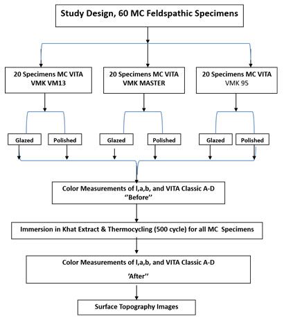
Study Design: Sixty feldspathic MC specimens were prepared for the in vitro study. The specimens were used to measure the effect of KE on the color stainability of glazed or polished feldspathic MC materials. Furthermore, the surface topography of the specimens after immersion in KE was evaluated using a white light interferometric microscope. Table 1 presents the materials and devices used in this study.
Table 1: Materials and devices used in the study
| Material or Device Type | Brand Name | Composition | Manufacturer |
| Nickel chromium casting alloy | Wiron(R) 99 | Ni 65%, Cr 22.5%, Mo 9.5%, Nb 1% Si1%, Fe 0.5%, Ce0.5%, Cmax 0.021 | BEGO, Germany |
| Feldspathic P/L ceramic | VITA VM(R)13 | Silicon dioxide, possesses a glassy matrix, and has assorted quantities of potassium, sodium, barium, or calcium | VITA Zahnfabrik, Bad Säckingen, Germany |
| Feldspathic P/L ceramic | VMK MASTER | Natural feldspar veneering ceramic for conventional bonding alloys | VITA Zahnfabrik, Bad Säckingen, Germany |
| Feldspathic P/L ceramic | VMK 95 | Pure-grade potash and albite feldspar materials | VITA Zahnfabrik, Bad Säckingen, Germany |
| Khat | C. edulis plant | Alkaloids, terpenoids, flavonoids, sterols, glycosides, tannins, amino acids, vitamins, and minerals | |
| Spectrophotometer | Vita Easy Shade Spectrophotometer | Device used for measuring wavelength transmitted from one object at a time without being affected by subjective interferences of color | VITA Zahnfabrik H. Rauter GmbH & Co. KG, Bad Sackingen, Germany |
| Surface roughness and topography tester | White light interferometric microscope | 3D printer of surface characteristics | Contour GT-K1, Bruker Nano GmbH, Berlin, Germany |
Specimen Preparation and Fabrication: Sixty metal specimens with 0.4 mm thickness and 12 × 12 mm dimension were prepared and constructed from green wax. All metal specimens were invested, burned, and casted with nickel–chromium dental casting alloy (Wiron(R) 99, Bego, Germany) using the conventional manner and according to manufacturer’s instructions. The casted specimens were divested, and the residual surface investment was removed by sandblasting with 250 μm aluminum oxide abrasion particles, finished by carborundum discs and metal trimmers. Finally, the specimens were adjusted to achieve a uniform thickness (i.e., 0.4 mm). The 60 metal disc specimens were divided into three equal groups on the basis of the type of feldspathic ceramic materials used.
The feldspathic ceramic of VITA VM(R) 13 (VITA, Zahnfabrik, Germany) was used to make 20 specimens of the first group. First, two coats of paste opaque were used, and a dentine layer, in which powder and liquid were mixed according to manufacturer’s instructions, was then applied over the specimens using a metallic jig. Subsequently, enamel layer was applied using the same technique for dentine layer, and the specimens were burned. Burning was performed according to manufacturer’s instructions, and specimens were grinded with a diamond bur to achieve the uniform thickness. Using the same technique, 20 specimens were constructed from each of VMK MASTER and VMK95 (VITA, Zahnfabrik, Germany). Lastly, the specimens were glazed. The opaque layer was 0.3 ± 0.1 mm, whereas the body porcelain was 2.0 ± 0.3 mm in thickness (Al Moaleem et al., 2012; Jan et al., 2013).
Samples Surface Treatments: The prepared specimens of the three groups were divided into two equal subgroups with 30 specimens each. The first subgroup specimens (10 samples from each group) were obtained from the laboratory with glazed layer and without any further treatment, whereas the specimens of the second subgroup (10 specimens from each group) were polished using a porcelain polishing kit according to manufacturer’s instructions to characterize the clinical condition of the prostheses. Each 10 feldspathic MC specimen was polished using a polishing kit with equal number of grits and in one direction under a constant load.
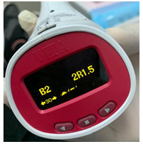
Color Measurements: The color of each specimen was recorded before and after thermocycling and immersion within KE mixture. The color of each specimen at both intervals was measured at the same position (i.e., center of the specimen) using a portable spectrophotometer (Vita Easy Shade, Vita Zahnfabrik H. Rauter GmbH & Co. KG, Bad Sackingen, Germany). A putty index was made around the tip of the device with a window of 4 mm diameter in the center to standardize the extent of color measurement (Haralur et al., 2019). The spectrophotometer was used to measure the CIE-Lab values to provide a numerical representation of 3D color measurements. Each time, the specimen color was measured twice for L*, a*, and b*, and the average value was then considered the color of the specimens before immersion, following the CIL Lab color system. The ΔE* values were calculated for the different specimens by assuming the formula: ∆E* = [(L1* − L2*)2 + (a1* − a2*)2 + (b1* − b2*)2]1/2, where ∆L* is the lightness of L*, ∆a* is the variation of a*, and ∆b* is the variation of b*. A low ∆E* was considered good shade matching, and a score of ≥3.5 was considered an acceptable color change (Alghazali et al., 2012; Alghazali et al., 2019). The Vita Easy Shade spectrophotometer recorded the basic color of the specimens according to the VITAPAN classical shade guide A1–D4 before the immersion (Figure 1).
Figure 1: Basic VITAPAN classical shade guide on the spectrophotometer
Specimen Immersion and Thermocycling: KE was prepared and presented by the Substance Abuse and Toxicology Research Center, Jazan University. It was prepared from fresh mincing khat leaves in 100% distilled water (V/W) and then finely minced. KE was kept in −80 °C ultralow-temperature freezer until further use. It was then mixed with NaOH until its pH was similar to that of the oral cavity. All specimens were immersed in KE for 10 days, as mentioned in previous in vitro studies (Al Anesi et al., 2019; Al Moaleem et al., 2020a). The same procedures were executed daily to obtain fresh KE. During the immersion time, an aging process was conducted using a thermocycling machine, where 100 cycles were accomplished every day in 5 °C cold water and then in 55 °C hot water (1000 cycles). All specimens were dipped in distilled water, followed by the removal from immersion media. Specimens were wiped dry with a tissue paper and left in place for complete dryness.
After 10 days of KE immersion and thermocycling, the colors of the specimens were remeasured with the same shade using the spectrophotometer, and the readings were registered as after immersion. The aforementioned equation was used to calculate the average color of L, b, and a after immersion. The reading of the basic shade for VITAPAN classical shade guide was measured again as the reading after immersion. All procedures for specimens’ preparation, fabrication, surface finishing and polishing, specimen immersion, and thermocycling were performed by the same operator. Color was measured for all assigned specimens by the same operator under the same settings and gray background.
Figure 2: Study design, steps for color measurement, and type of staining materials with thermocycling
Surface Topography Scanning : One specimen from each glazed or polished porcelain was scanned after KE immersion and thermocycling. The surface topography of the six specimens was characterized graphically using the white light interferometric microscope (Contour GT-K1, Bruker Nano GmbH, Berlin, Germany) under 50× magnification, with back scan and dimension parameters of 20 µm in VSI/VXI mode; this method was performed to obtain a 3D interpretation of the specimen surfaces. Vision 64 software (Bruker Nano GmbH, Berlin, Germany), which is part of the GT-K1 system, was used to replicate the surface topography constraints (Figures 3a–3f).
Data Statistical Analysis :The mean values of ΔE*, ∆L*, ∆a*, and ∆b* of the different feldspathic MC specimens (i.e., VMK VM13, VMK MASTER, and VMK 95) in forms of glazing and/or polishing were recorded and then compared before and after KE immersion. Changes in the basic color of VITAPAN classical shade were documented. Data were entered into Microsoft Excel 13 and analyzed using Statistical Package for Social Science version 22.0 (SPSS Inc., Chicago IL, USA) software. Descriptive statistics was intended for each parameter for the three groups. The ΔE* values were compared using one‑way ANOVA test, followed by a post hoc comparison by Bonferroni test to detect any significant difference between and within the groups at P > 0.05. Student’s t-test was used to detect the significances between each pair of the three groups.
RESULTS AND DISCUSSION
In the vitro study, 60 specimens were included, represented by letters a–f, in the analysis of results. The images were recorded using the white light interferometric microscope, in which a and b represent glazed and polished VMK VM 13, respectively; c and d denote the glazed and polished VMK MASTER, respectively; and e and f represent glazed and polished VMK 95 after KE immersion and thermocycling of the specimens. The ΔE* values were calculated using the aforementioned equation. High ΔE* values were observed for the polished subgroups of VMK MASTER, VMK 95, and VMK VM 13, followed by the glazed VMK 95 and VMK MASTER; whereas glazed VMK VM 13 had the lowest ΔE* of 0.348. The ΔE* values were significantly differed among all tested groups, either in glazed or polished forms, with P values of >0.05. Table 2 shows the significant differences in the ΔE* values using Student’s t-test. From the table, VMK MASTER had a significant P value of 0.005, whereas VMK VM13 had a nearly significant P value (0.053), and VMK95 had an insignificant P value (0.337; Table 3). Figure 3 shows the changes in the basic color of the VITAPAN classical shade guide (A1–D4) of the tested groups. The highest changes were observed for VMK MSSTER (90%), followed by 70% for VMK 95, and only less than half (40%) for VMK VM13 ceramic type.
Table 2. ΔE* after KE immersion and thermocycling for different groups in relation to surface type based on ANOVA, followed by Bonferroni tests
| Material Surface Type | Mean and SD of (ΔE) for Each Material | VMK 13 a,b | VMK MASSTER c,d | VMK 95 e,f | P value |
| Glazeda,c,e | 0.348 (0.115) VMK VM13 | — | 0.000 | 0.000 | 0.000* |
| 1.282 (0.259) VMK 95 | 0.000 | — | 0.000 | ||
| 2.881 (0.399) VMK MSSTER | 0.000 | 0.000 | —- | ||
| Polishedb,d,f | 2.963 (0.456) VMK VM13 | — | 0.000 | 0.000 | 0.000* |
| 7.256 (0.529) VMK 95 | 0.000 | — | 0.000 | ||
| 4.935 (0.569) VMK MSSTER | 0.000 | 0.000 | —- |
Table 3. Comparison among different groups in relation to the surface type using Student’s t-test
| Ceramic Type | Surface Type | Mean | SD | Sig. |
| VMK 13 | Glazed | 0.348 | 0.115 | 0.053 |
| Polished | 2.962 | 0.456 | ||
| VMK MASSTER | Glazed | 1.282 | 0.259 | 0.005* |
| Polished | 7.258 | 0.529 | ||
| VMK 95 | Glazed | 2.881 | 0.399 | 0.337 |
| Polished | 4.935 | 0.569 |
*Significant (P < 0.05)
Figure 3: Comparison between color changes in basic color shade of VITABAN classical shade for the tested specimens after KE immersion
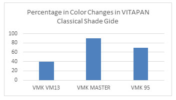
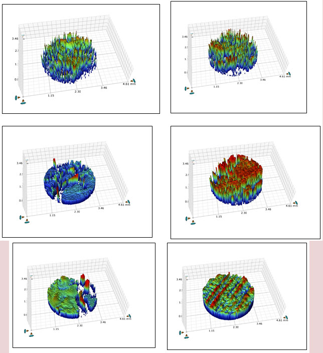
Figure 4 shows the representative white light interferometric microscopic images of the tested shades of ceramics after KE immersion and thermocycling. The red and blue areas represent the part of the surface with the highest (i.e., the peaks) and lowest (i.e., the valleys) heights, respectively. One specimen from each subgroup showed a different high pattern of peaks and valleys across the surfaces, with no identical pattern across each surface in either glazed or polished form. Furthermore, the microscopic images for VMK VM 13 specimens obtainable a nonuniform surface with distinct sharp projections, as shown by the dotted areas with pores at different sides originating from the center (Figures 4a and 4b). VMK MASTER showed a few pores located on the border for the glazed specimens (Figure 4c), whereas pores are all over the surface for the polished specimens (Figure 4d). VMK 95 specimens demonstrated a uniformly irregular surface pattern with heights and valleys, that is, narrow at the border, irregular, and superficial scratch areas at the border (Figure 4e); however, it is crossed the entire surfaces, which causes a uniformly irregular surface for all the specimens (Figure 4f).
Figure 4: Characteristic images of white light interferometric microscope of tested VMK 13 (a, b), VMK MASSTER (c, d), and VMK 95 (e, f) ceramic specimens obtained at 50× magnification
MC prostheses are still used as restorative materials in many countries due to their metal strength, aesthetic properties, color stability, biocompatibility, marginal integrity, and easy and familiar fabrication by dental technicians (Jain et al., 2013). Exogenous staining may occur due to the capability of prostheses to adsorb or absorb stains from khat chewing in the oral cavity, which may potentially affect color stainability.
This in vitro study investigated the effect of KE immersion with thermocycling on the stainability of different types of feldspathic MC specimens used for replacing missing/decayed coronal portions or missing tooth/teeth. The ΔE* values of glazed or polished surfaces were compared using a physical polishing set. The results of this study complement a wide range of adverse effect of KE on feldspathic MC specimens. The first null hypothesis of this study (i.e., KE and thermocycling can affect the color stainability of polished feldspathic MC specimens in comparison with glazed specimens) was accepted because the stainability of feldspathic MC material specimens in glazed or polished ceramic form were affected by KE immersion and the thermocycling. Nonetheless, the second hypothesis was at the margin because most of the tested specimens were clinically acceptable in the glazed form but mostly clinically unacceptable in the polished form.The most problematic issue that prosthodontists and dental clinicians face during their daily practice is shade selection. The reference tooth most often considered by dental clinicians is the neighboring, contralateral or the opposing tooth/teeth. However, other teeth should also be to obtain a clinically acceptable shade. Moreover, teeth should be polished with prophylaxis before selecting the appropriate shade.
The effect of KE solution and thermocycling on the color of tested materials was evident in all specimens with different degrees. Table 2 shows that the ΔE* value was the highest on polished specimens, with 2.963, 7.256, and 4.935 recorded for VMK VM 13, VMK MASTER, and VMK 95, respectively. This result agrees with the finding of Hill and Gibson., (1987), who stated that khat modifies the enamel surface of teeth, resulting in beverage collections on the surface and color change of the tooth after a period of time Al-Alimi et al., (2014), concluded that natural teeth become discolored because of the acidic and mechanical effect of KE on the tooth surfaces. Moreover, the use of large quantities of soft drinks or beverages and sugar tablets for a long period can result in cervical discoloration in the enamel and dentine, staining of teeth, attrition, and cervical caries at the chewing side (El-Wajeh and Thornhill., 2009; Al-Meshal et al., 1991).
The clinical precision of Vita Easy Shade spectrophotometer has been validated by several studies (Karagoz-Motro et al., 2012; ALGhazali et al., 2011). However, such devices may encounter problems while measuring curved surfaces because the measuring probe tip is flat. Edge loss errors are common due to the fact that the probe tip of the instrument cannot be in direct contact with the buccal surfaces of natural teeth or ceramic surfaces (Gupta et al., 2012). In addition, positioning errors of the probe tip cannot be excluded; such errors reduce the L* values recorded for such types of ceramic crown. The ΔE* values were assessed in terms of perceptibility and acceptability for small color differences because of their role as a guide control throughout the selection of ceramic materials for khat users. The ΔE* measurements using Vita Easy Shade spectrophotometer are more precise and accurate than those of other digital instruments and can thus be used in dental practice and research with some limitations (Kim-Pusateri et al., 2009; Alghazali et al. 2011). Also, the CIE Lab system was selected to evaluate the average color differences because it is well suited for the determination of small color differences (Alghazali et al., 2012; Sarikaya and Güler, 2011).
Prosthesis must not only have the dimensions, texture and contours of the teeth to be replaced but should also have similar light behavior. The color stability of the restoration is also critical for the long-term success of aesthetic restorations. Although the physicomechanical properties of ceramics have vastly improved, they remain susceptible to discoloration (Derafshi et al., 2017). Extrinsic factors, such as beverages, mouthwashes, acid solutions, toothbrushing, and high temperatures, are reported to induce surface degradation of ceramics (Kukiattrakoon et al., 2009).
Hmaidouc et al., (2014) and Sarikaya and Güler., (2010) found no significant difference between the surfaces of fine glazed and polished full-contour zirconia specimens and between feldspathic VMK 95 and Ceramco III porcelain groups, respectively. Lawson et al. (2014), recorded similar surfaces for glazed or polished feldspathic Ceramco III and lithium disilicate or zirconia. This result agrees with the findings of the present study, that is, no significant differences were observed between the glazed or polished test specimens (Table 3). By contrast, Sarikaya and Güler (2011) and Saba et al. (2017), reported that Ceramco III feldspathic porcelain demonstrated the highest ΔE* values with no significant difference among the other groups (i.e., Mark II, Matchmaker MC, and VMK 95). The results of the present study indicated that significant differences were observed among the three types of tested feldspathic materials (Table 2). These differences could be related to the type of feldspathic material used and the technique of construction of the specimens.
Alghazali et al., (2012), performed a clinical study and found that ΔE* of 2.8 is clinically acceptable. Alghazali et al., (2019); Abdalkadeer et al. (2019); Sarikaya and Güler (2011), concluded that ΔE* values between 1 and ≤3.3 can be detected by the human eyes and are clinically acceptable for feldspathic; moreover, low-fusing porcelain specimens immersed in coffee or Coca-Cola are color-stable, with average values of ΔE* at an acceptable level. Saba et al. (2017) confirmed that coffee may adversely affect color and may consequently compromise the aesthetics of feldspathic CAD/CAM blocks. All the tested specimens were clinically acceptable in ΔE* values ranging from 0.348 for glazed VMK VM13 to 2.963 for polished VMK VM13, which are clinically acceptable. The only unacceptable ΔE* values were for feldspathic specimens of VMK MASTER and VMK 95, and this result could be related to the composition of those ceramic materials (Tables 2 and 3).
Recdently, Alotaibi et al. (2019) have evaluated the crown color of extracted teeth after endodontic treatment with different endodontic sealers using a Vita Easy Shade Advance and the equation ΔE* = ([ΔL*]² + [Δa*]² + [Δb*]²)½ after the aging period reached 3 months. They recorded the ΔE* values ranging from 7.02 to 8.14 without significant difference between the groups. Greța et al (2020), performed a clinical study using Vita Classic shade guide with Vita Easy Shade Advance to measure the color They concluded that the color difference between the restoration and the reference tooth exceeded the perceptibility thresholds. The findings demonstrated high percentage in the color changes in relation to the basic Vita Classical shade guide and recorded 90%, 70%, and 30% changes for VMK MASTER, VMK 95, and VM VMK13, respectively (Figure 3).
Few studies have used white light interferometric microscopic images in examining the surface topography of ceramic materials after immersion in stained materials. In this study, the polished samples of VM VMK13 (Figure 4b) showed high surface topography alteration compared with other polished samples of VMK MASTER and VMK 95 (Figures 4d and 4f), indicating that the mechanical and acidic effect of KE was noticeable. KE is fibrous in nature, and this property may cause a mechanical effect on the surfaces of ceramics, especially those of polished specimens (Jorgensen et al., 2009). The glazed surfaces of VM VMK13 specimens replicated the lowest effect of KE on their surface topography (Figure 4a). The images obtained in this study were dissimilar compared with the images documented in an earlier study by Egilmez et al., (2018), but were in parallel of a previous finding in relation to KE and feldspathic samples (Al Moaleem et al., 2020b).
The limitations of the current in vitro study are as follows. Thermocycling was performed with water. This condition is different from the oral cavity environment during khat chewing, which is usually associated with soft drinks and smoking. Moreover, daily brushing of teeth by patients were not simulated during the immersion and thermocycling period. During aging, the immersion solution permitted staining on both sides of the samples However, in clinical conditions, the material is cemented to a tooth structure and is visible to KE and light on the exposed one.
CONCLUSION
The following conclusions can be drawn from this in vitro study. KE showed a significant effect on glazed and polished specimens of feldspathic MC materials. It showed an acceptable ΔE* values for all glazed specimens and polished VMK 13, with significant differences among all groups and subgroups. The effects were higher and unacceptable for VMK MASSTER, followed by polished VMK 95 specimens.
ACKNOWLEDGMENTS
The authors would like to thank the Deanship of Scientific Research, Jazan University, Kingdom of Saudi Arabia for funding this study in Future Scientists Program #V under Grant number FS5-038. The authors are also grateful for Substance Abuse Research Center, Jazan University for providing the khat extract.
Conflicts of Interest: The authors declare no conflict of interest.
REFERENCES
Abdalkadeer HK, Almarshedy SM, Al Ahmari NM, et al. Influence of the Coca-Cola Drinks on the Overall Color of Glazed or Polished Porcelain Veneers Fabricated from Different Materials and Thicknesses: An In Vitro Study. J Contemp Dent Pract 2020;21(1):56–61.
Ageely HM (2009). Prevalence of Khat chewing in college and secondary (high) school students of Jazan region, Saudi Arabia. Harm Reduct J. 6:18.
Al Moaleem MM Amit P, AlAhmari NM, et al (2020b). Evaluation of color changes and surface topography of different feldspathic ceramic materials after khat extract immersion. Curr Phar Biot. 21(3); DOI: 10.2174/1389201021666200121142300
Al Moaleem MM, Porwal A, Al Ahmari NM, Shariff M, et al (2020a). Khat Chewing Induces a Floral Shift in Dental Material-Associated Microbiota: A Preliminary Study. Med Sci Monit. 26: e918219
Al-Alimi KR, Razak AAA, Saub R (2014). Salivary caries parameters: Comparative study among Yemeni khat chewers and nonchewers. Journal of Dental Sciences. 9(4):328-31.
Al-Alimi, K. R.; Razak, A. A. A.; Saub, R. (2018). Is Khat chewing habit a risk factor for occlusal caries progression? African Health Sciences.1 8 (4), 1036-1045.
Al-Anesi WA, Madfa AA, Dubais MA and Albahari AA (2019). Effects of Khat Extract and Other Staining Media on Color Change of Composite Resins Subjected to Various Polishing Methods. Oral Biol Dent. 7:1-10
Alghazali N, Burnside G, Smith PW, Preston AJ, et al (2012). Assessment of perceptibility and acceptability of colour difference of denture teeth. J Dent. 40 supp: 10-7.
ALGhazali N, Burnside G, Smith PW, Preston AJ, Jarad FD (2011). Performance Assessment of Vita Easy Shade Spectrophotometer on Colour Measurement of Aesthetic Dental Materials. Eur. J. Prosthodont. Rest. Dent. 19(4); 168-174
Alghazali N, Hakami AA, AlAjlan GA, Alotaibi RM et al (2019).
Influence of the Arabic-Coffee on the Overall Color of Glazed or Polished Porcelain Veneers – In vitro Study. The Open Dentistry Journal. 13; 65-61.
Al-Meshal, I. A; Qureshi, S.; Ageel, A. M.; Tariq, M. (1991). The toxicity of Catha edulis (khat) in mice. Journal of substance abuse. 3 (1), 107-115
AL-Moaleem MM, Shah FK, Khan NS, Porwal A (2011): The effect of thermocycling on the bonding of different restorative materials to access opening through porcelain fused to metal restorations. J Adv Prosthodont. 4:186-189.
Alotaibi RM, Altuwaijri SM, Ahmed DR (2019). The degree of crown discoloration induced by a bioceramic and other conventional endodontic sealers. Saudi Endod J. 9:192‑7.
Derafshi, R.; Khorshidi, H.; Kalantari, M.; Ghaffarlou, I (2017). Effect of mouth rinses on color stability of monolithic zirconia and feldspathic ceramic: An in vitro study. BMC Oral Heal. 17:129-35.
Egilmez F, Ergun G, Cekic-Nag I, Vallittu PK, Lassila LV (2018). Comparative color and surface parameters of current esthetic restorative CAD/CAM materials. J Adv Prosthodont. 10:32-42
El-Wajeh, Y.; Thornhill, M. (2009). Qat and its health effects. British dental journal. 206 (1), 17.
Er AU, Turker SB, Kocak A, Aktepe E (2006). Effect of five staining solutions on the colour stabilityof two acrylics and three composite resins based provisional restorations. Eur J Prosthodont Restor Dent. 14: 121-125
Greța DC, Gasparik C, Colosi HA, Dudea D (2020). Color matching of full ceramic versus metalceramic crowns – a spectrophotometric study. MEDICINE AND PHARMACY REPORTS j. 93(1); 89-96.
Gupta R, Prakash H, Shah N, Jain V (2005). Spectrophotometric evaluation of color changes of various tooth colored veneering materials after exposure to commonly consumed beverages. J Indian Prosthodont Soc. 5:72-8.
Haralur SB, Alqahtani NRS, Mujayri FA (2019). Effect of Hydrothermal Aging and Beverages on Color Stability of Lithium Disilicate and Zirconia Based Ceramics. Medicina, 55, 749
Hill CM, Gibson A (1987). The oral and dental effects of q’at chewing. Oral Surg Oral Med Oral Pathol. 63(4):433-6.
Hmaidouch R, Mu ¨ller W_D, Lauer H-C, Weigl P (2014). Surface roughness of zirconia for full-contour crowns after clinically simulated grinding and polishing. International Journal of Oral Science. 6(4); 241-6.
Jain, C.; Bhargava, A.; Gupta, S.; Rath, R.; Nagpal, A.; Kumar, P (2013). Spectrophotometric evaluation of the color changes of different feldspathic porcelains after exposure to commonly consumed beverages. Eur J Dent. 7; 172–180.
Jorgensen, E.; Kaimenyi, J. T. (1990). The status of periodontal health and oral hygiene of Miraa (Catha edulis) chewers. East African medical journal. 67 (8), 585-90.
Karagoz-Motro P, Kursoglou P, Kazazoglu E (2012). Effects of different surface treatments on stainability of ceramics. J Prosthet Dent. 108:231-7
Kim-Pusateri S, Brewer JD, Davis EL and Wee AG (2009), Reliability and accuracy of four dental shade-matching devices, J Prosthet Dent. 101: 193-199.
Koseoglu, M., Albayrak, B., Gül, P. and Bayindir, F. (2019) Effect of Thermocycle Aging on Color Stability of Monolithic Zirconia. Open Journal of Stomatology 9; 75-80.
Kukiattrakoon,B.; Junpoom,P.; Hengtrakool, C (2009). Vicker’s micro hardness and energy dispersivex-rayanalysis of fluorapatite-leucite and fluorapatite ceramics cyclically immersed in acidic agents. J. Oral Sci. 51, 443–450.
Lawson N, Janyovula S, Syklawer S, McLaren EA, Burgess J (2014). Wear of enamel opposing zirconia and lithium disilicate after adjustment, polishing and glazing. J Dent. 42: 1586-91.
Raimondo RL Jr, Richardson JT, Wiedner B (1990). Polished versus autoglazed dental porcelain. J Prosthet Dent. 64(5):553-7.
Rosentritt M, Sawaljanow A, Behr M, Kolbeck C, Preis V (2015). Effect of tooth brush abrasion and thermo-mechanical loading on direct and indirect veneer restorations. Clin Oral Invest.19: 53-60.
Saba DA, Salama RA, Haridy R (2017). Effect of different beverages on the color stability and microhardness of CAD/CAM 53. hybrid vs feldspathic ceramic blocks: an in vitro study. Future Dent J. 3:61–66.
Sarikaya I, Güler AU (2010). Effects of different polishing techniques on the surface roughness of dental porcelains. J Appl Oral Sci. 18:10-6.
Sarikaya I, Güler AU (2011). Effects of different surface treatments on the color stability of various dental porcelainsy. J Dent Science. 6(2):65–70
Sarıkaya I, Yerliyurt K, Hayran Y (2018). Effect of surface finishing on the colour stability and translucency of dental ceramics. BMC Oral Health.. 18:40-47.
Singh K, Suvarna S, Agnihotri Y, Sahoo S, Kumar P (2015). Color stability of aesthetic restorative materials after exposure to commonly consumed beverages: A systematic review of literature. Eur J Prostho. 2(1): 15-22.
Stawarczyk B, Sener B, Trottmann A, Roos M, Ozcan M, Hammerle C (2012). Discoloration of manually fabricated resins and industrially fabricated CAD blocks versus glassceramic: effect of storage media, duration, and subsequent polishing. Dent Mater. 31(3):377-383
Turgut S, Bagis B. Color stability of laminate veneers (11): An in vitro study. J Dent. 39: 57-64.
Wabe NT (2011). Chemistry, pharmacology, and toxicology of khat (Catha edulis forsk): a review. Addict Health. 3:137-49.
Wright MD, Masri R, Driscoll CF et al (2004). Comparison of three systems for the polishing of an ultra-low fusing dental porcelain. J Prosthet Dent. 92(5): 486–490.
Yilmaz B, Karaagaclioglu L (2011). In vitro evaluation of color replication of metal ceramic specimens using visual and instrumental color determinations. J Dent. 105: 21-27.
Yılmaz, C, Korkmaz T, Demirk¨opr¨ul¨u H, Erg¨un G, ¨Ozkan Y (2008). Color Stability of Glazed and Polished Dental Porcelains. Journal of Prosthodontics. 17; 20–24.

