1Technology of Radiology and Radiotherapy Department, Allied Medical Sciences School, Tehran University of Medical Sciences, Tehran, Iran
2Molecular Medicine Department, Advanced Technologies in Medicine School, Tehran University of Medical Sciences, Tehran, Iran
3Medical Biotechnology Department, Advanced Technologies in Medicine School, Tehran University of Medical Sciences School, Tehran, Iran
Corresponding author Email: changizi@sina.tums.ac.ir
Article Publishing History
Received: 17/03/2018
Accepted After Revision: 13/06/2018
Today, radiotherapy is responsible for most of the therapies in a variety of cancers. However it could causes harm effects like bone deficiency. Bone reinforces human body. On the other side probiotics are living microorganisms that have been used in many studies to reduce the incidence of certain cancers or treat them. Therefore we decided to study if it is possible to use probiotics against bone lost. Male albino Wistar rats for four weeks were put under a diet of two types of probiotic Lactobacillus casei and Lactobacillus acidophilus. After the diet, the groups were subjected to radiation with a cobalt 60 apparatus. Then the bone marrow immediately was extracted and examined the expression of the osteogenic genes (RUNX-2, OCN, ALP) and CASPASE-3 (which is effective in apoptosis) by the Real Time PCR machine for the first time .In this study, we found that osteogenesis was much higher in groups with the probiotic diet than those without the probiotic diet. Lactobacillus acidophilus was also found to be more effective than Lactobacillus casei. Also, the use of these probiotics increases the expression of the CASPASE-3 gene in the common pathway of apoptosis, which means that probiotics increase apoptosis. This study showed probiotics could repair the harmful effects of ionizing radiation on bones.
Bone Marrow, Radiation, Probiotics, Lactobacillus Acidophilus, Lactobacillus Casei
Azadbakht O, Changizi V, Motevaseli E, Kolivand S, Khanzadeh P. Effects of Probiotic in Expression of RUNX-2, ALP, OCN and CASP-3 Genes in Wistar Albino Rat Receiving 2Gy Gamma Radiation. Biosc.Biotech.Res.Comm. 2018;11(2).
Azadbakht O, Changizi V, Motevaseli E, Kolivand S, Khanzadeh P. Effects of Probiotic in Expression of RUNX-2, ALP, OCN and CASP-3 Genes in Wistar Albino Rat Receiving 2Gy Gamma Radiation. Biosc.Biotech.Res.Comm. 2018;11(2). Available from: https://bit.ly/2ZcvWUi
Introduction
All living things, including humans, are constantly exposed to natural and human sources of ionizing radiation. Effective dose of the background is about 2.4 mSv per year. The major man-made origin of human exposure is radio diagnosis and Radiation therapy for cancer patients, (Wang, et al., 2016). Radiotherapy uses high-energy rays to kill cancer cells. This may be done alone or in mixture with other treatments such as surgery or chemotherapy, (Spyropoulos, et al., 2011). Radiation therapy is an important part of the treatment regimen in various human malignancies, and for many non-treatable pain management. It is estimated that 50-70% of all oncology patients are treated with radiation therapy, or a combination of chemotherapy and radiation therapy programs, (Michelin et al., 2004). To eliminate most of the malignant tumors, ionizing radiation requires approximate dose regimens near tolerance to adjacent tissues. On the other hand tissues that contain renewal cells, such as bone marrow and gastric mucosal mucosa, require fast cell proliferation and there are more susceptible to toxic effects of ionizing, (Michelin, et al., 2004). Bone damage in due to radiotherapy has been confirmed in epidemiological and animal studies. Bone is one of the most common natural tissues, and would have harmful effects a like fractures and loss of bone marrow function after radiotherapy, (Mego et al., 2013) osteogenesis associated genes are, RUNX-2, ALP, OCN and adipogenesis associated genes are PPAR-ã and C/EBPá, (Mansouri-Tehrani et al., 2015).
It should be noted that apoptosis is a cell death mechanism with various physical and biological causes. It plays a major role in many natural and physiological processes, as well as in the pathogenesis of various diseases, (Liu et al., 2013). Apoptosis can be activated through the extrinsic and mitochondrial dependent pathway. All paths eventually lead to caspase activation. (Jilka, et al., 1998) on the other hands probiotics are living microorganisms that help to preserve the beneficial microbial balance in humans or other hosts as drugs or dietary supplements. Most probiotics belong to a group of lactic acid producing bacteria (Lactobacilli, Streptococci, and Bifidobacterium). Some of the inhumane strains are used in the fermentation of dairy products, while others are human intestinal biochemistry, (Weiss
et al., 2011). Lactobacilli and Bifidobacterium are generally known as probiotics due to their beneficial effects on health and include various effects, such as deprivation and inhibition of pathogens in the intestine, increased integration of intestinal epithelium and modulation of the host immune system both locally and systemically, (Dobrzyn´ska et al., 2015). So, based on the above, we decided to use probiotics as a diet to reduce the effects of radiation on the bone marrow and reduce apoptosis.
Material And Methods
36 male Wistar rats with weighting of 220 ± 220 g were purchased from the Tehran University of Medical Sciences Pharmaceutical Research Center in 6 groups at the animal house for four weeks prior to exposure to radiation. The groups were kept in special cages under constant ambient conditions at 22 ± 2 ° C and the light was adjusted for 12 hours of light and 12 hours of darkness. Water and special food were provided to animals without restrictions, except during tests. All experiments were conducted on the basis of ethical standards for animal behavior.
There Were Six Rats In Each Group:
- Non-radiation and non-probiotic group (control group)
- Non-radiation group with Lactobacillus casei probiotics
- Without radiation and with probiotic consumption of Lactobacillus acidophilus
- Group with irradiation and no probiotic consumption
- Radiation and probiotic Lactobacillus caseigroup
- Radiation and probiotic group Lactobacillus acidophilus
Gavage
0.1 g of each bacterium is equivalent to 1010 CFU / g, the amount of each serving was set for each group. Then for each rat, the calculated amount of each drug was dissolved in one ml of PBS buffer (pH 7.2) and Daily, once for Lactobacillus acidophilus and three times for Lactobacillus casei, the calculated data was fed to a stomach rat with a gavage needle. Also, control groups received 1 ml of PBS buffer per day.
Irradiation
The mice were anesthetized with ethical standards. At the center of radiotherapy at Imam Khomeini Hospital in Tehran, 60 cobalt irradiation devices were exposed to 2Gy and a dose rate of 100 cGy /min in a field with a size of 34.8 cm in 34.8 cm and an SSD of 80 cm were placed. Mice were sacrificed by displacement of the neck. The animal skeletal was washed in 70% ethanol. We discrete the muscle and cut the two ends of the thigh bone. 10 ml syringe by a 27-degree needle was injected from one end of the thigh bone and was poured from the other end of thigh bone into the test tube. Using centrifuge at 1000 rpm for seven minutes, the solvent phase was discarded. These test tubes were placed in a freezer at a temperature of minus 80 (-80) degrees.
Total Rna Extraction
We extract a 5mm3 piece of bone marrow tissue and added 1 ml of RNA extraction solution and then homogenized the mixture with the homogenizer. 200 μl of cold chloroform was added to the solution and the tubes were shaken vigorously for 15 seconds. Then the tubes were incubated for 5 minutes in ice. The tubes were centrifuged for 15 min at 4 ° C and 12,000 rpm. After centrifugation, three layers were formed in each vial from up to down including the aqueous phase RNA, the protein phase in the middle with white color, and the green phenolic phase at the bottom. The upper phase was slowly detached and transferred to a new 1.5-ml sterile tube. Equilibrium solution was added to cold isopropanol and incubated after mixing for 10 min at -20° C, then centrifuged for 15 min at 4 ° C and 12,000 rpm. To remove any impurities, the superfluous solution was discarded. The RNA precipitate was rinsed gently with a milliteratanol; after adding ethanol 80%, the tubes were slowly gutted several times and then centrifuged for 10 min at 4 ˚C and 12,000 rpm. The supernatant was removed slowly and the sediment was placed for 10-15min at the laboratory temperature to dry. RNA deposition added to twenty microliters of treated water with DEPC. The solution was placed on a hot plate at 50-55 ° C for 5 minutes to solve the RNA, then the tubes were kept in – 80 ° C freezer.
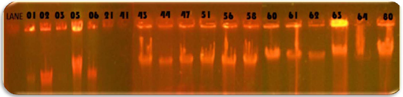
Evaluate Rna Quality
To evaluate RNA quality, electrophoresis gel was used. Before electrophoresis, all devices were treated with DEPC water. Electrophoresis was performed in 1% agarose gel. The gels were stained with ethidium bromide.
Determine The Concentration Of Extracted Rna
To determine the concentration of RNA, the BioTek Nano Drop device was used. The OD= 260/280 expresses the purity of the extracted RNA and has an inverse relationship with RNA contamination with the protein. The aforementioned ratio close to 2 represents the absence of contamination with the protein.
The ratio 260/230 was also used to check out the amount of RNA contaminated with the materials used for extraction. It is desirable that this number is also close to 2.
Synthesis Of Cdna
The master mix was made and added to each tube (all the work was done on ice). One microgram of the extracted RNA was used to synthesize cDNA. As a result, the amount of the required RNA was calculated based on its concentration in the sample and added to the tube. The tubes were transmitted to the thermocycler, and the cDNA synthesis reaction was performed according to the following procedure.
Finally, the cDNA was maintained at -20 ° C.
Primer
Primers used in these experiments were designed and tested using NCBI and Gene Runner software. By the company Sinoclon with OD about 2 was made as freeze-dried. The table 3 shows the characteristics of the primers used in this thesis.
| Table 1: Materials for synthesis of cDNA | |
| ingredients | Quantity |
| RT Buffer (x5) | µl2 |
| primer(50µM) oligo dT | µl0.5 |
| Primer:Random Hexamer(100µM) | µl5/0 |
| Reverse TranscriptaseEnzyme | µl0.5 |
| Sterilized water treated with DEPC | The final volume is 10 μL |
| Table 2: Thermosilocera device program for reverse transcription reaction | ||
| time | Temperature (°c) | The type of reaction |
| 15 min | 37 | Synthesis of single-stranded cDNA |
| 5 s | 85 | Enzyme inactivation |
Real-Time Pcr Steps
All ingredients were removed from the freezer and let to melt gently. Master Mix was kept in aluminum foil to protect it against light. According to the table 4, a mixture of the desired materials was prepared to do real time PCR with final volume of 20 microliter. All reactions were repeated twice. Special microcircuits were put in a cold box with pins and the ingredients were added to each of them according to the instructions below. All the microtubules completely were sterilized to avoid error in results. To control the contamination of the reaction during the test, one sample without cDNA was considered for each gene, as a template called the NTC.
| Table 3: Characteristics of primers | ||||
| OD | length | Tm | Sequences | primer |
| 2 | 22 | 59.2 | F:CAGACCTAGCAGACACCATGAG | OCN |
| 2 | 22 | 58.5 | R:GGACATGAAGGCTTTGTCAGAC | |
| 2 | 20 | 59 | F:CGTTTTCACGTTTGGTGGCT | ALP |
| 2 | 20 | 58.9 | R:ACCGTCCACCACCTTGTAAC | |
| 2 | 20 | 58.1 | F:GGCCACTTACCACAGAGCTA | RUNX-2 |
| 2 | 20 | 58.3 | R:AGGCGGTCAGAGAACAAACT | |
| 2 | 20 | 59.35 | F:AGCTGGACTGCGGTATTGAG | CASPASE-3 |
| 2 | 20 | 57.3 | R:ATGGCGCAAAGTGACTGGAT | |
| Table 4: Materials for Real Time PCR | ||
| Materials | Volume | Final concentration |
| Master Mix 2X | µl10 | X1 |
| cDNA | µl2 | ng/reaction100≥ |
| μM10forward primer | μl 8/0 | μM4/0 |
| μM 10 reverse primer | μl 8/0 | μM4/0 |
| water RNase- Free | µl4/6 | – |
Ingredients For Real Time Pcr
The results of Real Time PCR were obtained from the Corbett-6000 device.
Data analysis
the results of the Real Time were analyzed through a Fafel test. After analysis, one-way analysis of variance (ANOVA) was performed. Because there was a meaningful difference between the different groups, we set up the Tukey HSD Post-hoc test and compared the different groups.
Results aand Discussion
Data analysis results for RUNX-2 gene between different research groups are shown in figure 1. It can be seen that all the groups had a higher expression than the control group (first group), while only the second group had no significant difference compared to the control group. In groups three, four, five and six, there is a significant increase compared to the second group. The fourth group is also the only group that has a significant difference compared to the third group, the difference is also significant. The last significant difference was observed in this chart is for groups five and four, which expresses the decline of group five compared to group four. Data analysis results for OCN gene between different research groups are revealed in figure 2 .
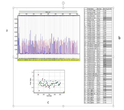
Figure 2 shows a significant increase in the expression of the three, four, five, and six groups relative to group one. And all groups except group 1 have a significant increase compared to the second group.
Figure 3 shows data analysis for ALP gene between different research groups.
The four, five, and six groups showed a significant increase compared to the control group in Figure 3. It can also be seen that groups four and five have a significant increase compared to the second group. The last result indicated in this chart is the increase in ALP gene expression in groups four, five and six compared to the third group.
Figure 4 reveals data analysis for CASPASE-3 gene between different research groups. Increasing the expression of groups 3, 4 , 5 and 6 was significantly different from that of the control group. Fourth and sixth groups also had a significant increase compared to the second group. The fourth group is the only group that has significantly increased relative to the third group. The fifth and sixth groups also had a significant decrease compared to the fourth group, which is visible on the chart.
In order to better understand the conclusions and conclusions about the results of the study, the mechanisms of the effect of probiotics should be considered first. The mechanism of action of these probiotics includes the production of inhibitor compounds, competition for binding sites, competition for food, elimination of tox Enhances the immune system by increasing the level of cytokines, immunoglobulins activating macrophages and mononucleosis, and increasing the activity of natural killer cells, self-immune modulation and the production of TNFá and interleukin 6.in receptors and ultimately boosting the immune system. The tumor necrosis factor effect has three pathways: one way reaches to caspase-3, and the second can be terminated into three different functions, namely pre-apoptosis, proliferation and cellular differentiation, and the third pathway is the anti-inflammatory route .
Caspase-3
All groups under the probiotic diet were more likely to increase than the control group because of the mechanism of probiotic effects on TNF alpha, which increases the expression of CASPASE-3. Groups under the Lactobacillus acidophilus probiotic diet, in the presence, and in the absence of radiation, gave rise to the expression of the CASPASE-3 gene in comparison to the only radiation group, since probiotics express the expression of CASPASE-3 in accordance with the above mentioned mechanism. Lactobacillus acidophilus group has increased expression in lactose-bacillus casei in the presence and absence of radiation, which probably indicates a greater effect of Lactobacillus acidophilus than Lactobacillus casei. The Lactobacillus acidophilus group has a greater expression than the Lactobacillus acidophilus group with radiation, which indicates radiation and Lactobacillus acidophilus neutralize each other. Perhaps radiation in the bone marrow may produce an immunological status that is more favorable than the pathway of differentiation and proliferation cells arrive.
RUNX-2
All groups under the probiotic diet were more likely to be exposed than the control group and the radiation group, as probiotics increase the cellular differentiation and multiply their pathways. The Lactobacillus acidophilus group has been shown to have a higher expression than Lactobacillus casei (both in the presence and in the absence of radiation), indicating a greater effect of Lactobacillus acidophilus.
ALP
Groups that have taken Lactobacillus acidophilus (both in the presence of radiation and in the absence of radiation) have a greater expression than the control group, which is probably due to the effect of Lactobacillus acidophilus. Lactobacillus casei group has a significant increase in expression in comparison with the control group, probably due to the synergistic effect of the radiation and probiotics. Because according to the radiation signal path and the osteoblastic signal path, radiation can ultimately cause cellular sensitivity so that the cell shows the counter-effects of radiation. This issue of the effects of signals can be investigated in the future. Groups that have taken Lactobacillus acidophilus (both in the presence of radiation and in the absence of it) have a higher expression than the Lactobacillus casei group; it indicates the greater effect of Lactobacillus acidophilus. The Lactobacillus casei group (in the presence of radiation) has a greater expression than Lactobacillus casei which is due to synergistic effect.
OCN
All groups under the probiotic diet have increased expression in both the control group and the radiation group because probiotic activates the second alpha-TNF pathway, resulting in differentiation and proliferation.
Conclusion
This study shows, the use of probiotics increase the expression of osteogenic genes and apoptosis, except in cases where this increase was not significantly expressed. According to the results and analysis, probiotic Lactobacillus acidophilus has a greater effect on Lactobacillus casei. That is, this probiotic increased the expression of the two groups of radiation and control. Although it also expands the gene for apoptosis. In general, the use of probiotics in this study increased the expression of osteogenic genes and apoptosis, except in cases where this increase was not significantly expressed. In some studies, we observed that radiation combined with two probiotics has different effects of synergistic and inhibitory effects for two probiotics, which is probably due to the effect of various probiotics in their signal paths, which is not exactly clear and can be tracked and researched.
Acknowledgments
This study has been supported by Tehran University of Medical Sciences.
Conflict of interest: There is no conflict of interest.
Funding: This study was funded by Tehran University of Medical Sciences (grant number: 33517).
Ethical approval: All applicable international, national, and/or institutional guidelines for the care and use of animals were followed.
References
Alwood, J. S., Shahnazari, M., Chicana, B., Schreurs, A., Kumar, A., Bartolini, A., Globus, R. K. (2015). Ionizing radiation stimulates expression of pro-osteoclastogenic genes in marrow and skeletal tissue. Journal of Interferon & Cytokine Research, 35(6), 480-487.
Britton, R. A., Irwin, R., Quach, D., Schaefer, L., Zhang, J., Lee, T., McCabe, L. R. (2014). Probiotic L. reuteri treatment prevents bone loss in a menopausal ovariectomized mouse model. Journal of cellular physiology, 229(11), 1822-1830.
Cai, S., Kandasamy, M., Rahmat, J. N., Tham, S. M., Bay, B. H., Lee, Y. K., & Mahendran, R. (2016). Lactobacillus rhamnosus GG activation of dendritic cells and neutrophils depends on the dose and time of exposure. Journal of immunology research, 2016.
Chen, M., Ona, V. O., Li, M., Ferrante, R. J., Fink, K. B., Zhu, S., Hersch, S. M. (2000). Minocycline inhibits caspase-1 and caspase-3 expression and delays mortality in a transgenic mouse model of Huntington disease. Nature medicine, 6(7), 797.
Ciorba, M. A., & Stenson, W. F. (2009). Probiotic Therapy in Radiation‐Induced Intestinal Injury and Repair. Annals of the New York Academy of Sciences, 1165(1), 190-194.
Ciorba, M. A., Riehl, T. E., Rao, M. S., Moon, C., Ee, X., Nava, G. M., Stenson, W. F. (2012). Lactobacillus probiotic protects intestinal epithelium from radiation injury in a TLR-2/cyclo-oxygenase-2-dependent manner. Gut, 61(6), 829-838.
Collins, F. L., Irwin, R., Bierhalter, H., Schepper, J., Britton, R. A., Parameswaran, N., & McCabe, L. R. (2016). Lactobacillus reuteri 6475 increases bone density in intact females only under an inflammatory setting. PLoS One, 11(4), e0153180.
Dasika, G. K., Lin, S.-C. J., Zhao, S., Sung, P., Tomkinson, A., & Lee, E. Y. P. (1999). DNA damage-induced cell cycle checkpoints and DNA strand break repair in development and tumorigenesis. oncogene, 18(55), 7883.
Demers, M., Dagnault, A., & Desjardins, J. (2014). A randomized double-blind controlled trial: impact of probiotics on diarrhea in patients treated with pelvic radiation. Clinical Nutrition, 33(5), 761-767.
Deng, H., Li, Z., Tan, Y., Guo, Z., Liu, Y., Wang, Y., Bai, Y. (2016). A novel strain of Bacteroides fragilis enhances phagocytosis and polarises M1 macrophages. Scientific reports, 6, 29401.
Dobrzyn´ska, M. M., Gajowik, A., & Radzikowska, J. (2015). The effect of in vivo resveratrol supplementation in irradiated mice on the induction of micronuclei in peripheral blood and bone marrow reticulocytes. Mutagenesis, 31(4), 393-399.
Fernandes, K. R., Ribeiro, D. A., Rodrigues, N. C., Tim, C., Santos, A. A., Parizotto, N. A., Renno, A. C. (2013). Effects of low-level laser therapy on the expression of osteogenic genes related in the initial stages of bone defects in rats. Journal of biomedical optics, 18(3), 038002.
Gamian, A. (2014). Distinct immunomodulation of bone marrow-derived dendritic cell responses to Lactobacillus plantarum WCFS1 by two different polysaccharides isolated from Lactobacillus rhamnosus LOCK 0900. Applied and environmental microbiology, 80(20), 6506-6516.
Gaur, U., & Aggarwal, B. B. (2003). Regulation of proliferation, survival and apoptosis by members of the TNF superfamily. Biochemical pharmacology, 66(8), 1403-1408.
Gerbitz, A., Schultz, M., Wilke, A., Linde, H.-J., Schölmerich, J., Andreesen, R., & Holler, E. (2004). Probiotic effects on experimental graft-versus-host disease: let them eat yogurt. Blood, 103(11), 4365-4367.
Giralt, J., Regadera, J. P., Verges, R., Romero, J., de la Fuente, I., Biete, A., Guarner, F. (2008). Effects of probiotic Lactobacillus casei DN-114 001 in prevention of radiation-induced diarrhea: results from multicenter, randomized, placebo-controlled nutritional trial. International Journal of Radiation Oncology Biology Physics, 71(4), 1213-1219.
González Pereyra, M., Dogi, C., Torres Lisa, A., Wittouck, P., Ortíz, M., Escobar, F., Torres, A. (2014). Genotoxicity and cytotoxicity evaluation of probiotic Saccharomyces cerevisiae RC016: a 60‐day subchronic oral toxicity study in rats. Journal of applied microbiology, 117(3), 824-833.
Górska, S., Schwarzer, M., Jachymek, W., Srutkova, D., Brzozowska, E., Kozakova, H., &
Hall, E., & Giaccia, A. (2006). Radiobiology for the Radiologist Lippincott Williams & Wilkins: Philadelphia.
Hayashi, M., Morita, T., Kodama, Y., Sofuni, T., & Ishidate Jr, M. (1990). The micronucleus assay with mouse peripheral blood reticulocytes using acridine orange-coated slides. Mutation Research Letters, 245(4), 245-249.
Huang, B., Guang, M., Ye, J., Gong, P., & Tang, H. (2015). Effect of increasing doses of ã-radiation on bone marrow stromal cells grown on smooth and rough titanium surfaces. Stem cells international, 2015.
Ji, J., Hu, S.-L., Cui, Z.-W., & Li, W.-F. (2013). Probiotic Bacillus amyloliquefaciens mediate M1 macrophage polarization in mouse bone marrow-derived macrophages. Archives of microbiology, 195(5), 349-356.
Jilka, R. L., Weinstein, R. S., Bellido, T., Parfitt, A. M., & Manolagas, S. C. (1998). Osteoblast programmed cell death (apoptosis): modulation by growth factors and cytokines. Journal of Bone and Mineral Research, 13(5), 793-802.
Khoshtabiat, L., & Mahdavi, M. (2015). The Role of Oxidative Stress in Proliferation and Cell Death. Journal of Mazandaran University of Medical Sciences, 25(127), 130-145.
Li, J., Yan, M., Wang, Z., Jing, S., Li, Y., Liu, G., Fan, Z. (2014). Effects of canonical NF-êB signaling pathway on the proliferation and odonto/osteogenic differentiation of human stem cells from apical papilla. BioMed research international, 2014.
Liu, C., Zhang, C., Mitchel, R. E., Cui, J., Lin, J., Yang, Y., Cai, J. (2013). A critical role of toll-like receptor 4 (TLR4) and its’ in vivo ligands in basal radio-resistance. Cell death & disease, 4(5), e649.
Mansouri-Tehrani, H.-A., Rabbani-Khorasgani, M., Hosseini, S. M., Mokarian, F., Mahdavi, H., & Roayaei, M. (2015). Effect of supplements: Probiotics and probiotic plus honey on blood cell counts and serum IgA in patients receiving pelvic radiotherapy. Journal of research in medical sciences: the official journal of Isfahan University of Medical Sciences, 20(7), 679.
Mariman, R., Kremer, B., Koning, F., & Nagelkerken, L. (2014). The probiotic mixture VSL# 3 mediates both pro-and anti-inflammatory responses in bone marrow-derived dendritic cells from C57BL/6 and BALB/c mice. British Journal of Nutrition, 112(7), 1088-1097.
Mego, M., Holec, V., Drgona, L., Hainova, K., Ciernikova, S., & Zajac, V. (2013). Probiotic bacteria in cancer patients undergoing chemotherapy and radiation therapy. Complementary therapies in medicine, 21(6), 712-723.
Meltz, M. L., Reiter, R. J., Herman, T. S., & Kumar, S. (1999). Melatonin and protection from whole-body irradiation: survival studies in mice. Mutation Research/Fundamental and Molecular Mechanisms of Mutagenesis, 425(1), 21-27.
Michelin, S., del Rosario Perez, M., Dubner, D., & Gisone, P. (2004). Increased activity and involvement of caspase-3 in radiation-induced apoptosis in neural cells precursors from developing rat brain. Neurotoxicology, 25(3), 387-398.
Mozdarani, H., & J Vessal, N. (1993). Cimetidine Can Modify The Effects Of Whole Body ‘y Irradiation On Lymphohematopoietic System. Medical Journal of The Islamic Republic of Iran (MJIRI), 7(2), 95-99.
Norizadeh Tazehkand, M., Topaktas, M., & Yilmaz, M. B. (2016). Assessment of chromosomal aberration in the bone marrow cells of Swiss Albino mice treated by 4-methylimidazole. Drug and chemical toxicology, 39(3), 307-311.
Porter, A. G., & Jänicke, R. U. (1999). Emerging roles of caspase-3 in apoptosis. Cell death and differentiation, 6(2), 99.
Rodríguez, M. L., Martín, M. M., Padellano, L. C., Palomo, A. M., & Puebla, Y. I. (2010). Gastrointestinal toxicity associated to radiation therapy. Clinical and Translational Oncology, 12(8), 554-561.
Salva, S., Marranzino, G., Villena, J., Agüero, G., & Alvarez, S. (2014). Probiotic Lactobacillus strains protect against myelosuppression and immunosuppression in cyclophosphamide-treated mice. International immunopharmacology, 22(1), 209-221.
Seal, M., Naito, Y., Barreto, R., Lorenzetti, A., Safran, P., & Marotta, F. (2007). Experimental radiotherapy‐induced enteritis: A probiotic interventional study. Journal of digestive diseases, 8(3), 143-147.
Spyropoulos, B. G., Misiakos, E. P., Fotiadis, C., & Stoidis, C. N. (2011). Antioxidant properties of probiotics and their protective effects in the pathogenesis of radiation-induced enteritis and colitis. Digestive diseases and sciences, 56(2), 285-294.
Spyropoulos, B. G., Theodoropoulos, G., Misiakos, E. P., Stoidis, C. N., Zapatis, H., Diamantopoulou, K., Machairas, A. (2013). The effect of synbiotics on acute radiation-induced diarrhea and its association with mucosal inflammatory and adaptive responses in rats. Digestive diseases and sciences, 58(9), 2487-2498.
Steeve, K. T., Marc, P., Sandrine, T., Dominique, H., & Yannick, F. (2004). IL-6, RANKL, TNF-alpha/IL-1: interrelations in bone resorption pathophysiology. Cytokine & growth factor reviews, 15(1), 49-60.
Vejdani, R. , Zali, M. R. (2003). Probiotics and the mechanism of their effect on the prevention and treatment of Human diseases. Shahid Beheshti University of Medical Sciences.Tehran.Iran, 27(4) 319-330.
Wang, Y., Zhu, G., Wang, J., & Chen, J. (2016). Irradiation alters the differentiation potential of bone marrow mesenchymal stem cells. Molecular medicine reports, 13(1), 213-223
Weiss, G. , Christensen, H. R. , Zeuthen, L.H. , Vogensen, F.K. , & Jakobsen, M. (2011). Lactobacilli and bifidobacteria induce differential interferon-b profiles in dendritic cells. Cytokine, 56(2), 520–530.
Yavropoulou, M. P., & Yovos, J. G. (2007). The role of the Wnt signaling pathway in osteoblast commitment and differentiation. Hormones-Athens-, 6(4), 279.

