1Bansal Institute of Engineering and Technology, Lucknow, Uttar Pradesh, India
2Noida Institute of Engineering and Technology, Noida, Uttar Pradesh, India
3Indian Institute of Technology, Delhi, India
Corresponding author email: ashishbiotech63@gmail.com
Article Publishing History
Received: 10/07/2020
Accepted After Revision: 24/09/2020
An attempt was made to synthesize biocompatible silver nanoparticles by using V. serpens plant extract, which can be used in different biomedical applications. The synthesis of silver nanoparticles was optimized in various physicochemical conditions and highly stable silver nanoparticles were synthesized with 8.0 mL of V. serpens leaf extract, pH 7.0, 1.0 mM AgNO3 and 37 oC. The synthesized silver nanoparticles were characterized by UV-vis spectrophotometer, Ph analysis, FE-SEM, XRD and FTIR analysis. The average size of synthesized silver nanoparticles was 60-70 nm. The findings from our study showed that the aqueous extract of V. serpens and silver nitrate solution did not show significant inhibiting activity against Enterococcus faecalis bacteria in tested concentrations but the green synthesized silver nanoparticles, inhibited the growth of Enterococcus faecalis, which is responsible for many human diseases like urinary tract infection (UTI). In addition, synthesized silver nanoparticles showed a synergetic effect on the antimicrobial activity of the standard antibiotic levofloxacin against Enterococcus faecalis bacteria under this study. These findings indicate that green synthesized silver nanoparticles can be used to control Enterococcus faecalis-related human diseases as effective growth inhibitors.
Silver Nanoparticles; Antibacterial Activity; Scanning Electron Microscopy, FTIR, XRD.
Singh A, Rai A. K, Mishra A, Nand V, Tripathi G. D, Gupta N, Saxena V, Singh V. Eco-Friendly Synthesis of Silver Nanoparticles Using V. serpens Plant, and Evaluation of Their Antibacterial Activity Against Enterococcus faecalis. Biosc.Biotech.Res.Comm. 2020;13(3).
Singh A, Rai A. K, Mishra A, Nand V, Tripathi G. D, Gupta N, Saxena V, Singh V. Eco-Friendly Synthesis of Silver Nanoparticles Using V. serpens Plant, and Evaluation of Their Antibacterial Activity Against Enterococcus faecalis. Biosc.Biotech.Res.Comm. 2020;13(3). Available from: https://bit.ly/2CrAiBL
Copyright © Singh et al., This is an open access article distributed under the terms of the Creative Commons Attribution License (CC-BY) https://creativecommons.org/licenses/by/4.0/, which permits unrestricted use distribution and reproduction in any medium, provide the original author and source are credited.
INTRODUCTION
Research and development in nanotechnology are a fast-growing field worldwide (Ahmed et al., 2016). Nanobiotechnology is currently one of the vibrant research disciplines of contemporary material sciences in which plant and plant products find an imperative use in nanoparticles synthesis (Banerjee et al., 2014; Terra et al., 2019). Such particles exhibit entirely new characteristics such as size scale, morphology and distribution compared to the larger particles of the mass material from which they were prepared (Raju et al., 2013; Elemike et al., 2020). Silver nanoparticles (AgNPs), with a broad number of applications including its antibacterial catalytic properties and its non-toxicity towards human health, are one of the most commonly used varieties of NPs (Amooaghaie et al., 2015; Ferdous et al., 2020).
Genes that couple with silver nanoparticles can inhibit the progression of the cell cycle, DNA damage and apoptosis in human cells at non-cytotoxic doses (Asharani et al., 2009; Carvalho et al., 2018). Mechanisms for toxicity caused by AgNP include genetic material, mitochondria and cell membranes. As the silver ions enter the bacterial cell, the DNA molecule is condensed and loses its ability to replicate causing bacterial cell death (Feng et al., 2000; Rajoka et al., 2020). Many techniques for synthesizing AgNPs, including physical, chemical and, most recently, biological methods have been investigated Nanoparticles are quickly synthesized in chemical synthesis which uses harmful chemical materials such as sodium borohydride, hydroxylamine, hydrazine, and ethanol (Negm et al., 2015, Amooaghaie et al., 2015; Bilal et al., 2019).
In addition, the aggregation, growth, and stability of particles are difficult to control with chemical synthesis and capping agents are required for the stabilization of nanoparticles size (Saha et al., 2017). Recently great attention has been paid to green synthesis using different parts of plant extracts for nanoparticles synthesis (Bilal et al., 2017; Hemmati et al., 2019). Biosynthetic green metal nanoparticles are increasingly being used as a result of their usability, non-toxicity and availability for large-scale processing (Nagajyothi et al., 2012; Sharma et al., 2019).
This method is superior to traditional synthesizing because it is an environmentally sustainable, inexpensive, single-step process, and simple to apply to large-scale manufacturing and also does not require high pressures, temperature, resources or toxic chemicals. Much research has been done on the green synthesis of silver nanoparticles using bacteria, fungi and various plant extracts; due to their antioxidant properties, they are able to reduce metal compounds to their respective nanoparticles (Omidi et al., 2018; Manosalva et al., 2019).
Plant extracts provide the best capping content for silver nanoparticles’ stabilization (Ahmed et al., 2016; Alfuraydi et al., 2019). Medicinal plants have been used in India since ancient times for their ability to function against both infectious and non-infectious diseases and have been properly referred to in Ayurveda. Traditional medical practitioners also increased the use of medicinal plants for the treatment of various diseases (Azaizeh et al., 2003).
Viola serpens is a plant belonging to the Violaceae family. For preparation of antibacterial medicinal plant extract, the different sections of different viola species are used. Several species within the Violaceae family are rich in cyclotides and contain phytochemical products (Gerlach et al., 2010; Chand et al., 2019). The biological synthesis of silver nanoparticles has yet to show bioreduction mechanisms through the use of V. serpens extract, though this plant is commonly used in medical usage (Gerlach et al., 2010; Chand et al., 2019).
The V. serpens extract as a bioreduction reducer which transforms silver ions into silver nanoparticles has been shown in this study. Biologically synthesized silver nanoparticles were characterized by UV-vis spectroscopy, Field emission scanning electron microscopy (FE-SEM), Fourier transform infrared spectroscopy and X-ray diffraction techniques (Chand et al., 2019). The present investigation notes the simple green synthesis of AgNPs using V. serpens leaf extracts and their anti-microbial action against Enterococcus faecalis. The method of well diffusion has been used to determine the antibacterial ability of green synthesized AgNPs (Chand et al., 2019).
MATERIAL AND METHODS
Collection of plant materials and preparation of extract: In August 2019, V. Serpens fresh mature leaves were collected from the Dehradun hilly region for plant extract preparations, green synthesis of silver nano particles and antibacterial analysis of green synthesized silver nanoparticles. The plant leaves were then washed carefully three times with water, followed by distilled water to remove dirt and dust on the leaf surface. These leaves were then dried and finely made powder with pestle and morter. Until the extraction, in a separate case, the powdered product was stored. Weighed and mixed 10 g of powdered plant material with 80 mL of deionized water and boiled it for 20 minutes. Then the material was filtered through the paper filter (Whatman No. 1). For further investigations, the prepared extract was kept at 4 °C.
Synthesis of silver nanoparticles by using the V. serpens extract: V. serpens extract was used for the synthesis of silver nanoparticles. In order to achieve the best values, it was studied the effects of the extract quantity and the silver nitrate solution concentration. By adjusting the amount of extract, the best concentration was achieved. The different concentration levels of silver nitrate solutions were then added to different amounts of 1-15 mL extract. The plant extract volume of 8 ml and silver nitrate solution concentration of 1 mmol / L were both optimally chosen. Drop by drop, eight mL plant extracts have been applied in 1 mmol / L silver nitrate solution for 40 mL. Colorless to dark gray, the silver nitrate solution revealed the development of silver nanoparticles. The solution containing silver nanoparticles was centrifuged to removal of silver nanoparticles from the other composition of a solution at 9000 RPM for 15 minutes and the deposit was prepared for the appropriate analysis.
Characterization of green synthesized silver nanoparticles: Silver nanoparticles can be characterized in a number of ways. The first and most common approach is the color changing of the solution. We used the Fourier transform infrared spectroscopy and UV-vis spectrophotometer for determination of optical properties of green synthesized silver nanoparticles. A Wensar UV-vis spectrophotometer was used for UV-vis spectral analysis and at a 300 nm / min scan speed the test sample had been scanned between 200 to800 nm. As a blank point of reference was the deionised water.
Initially pH 3.9 was recorded for 1 mmol/L aqueous solution of silver nitrate and pH changes were observed to indicate the synthesis of silver nanoparticles with plant extracts. Digital pH meter of Wensar were used to determine the pH. A high-resolution FE-SEM analysis was used for the morphological characterization of the synthesis of silver nanoparticles. The sample was made using a simple drop cover with a synthesized silver nanoparticle suspension in a clean electric glass to evaporate the solvent. At room temperature, the sample was allowed to dry. With the use of phase scan and Cu-Ka radiation (1,500 Å, 40 kV, 30 mA) in 1-2 h, X-Pert Pro diffractometer obtained the results for x-ray diffraction. The glass slide was filled with samples of biosynthesised silver nanoparticles accompanied by drying and an X-ray diffractometer eventually examined.
Antibacterial assays of synthesized silver nanoparticles: Agar well diffusion test was used to examine the antimicrobial activity of green synthesized silver nanoparticles. With the help of sterile cotton swab the tested bacteria were swabbed uniformly on two nutrient agar plates, then wells of 6 mm diameter were created by using sterile cork borer. In one plate the corresponding well has been covered by twenty-five microliters of synthesized silver nanoparticles with varying concentration (1.0 and 2.0 mM) and in other plate well has been covered by silver nitrate solution as control and plant extract.
In order to determine the combined effect of standard antibiotic (levofloxacin) and the synthesized silver nanoparticles, 15 mL of 0.5 mM antibiotic solution were mixed with 15 mL of synthesized silver nanoparticle solution (1 mM) and placed with the microorganisms tested (Enterococcus faecalis) in the corresponding agar plate well. The plates were then incubated for 24 h at 370C. The inhibition zone of synthesized silver nanoparticles, plant extract, silver nitrate solution, levofloxacin and synthesized silver nanoparticles with levofloxacin antibiotic was measured and compared individually.
RESULTS AND DISCUSSION
The biological and environmental safety of the production process in many sectors of the economy is becoming an increasing concern while metal nano-particles are being more and more used. Key methods for production of nanoparticles are chemical and physical strategies that are costly, and potentially environmentally damaging. The risks to human health are getting more severe with antimicrobial and antibiotic resistances. It must be overcome with nature’s support. Because of the urgent need for antimicrobial drugs, research on the chemistry of medicinal plants is increasingly concerned (Meenambigai et al., 2018).
Medicinal plants have been a potentially therapeutic source for thousands of years. There are a significant number of modern medicines coming from natural sources such as plants which are known for thousands of years as part of the advancement of human health care (Patil et al., 2019). In the past decade, furthermore, many organic systems such as plants have been shown to transform metal ions into metal nanoparticles through their reductive metabolite capacity (Usmani et al., 2019). Silver has been widely used in human culture for many years. It is the only element that can be tuned with its plasmon resonance at any wavelength of the visible spectrum. This element has a large number of applications, including its disinfectant property. Silver nanoparticles have shown antibacterial properties with a close connection to the microbial cell of the nanoparticles and activity based on their dimension (Ahmad et al., 2019; Fahimmunisha et al., 2020).
Basic studies showed the valuable property of silver nanoparticles, namely the special optical characteristics of catalytic operations, resonance of surface plasmon, high electric double-layer efficiency and so on, are rarely combined. Nano silver particles were commonly used as an antibacterial agent in the healthcare industry, for preserving food, cloth coatings and in specific environmental applications (Thomas et al., 2019). In this respect the synthesis of silver nanoparticles by V. serpens extracts was carried out using an efficient and versatile process. In order to achieve the best values, it was studied the effects of the extract quantity and the silver nitrate solution concentration. By adjusting the amount of extract, the best concentration was achieved. Drop by drop, plant extracts have been applied in 1 mmol / L silver nitrate solution. Colorless to dark gray, the silver nitrate solution revealed the development of silver nanoparticles.
The solution containing silver nanoparticles was centrifuged to removal of silver nanoparticles from the other composition of a solution at 9000 RPM for 15 minutes (Thomas et al., 2019).
Then the deposit was prepared for the appropriate analysis. For the detection of the optical properties of silver nanoparticles, we used UV-vis and FTIR spectrums. The surface plasmon resonance of biosynthesized silver nanoparticles displayed a peak at the UV-vise range of almost 430 nm, which correlates to the absorbance of silver nanoparticles. We compared the antibacterial properties of biosynthesized silver nanoparticles, aqueous V. serpens sample extract, standard antibiotic levofloxacin and biosynthesized silver nanoparticles with biosynthesized silver nanoparticles (Fahimmunisha et al., 2020).
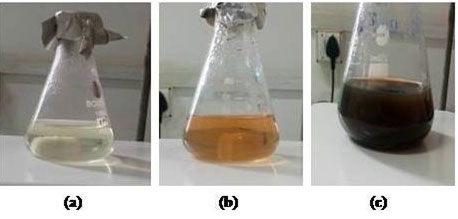
Colour change and UV–vis spectroscopy: The primary indication of the formation of silver nanoparticles is the color change of the reaction solution to dark brown (Behravan et al., 2019). Addition of silver nitrate to V. serpens leaf extract produced and instantaneous colour change from an initial, light yellow solution to dark brown solution within 45 min of reaction time (Figure 1) (Fahimmunisha et al., 2020).
Figure 1: Synthesis of silver nanoparticles by using V. serpens plant extract (a) Solution of silver nitrate and V. serpens plant extract (after 2min); (b) Solution of silver nitrate and V. serpens plant extract (after 15 min); (c) Solution of silver nitrate and V. serpens plant extract (after 45 min).
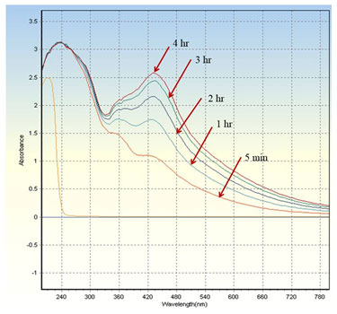
The silver nanoparticles surface plasmon resonance showed a peak centered close to 430 nm which corresponds to the absorption of silver nanoparticles (Figure 2) (Roy et al., 2019). The intensity of the reaction time increased until the fourth hour without a change in the peak wavelength, as illustrated in Figure 2.
Figure 2: UV-vis absorption spectra of green synthesized silver nanoparticles at different incubation time.
pH analysis: When a V. serpens plant’s extract was applied dropwise to the aqueous silver nitrate solution, immediately a color shift was observed, resulting in a pH reduction that can be a sign of silver nanoparticles synthesis. In this study the pH from high acidic to low acidic was observed.
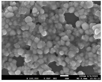
FE –SEM analysis: For size distributions and morphological characterizations of synthesized silver nanoparticles, high-resolution FE-SEM images are used. FE-SEM study showed clearly that average size and shape of silver nanoparticles was 60-70 nm and spherical, respectively (Figure 3).
Figure 3: FE-SEM image of green synthesized silver nanoparticles.
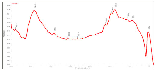
FTIR analysis: FTIR experiment is used to describe surface chemistry of synthesiszed nanoparticle. FTIR finds organic functional groups such as OH, C=O, associated with the surface of nanoparticles (Pirtarighat et al., 2019). The solution that contains synthesized silver nanoparticles was centrifuged at 8,000 rpm for 10 min to isolate silver nanoparticles from other compounds and a deposit for FTIR analysis was prepared. The FTIRspectra of the green synthesized silver nanoparticles from V. serpens are shown in Figure 4 (Transmission) and Figure 5 (Absorbance). The FTIR peaks in the range of 1640 cm-1 that are applicable to C=O bond of the carbonyl group and the stretching vibrations of different amides also appeared in this range. The FTIR peaks in the range of 3200 to 3500 cm-1 were allocated as –OH stretching in phenolics and alcohol compounds with strong hydrogen bonds. Results showed the FTIR bands at 3894.7cm-1, 3418.5 cm-1, 2923.2cm-1, 2566.4 cm-1, 2321.0 cm-1, 1622.6 cm-1, 1467.1 cm-1, 1374.3 cm-1, 1063.4 cm-1, 946.5cm-1 and 537.4cm-1. This demonstrated various functional groups linked to synthesized nanoparticles surface.
The existence of these peaks indicated that synthesized silver nanoparticles were covered by functional groups such as carboxylic acid, aldehyde, ketone, with plant secondary metabolites such as tannins, phenols, glycosides, flavonoids, terpenoids and others. The stability of the nanoparticles is responsible for the existence of these groups.
Figure 4: FTIR spectra of capped silver nanoparticles synthesized using V. serpens (Transmission).
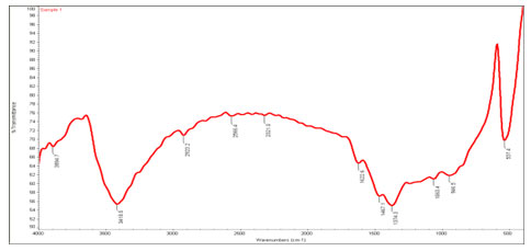
Figure 5: FTIR spectra of capped silver nanoparticles synthesized using V. serpens (Transmission).
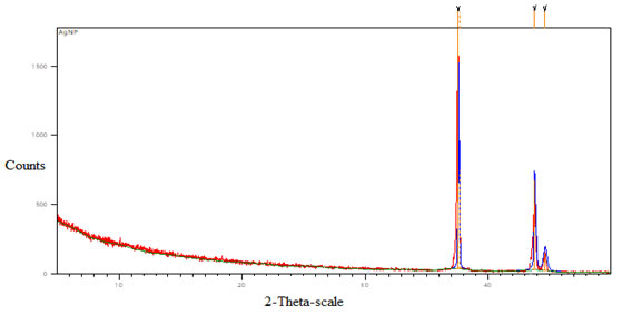
XRD analysis: Figure 6 shows the XRD pattern gained for the green synthesized silver nanoparticles using the extract of V. serpens. The XRD analysis determined the crystalline nature, phase identification, and size of the silver nanoparticles. Comparison of the XRD spectrum with the Standard confirmed that our experiments made silver particles into nanocrystals, as evidenced by the 2θ peak value of 37.5873, 37.6909, 43.7941, 44.629, corresponding to (111), (200), (220) and (311) planes for silver, respectively. Unallocated peaks may be caused by the bio-organic crystallization on the nanoparticle surface.
Figure 6: XRD pattern of silver nanoparticles synthesized by using extract of V. serpens.
Antibacterial activity of silver nanoparticles: The results of our analysis have shown that V. serpens aqueous extract and silver nitrate solution showed no substantial inhibition of Enterococcus faecalis bacteria (Figure 7a) but the silver nanoparticles synthesized by green synthesis, inhibited the growth of Enterococcus faecalis (Figure 7b). In addition, synthesized silver nanoparticles showed a synergetic effect on the antimicrobial activity of the standard antibiotic levofloxacin against Enterococcus faecalis bacteria under this study (Figure 7b) (Table 1).
Figure 7: Antimicrobial Activity of (a) Plant extract and silver nitrate solution against Enterococcus faecalis; (b) AgNP (1 mM), AgNP (2 mM), standard antibiotic levofloxacin and synthesized AgNP with standard antibiotic levofloxacin against Enterococcus faecalis.
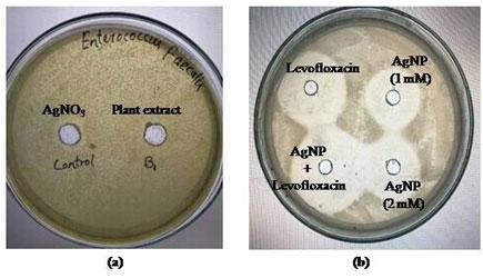
Table 1. Diameter of inhibition zone (mm)against Enterococcus faecalis.
| Variables | Diameter of inhibition zone (mm) |
| Plant extract | 10 |
| AgNO3 | 12 |
| AgNP (1 mM) | 18 |
| AgNP (2 mM) | 20 |
| Levofloxacin (0.5 mM) | 24 |
| AgNP (1 mM) + Levofloxacin (0.5 mM) | 28 |
CONCLUSIONS
The present study demonstrated an innovative way to use natural products to synthesize silver nanoparticles, which can be used in different biomedical applications. The synthesis of silver nanoparticles using V. serpens extracts as a green method without using any chemical stabilizer or reducer was reported. The findings from our study showed that the aqueous extract of V. serpens and silver nitrate solution did not show significant inhibiting activity against Enterococcus faecalis bacteria in tested concentrations but the green synthesized silver nanoparticles, inhibited the growth of Enterococcus faecalis. In addition, synthesized silver nanoparticles showed a synergetic effect on the antimicrobial activity of the standard antibiotic levofloxacin against Enterococcus faecalis bacteria under this study. Biosynthesized silver nanoparticles demonstrated a large range of antimicrobial susceptibility and thus are promising antimicrobial agents with potential biomedical uses.
ACKNOWLEDGMENTS
This research was supported by a grant from the Collaborative research and Innovative Program (CRIP) funding through TEQUIP-III, of Abdul Kalam Technical University (File no. AKTU/Dean-PGSR/2019/CRIP/27) Lucknow, Uttar Pradesh. Authors acknowledged the technical supported by Mr. Kuldeep Srivastava, Miss. Astha Pandey and Mr. Rohit Kumar (Lab Technician, Department of Biotechnology, BIET, Lucknow), Authors also highly thankful to Editor of this Journal for their valuable suggestions for strengthen this paper.
Conflicts of Interest: The authors declare no conflict of interest.
REFERENCES
Ahmad S Munir S Zeb N Ullah A Khan B Ali J and Ali S (2019) Green nanotechnology: a review on green synthesis of silver nanoparticles—an ecofriendly approach International journal of nanomedicine Vol 14 Pages 5087.
Ahmed S Ahmad M Swami B L and Ikram S (2016) A review on plants extract mediated synthesis of silver nanoparticles for antimicrobial applications: a green expertise Journal of advanced research Vol 7 No 1 Pages 17-28.
Ahmed S Saifullah Ahmad M Swami B L and Ikram S (2016) Green synthesis of silver nanoparticles using Azadirachta indica aqueous leaf extract Journal of radiation research and applied sciences Vol 9 No 1 Pages 1-7.
Alfuraydi A A Devanesan S Al-Ansari M AlSalhi M S and Ranjitsingh A J (2019) Eco-friendly green synthesis of silver nanoparticles from the sesame oil cake and its potential anticancer and antimicrobial activities Journal of Photochemistry and Photobiology B: Biology Vol 192 Pages 83-89.
Amooaghaie R Saeri M R and Azizi M (2015) Synthesis characterization and biocompatibility of silver nanoparticles synthesized from Nigella sativa leaf extract in comparison with chemical silver nanoparticles Ecotoxicology and Environmental Safety Vol 120 Pages 400-408.
Asharani P V Hande M P and Valiyaveettil S (2009) Anti-proliferative activity of silver nanoparticles BMC cell biology Vol 10 No 1 Pages 65.
Azaizeh H Fulder S Khalil K and Said O (2003) Ethnobotanical knowledge of local Arab practitioners in the Middle Eastern region Fitoterapia 74 No 1-2 Pages 98-108.
Banerjee P Satapathy M Mukhopahayay A and Das P (2014) Leaf extract mediated green synthesis of silver nanoparticles from widely available Indian plants: synthesis characterization antimicrobial property and toxicity analysis Bioresources and Bioprocessing Vol 1 No 1 Pages 3.
Behravan M Panahi A H Naghizadeh A Ziaee M Mahdavi R and Mirzapour A (2019) Facile green synthesis of silver nanoparticles using Berberis vulgaris leaf and root aqueous extract and its antibacterial activity International journal of biological macromolecules Vol 124 Pages 148-154.
Bilal M Rasheed T Iqbal H M Li C Hu H and Zhang X (2017) Development of silver nanoparticles loaded chitosan-alginate constructs with biomedical potentialities International journal of biological macromolecules Vol 105 Pages 393-400.
Bilal M Zhao Y Rasheed T Ahmed I Hassan S T Nawaz M Z and Iqbal H (2019) Biogenic nanoparticle‒chitosan conjugates with antimicrobial antibiofilm and anticancer potentialities: Development and characterization International journal of environmental research and public health Vol 16 No 4 Pages 598.
Carvalho P M Felício M R Santos N C Gonçalves S and Domingues M (2018) Application of light scattering techniques to nanoparticle characterization and development Frontiers in chemistry Vol 6 Pages 237.
Chand K Abro M I Aftab U Shah A H Lakhan M N Cao D and Mohamed A M A (2019) Green synthesis characterization and antimicrobial activity against Staphylococcus aureus of silver nanoparticles using extracts of neem onion and tomato RSC advances Vol 9 No 30 Pages 17002-17015.
Elemike E Onwudiwe D C and Ekennia A C (2020) Eco-friendly synthesis of silver nanoparticles using Umbrella plant and evaluation of their photocatalytic and antibacterial activities Inorganic and Nano-Metal Chemistry Vol 50 No 5 Pages 389-399.
Fahimmunisha B A Ishwarya R AlSalhi M S Devanesan S Govindarajan M and Vaseeharan B (2020) Green fabrication characterization and antibacterial potential of zinc oxide nanoparticles using Aloe socotrina leaf extract: A novel drug delivery approach Journal of Drug Delivery Science and Technology Vol 55 Pages 101465.
Feng Q L Wu J Chen G Q Cui F Z Kim T N and Kim J O (2000) A mechanistic study of the antibacterial effect of silver ions on Escherichia coli and Staphylococcus aureus Journal of biomedical materials research Vol 52 No 4 Pages 662-668.
Ferdous Z and Nemmar A (2020) Health Impact of Silver Nanoparticles: A Review of the Biodistribution and Toxicity Following Various Routes of Exposure International Journal of Molecular Sciences Vol 21 No 7 Pages 2375
Gerlach S L Burman R Bohlin L Mondal D and Göransson U (2010) Isolation characterization and bioactivity of cyclotides from the Micronesian plant Psychotria leptothyrsa Journal of natural products Vol 73 No 7 Pages 1207-1213.
Hemmati S Rashtiani A Zangeneh M Mohammadi P Zangeneh A and Veisi H (2019) Green synthesis and characterization of silver nanoparticles using Fritillaria flower extract and their antibacterial activity against some human pathogens Polyhedron Vol 158 Pages 8-14.
Manosalva N Tortella G Diez M C Schalchli H Seabra A B Durán N and Rubilar O (2019) Green synthesis of silver nanoparticles: effect of synthesis reaction parameters on antimicrobial activity World Journal of Microbiology and Biotechnology Vol 35 No 6 Pages 88.
Meenambigai K Kokila R and Kumar A N (2018) Nilgirianthus ciliatus mediated environment friendly extracellular synthesis of AgNps to exact its potential against Dengue vector Aedes aegypti and Staphylococcus aureus Journal of Entomological and Acarological Research Vol 50 No 1.
Nagajyothi P C Lee S E An M and Lee K D (2012) Green synthesis of silver and gold nanoparticles using Lonicera japonica flower extract Bull Korean Chem Soc Vol 33 No 8 Pages 2609-2612.
Negm N A Tawfik S M and Abd-Elaal A (2015) Synthesis characterization and biological activity of colloidal silver nanoparticles stabilized by gemini anionic surfactants Journal of Industrial and Engineering Chemistry Vol 21 Pages 1051-1057.
Omidi S Sedaghat S Tahvildari K Derakhshi P and Motiee F (2018) Biosynthesis of silver nanocomposite with Tarragon leaf extract and assessment of antibacterial activity Journal of Nanostructure in Chemistry Vol 8 No 2 Pages 171-178.
Patil M P Bayaraa E Subedi P Piad L L A Tarte N H and Kim G D (2019) Biogenic synthesis characterization of gold nanoparticles using Lonicera japonica and their anticancer activity on HeLa cells Journal of Drug Delivery Science and Technology Vol 51 Pages 83-90.
Pirtarighat S Ghannadnia M and Baghshahi S (2019) Green synthesis of silver nanoparticles using the plant extract of Salvia spinosa grown in vitro and their antibacterial activity assessment Journal of Nanostructure in Chemistry Vol 9 No 1 Pages 1-9.
Rajoka M S R Mehwish H M Zhang H Ashraf M Fang H Zeng X and He Z (2020) Antibacterial and antioxidant activity of exopolysaccharide mediated silver nanoparticle synthesized by Lactobacillus brevis isolated from Chinese koumiss Colloids and Surfaces B: Biointerfaces Vol 186 Pages 110734.
Raju D Hazra S and Mehta U J (2013) Phytosynthesis of silver nanoparticles by Semecarpus anacardium L leaf extract Materials Letters Vol 102 Pages 5-7.
Roy A Bulut O Some S Mandal A K and Yilmaz M D (2019) Green synthesis of silver nanoparticles: biomolecule-nanoparticle organizations targeting antimicrobial activity RSC advances Vol 9 No 5 Pages 2673-2702.
Saha J Begum A Mukherjee A and Kumar S (2017) A novel green synthesis of silver nanoparticles and their catalytic action in reduction of Methylene Blue dye Sustainable Environment Research Vol 27 No 5 Pages 245-250.
Sharma V Kaushik S Pandit P Dhull D Yadav J P and Kaushik S (2019) Green synthesis of silver nanoparticles from medicinal plants and evaluation of their antiviral potential against chikungunya virus Applied microbiology and biotechnology Vol 103 No 2 Pages 881-891.
Swamy M K Akhtar M S Mohanty S K and Sinniah U R (2015) Synthesis and characterization of silver nanoparticles using fruit extract of Momordica cymbalaria and assessment of their in vitro antimicrobial antioxidant and cytotoxicity activities Spectrochimica Acta Part A: Molecular and Biomolecular Spectroscopy Vol 151 Pages 939-944.
Terra A L M Kosinski R D C Moreira J B Costa J A V and Morais M G D (2019) Microalgae biosynthesis of silver nanoparticles for application in the control of agricultural pathogens Journal of Environmental Science and Health Part B Vol 54 No 8 Pages 709-716.
Thomas B Vithiya B Prasad T Mohamed S B Magdalane C M Kaviyarasu K and Maaza M (2019) Antioxidant and photocatalytic activity of aqueous leaf extract mediated green synthesis of silver nanoparticles using Passiflora edulis f flavicarpa Journal of Nanoscience and Nanotechnology Vol 19 No 5 Pages 2640-2648.
Usmani A Mishra A Jafri A Arshad M and Siddiqui M A (2019) Green Synthesis of Silver Nanocomposites of Nigella sativa Seeds Extract for Hepatocellular Carcinoma Current Nanomaterials Vol 4 No 3 Pages 191-200.

