Department of Electronics and Communication Engineering, M. Kumarasamy College of Engineering, Karur, Tamilnadu, India
Corresponding author Email: abiramit.ece@mkce.ac.in
Article Publishing History
Received: 22/12/2018
Accepted After Revision: 03/03/2019
Now a days most of the females are facing the problems of infertility in the age group of 22 to 35. In order to analyze and classify the problems, one can start with making the decision of comparing the normal ovary and affected ovary structurally using advanced techniques. The two images have to be taken and can be compared using the accurate images for detecting the affected ovary or its areas. Using the feature extraction method, the image can be further diagnostically extracted. Then the image can be processed by segmentation and finally it can be detected and subjected to a diseased degree classification. The method used here is Support Vector Machine (SVM) with Fuzzy Logic. SVM algorithm is used to classify and regression analysis. It works on the concept of partial truth, where the given value might range between absolutely true and absolutely false. Histogram adjustment calculation is connected to get a complexity improved picture. The wiener channel is the greatest appropriate decision for diminishment of clamor in ovarian using the ultrasound picture. The results are distinguished and then taken for physical request from the experts. Exactness of system can be approximately 95%. Subsequently, this figuring can be customized by screening of Poly Cystic Ovarian Syndrome problems.
Polycystic Ovarian Detection (Pcod), Body Mass Index (Bmi), Follicle-Stimulating Hormone (Fsh) And Luteinizing Hormone (Lh)
Abirami T, Rajan S. P. Detection of Poly Cystic Ovarian Syndrome (PCOS) Using Follicle Recognition Techniques. Biosc.Biotech.Res.Comm. 2019;12 (1).
Abirami T, Rajan S. P. Detection of Poly Cystic Ovarian Syndrome (PCOS) Using Follicle Recognition Techniques. Biosc.Biotech.Res.Comm. 2019;12(1). Available from: https://bit.ly/2MBcrEf
Copyright © Abirami and Rajan, This is an open access article distributed under the terms of the Creative Commons Attribution License (CC-BY) https://creativecommns.org/licenses/by/4.0/, which permits unrestricted use distribution and reproduction in any medium, provide the original author and source are credited.
Introduction
Many females of reproductive age suffer with the disorders Polycystic Ovarion Syndrome commonly known as PCOS. It is a heterogenous endocrine disorder and is most common among the young generation. Though the exact cause for this disorder is unknown factors, like hormonal imbalance, central or overall obesity and having Body Mass Index greater than 24 can be considered as some of its causes (Abirami et al. 2018). This may result is abnormal ovulation, with absence of or irregular menstrual cycle, infertility conditions called as called as amenorrhea, (Vijayprasath et al. 2015). It also results in release of additional quantity of androgenic hormones and it’s effects are frustrating. This leads to acne and insulin resistance which is often accompanied by obesity, high cholesterol levels and type 2 diabetes. The diagnosis procedures usually followed are the ovarian ultrasound images which show ovaries with the disorder having multiple cysts. But it cannot be stated as the only symptom for the disorder. Huge variety in the side effects and seriousness of the problem has been seen in some females, (Annakamatchi et al. 2018).
Few methodologies are used to identify the PCOD problems. These are medical, biological, and the ovarian ultrasound imaging which are used to fi nd the problem. A patient can be examined as affliction from PCOD or not, if the any of two from the three situations are given below: (1) Prolonged ovulation or portrayed by sporadic menses series, (2) Additional measures which are caused by membrane breakdown, besides raised serum chemicals and (3) Occurrence of polycystic ovaries realized by the image of gynecologic ultrasounds, (Choudhary et al. 2015).
For an ordinary and sound ovary just a single follicle develops in a size of 20mm in width, develops and is prepared for ovulation, under right levels of FSH and LH hormones1. But in the case of PCOS affected ovary many small follicles of size 12 or more than that and about 2-9 mm in diameter is found dispersed along the periphery of the ovary which is medically termed as ‘necklace formation’. Hence the volume of ovary will raise above 10 cm3 in PCOS affected patients (Dinesh et al. 2015a). Figure 1 indicates ultrasound picture of typical ovary demonstrating just a single follicle prepared for ovulation and fi gure 2 depicts the scenario in which various little follicles are found in the edge of ovary (Dinesh et al. 2015b).
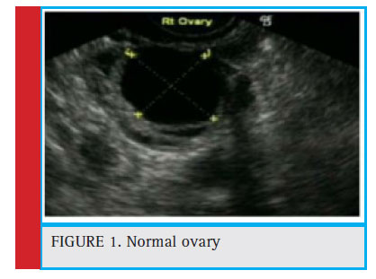 |
Figure 1: Normal ovary |
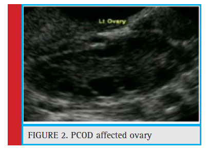 |
Figure 2: PCOD affected ovary |
In this paper, the images have been tested and affected by PCOS tended to by all the three techniques. Ovarian ultrasound pictures have been gathered for around 20 patients either with or without PCOS side effect alongside values for the measuring parameters of PCOS (Essam et al. 2018). This process is carried out with a 6MHz ultrasound transducer. Techniques such as enhancement of contrast and then filters are used to increase the excellence of the images can be obtained. Highlight extraction incorporates were done by removing dull or splendid highlights from the fi rst picture. Divisional identification was carried from which the coveted areas are chosen utilizing qualities such as size, area, compactness and so forth, (Kavitha et al. 2017). The facts were utilized for the compactness which was constructed from the count of follicles acquired in the overhead process and medical and biological information was gathered from specialists, characterization was completed by the algorithm following the method of Keerthi et al. (2018).The Process Flow of PCOS detection is shown in figure.3
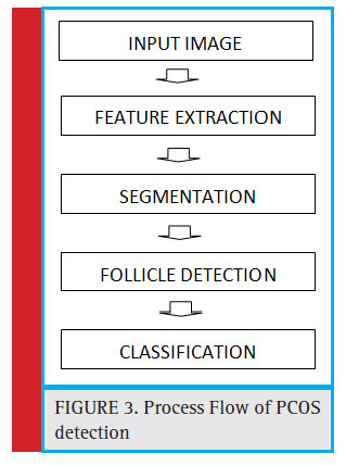 |
Figure 3: Process Flow of PCOS detection |
The outcomes are noted and it is contrasted with finding the manual location with figure exactness of the proposed calculation. The stream of process is given beneath in the figure.3. Medical ultrasonography produces poor quality images of high noise content and low contrast. For upcoming processes such as feature extraction, segmentation and follicle detection high quality images are required (Vijayprasath et al. 2012). Hence it is necessary to remove the noise and improve the contrast of images (Rajan 2014). The preprocessing technique involves improving the quality of images with the help out of distinction improvement and filtering methodologies. Contrast enhancement which was the first step in preprocessing improves the contrast of ovarian ultrasound images using different global and local techniques (Rajan 2015). This paper focuses to contrast enhanced image and performances are calculated based on Signal-to-Noise ratio (SNR) given in equation 1 where is the variance of original image and is the variance of enhanced image.

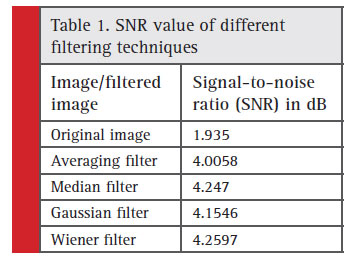 |
Table 1: SNR value of different filtering techniques |
In table 1, the signal to noise ratio of different filtering methods are illustrated. From the table original image is a ultrasound image. If processing of original image may give wrong results. So that the original image can be processed for filtering to remove the noise and enhance the contrast for further processing. Here different filtering methods are carried out. Signal to Noise Ratio (SNR) is calculated for various methods of filtering. Examples Median Filtering, Averaging Filtering, Gaussian Filtering, Wiener Filtering. The output of preprocessing stage is given the figure
In the ovarian image using the ultrasound picture, aside since follicles, endometrial veins, lymph hubs, nerve strands and stroma is seen (Ramakrishnan et al. 2018). Hence, it is to be reduced the incorrect discovery, include removals are finished through utilizing Multistage morphological method, for the situation Top-cap change. White and Dull highlights dismiss be removed after the picture and later the subsequent picture is deducted from sole picture to become differentiate improved picture on (Sukanesh et al. 2013). In the calculation the change assistances by the extraction of brilliant highlights since the foundation by means of utilizing morphological initial and finish task in an organizing component individually5. The organizing component utilized as a part of this case is a plate organizing component (Ramesh et al. 2018). The splendid best cap change is characterized as the contrast between the information picture and its opening by the basic component. Then again, dark best cap change is characterized as the contrast between shutting by same basic component and information picture6. On the off chance that is an organizing component, at that point splendid and dull best cap changes can be given as in equation 2 and 3 individually (Rajan et al. 2013).

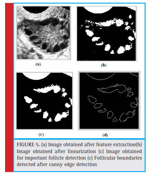
Picture division is utilized to distinguish wanted highlights with the goal that the consequential picture being less demanding to investigate7 (Ribana et al. 2018). The different methodologies have been suggested and second-hand for picture division like edge area, edge, etc., various edge distinguishing proof practices can be utilized for the division of ovarian picture. In the edge identification method, focuses in picture are documented where picture brilliance changes pointedly or consumes discontinuities, in the picture, get after linearization8. In binaries picture, pixel esteems at all the specific focuses or the areas alteration all of a sudden from 0 to 255 (i.e. dull to brilliant) and the other way around. Every one of these focuses is sorted out into an arrangement of bended line sections to recognize the edges (Rajan et al. 2012).
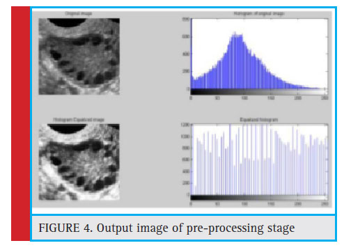 |
Figure 4: Output image of pre-processing Stage |
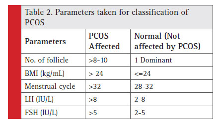 |
Table 2: Parameters taken for classifi cation of PCOS |
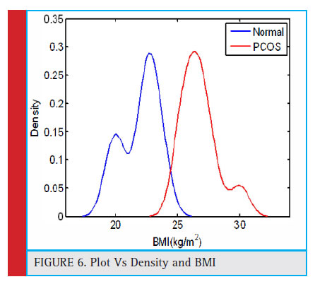 |
Figure 6: Plot Vs Density and BMI |
Vast number of highlights is recognized after binarization what’s more, edge identification (Sivagurunathan et al. 2018). To isolate out imperative follicles from identified highlights, criterion for example highest and lowest amount of follicles, zone, eroticism as well as smallness must be additionally taken for the calculation. Consider the follicles are round fi t as a fiddle, isolate locales will associate with 4-80 mm2 for PCOS ovary and around 314 mm2 for common place ovarian follicle. Although it is accredited with the purpose of the follicles are circuitous alive and well, whim is generally comparable to 1 (Sivaranjani et al. 2018). Thinking about every one of these qualities for edge, region of each identified area or highlight is fi gured and number of essential follicles identifi ed is discovered. In Figure 4, the Image can be obtained after all the process image extraction (Sukanesh et al. 2010a).
The nu mber of ovarian follicles is not an only matter for the problem of PCOS and also it has to measure the parameters of BMI, cycle length, post menstrual FSH and LH10 (Sukanesh et al. 2010b). In every image of the parameter is close at hand of number of follicles is measured and it shown patients are having the problem otherwise it is not showing it to the maximum level14. Here comparing the parameters of Normal Patient with the PCOS affected patients are given in table 1. The parameters were No. of follicle, BMI (kg/mL), Period Cycle, LH (lU/L), FSH (lU/L). The follicle may present in normal patient also but the number should be only one, and value of BMI (Body Mass Index) is about to be less than or equal to 2415. The menstrual cycle also taken as 28 days to 32 days if it is more than 32 days three will be a problem of PCOS. The value of LH and also FSH
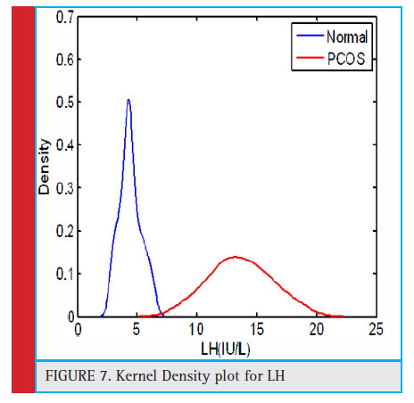 |
Figure 7: Kernel Density plot for LH |
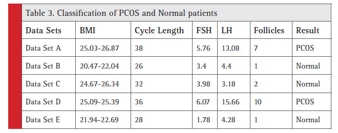 |
Table 3: Classification of PCOS and Normal patients |
are also compared with PCOs and the Normal patients. With these above parameters are helped to classify the problems to be taken 16.
The different set of images are monitored and preliminary 15 number of data’s is used for setting up for computation, course of action of subsequently 5 instructive lists is finished. The system is frequently for instructive record of the impressive number of patients. It is used for planning the data sets and gathering of all the information. Outcomes acquired be contrasted and individuals gotten by physical grouping and commencing to specialists. In the figure 6, it shows the graph between BMI vs density. It is used to predict PCOS ovary or normal ovary.
The both information’s are taken into account and number of follicles are identified and in the above process are utilized for order utilizing in SVM calculation. Motion to-commotion proportions (SNR) are computed for unique picture and for each separated picture and outcomes are gotten by way of appeared in the table 1. From this, it can be derived that wiener channel is the greatest appropriate decision for diminishment of clamor in ovarian using the ultrasound picture. In the figure 7, it shows the graph between kernel densities with LH. The Wiener sifted picture is utilized for additional processing.
In the given table 3, already the parameter taken for classification of PCOS illustrated in Table 2. 5 different data sets are taken to find the values of BMI, Menstrual Cycle, FSH, LH, No of Follicles. After finding the values of all the parameters comparing with the table No:2 and it will help to decide or classify the Normal patient or PCOS affected patients. Training sets are available to make decision of PCOS affected or not.
At last it has been concluded with a robotized procedure for Polycystic Ovarian Syndrome area using follicle affirmation as well as portrayal by means of Support Vector Machine. Picture is to be preprocessed and it is (differentiate upgrade and sifting) utilized for enhancing nature of the picture. Highlights are removed utilizing Multiscale morphological approach and brilliant best cap change. The picture is binarized and portioned utilizing watchful edge discovery method. Critical follicles are isolated from different areas utilizing region and unpredictability limit. Grouping of the considerable number of information is fi nished utilizing the SVM algorithm for calculation. The results have been achieved and the accuracy is about 95%. In the same way, this estimation has been done by enough number of modified screenings of PCOS.
References
Abirami T,Ramesh L (2018) Bio-Nano Things: Organically stimulated Bio-Cyber Interface Architecture, International Journal of Pure and Applied Mathematics 119(15), 3257-3262
Annakamatchi M, Keralshalini V (2018) Design of Spiral Shaped Patch Antenna for Bio-Medical Applications. International Journal of Pure and Applied Mathematics 118(11): Pages 131-135.
Choudhary, J. Chowdhary, M.L. Swarankar, et al., (2015) Predictive value of subendometrial–endometrial blood flow assessment by transvaginal 3D power doppler on the day of HCG on clinical outcome of IVF cycles, Int. J. Res. Med. Sci. 3 3114.
Dinesh T, Palanivel S (2015a) Systematic Review on Wearable Driver Vigilance System with Future Research Directions. International Journal of Applied Engineering Research 2(2): Pages 627-632.
Dinesh T, Palanivel S (2015b) Statistical Investigation of EEG Based Abnormal Fatigue Detection using LabVIEW. International Journal of Applied Engineering Research 10(43): Pages 30426-30431
Essam R. Othman, Karim S.Abdullah, Ahmed M.Abbas, Mostafa Hussein(2018), Elwany Elsnosy, Ihab H.EI-Nashar, “Evaluation of endometrial and subendrometrial vascularity in obese women with polycystic ovarian disease” Middle East Fertility Society Journal 23, pages 324–330
Kavitha V, Palanivel Rajan S (2017) Diagnosis of Cardiovascular Diseases using Retinal Images through Vessel Segmentation Graph. Current Medical Imaging Reviews 13(4).
Keerthi S, Dhivya S (2017) Comparison of RVM and SVM Classifier Performance in Analysing the Tuberculosis in Chest X Ray. International Journal of Control theory and Applications 10(36): Pages 269-276.
Palanivel Rajan S (2014) A Signifi cant and Vital Glance on “Stress and Fitness Monitoring Embedded on a Modern Telematics Platform. Telemedicine and e-Health Journal 20(8): Pages 757-758.
Palanivel Rajan S (2015) Review and Investigations on Future Research Directions of Mobile Based Tele care System for Cardiac Surveillance. Journal of Applied Research and Technology 13(4): Pages 454-460.
Ramakrishnan P, Sivagurunathan P T (2018) Wireless Patient Monitoring systems. International Journal of Pure and Applied Mathematics 118(11): Pages 761- 766.
Ramesh L, Abirami T (2018) Segmentation of Liver Images Based on Optimization Method. International Journal of Pure and Applied Mathematics 118 (8) : Pages 401-405.
Rajan S P, Sukanesh R (2013) Viable Investigations and Real Time Recitation of Enhanced ECG Based Cardiac Tele-Monitoring System for Home-Care Applications: A Systematic Evaluation. Telemedicine and e-Health Journal 19(4): Pages 278-286.
Rajan S P, Sukanesh R, Vijayprasath S (2012) Performance Evaluation of Mobile Phone Radiation Minimization through Characteristic Impedance Measurement for Health-Care Applications. IEEE Digital Library Xplore.
Ribana K, Pradeep S (2018) Contrast Enhancement Techniques for Medical Images. International Journal of Pure and Applied Mathematics 118: Pages 695-700.
Sivagurunathan P T, Ramakrishnan P (2018) A Survey on Wearable health Sensor– Based System Design. International Journal of Pure and Applied Mathematics 118(08): Pages 383-385.
Sivaranjani S, Kaarthik K (2018) Medical Imaging Technique to Detect Tumor Cells. International Journal of Pure and Applied Mathematics, 118(11): Pages 399 – 404.
Sukanesh R, Gautham P, Rajan S P, Vijayprasath S (2010a) Cellular Phone based Biomedical System for Health Care. IEEE Digital Library Xplore: Pages 550-553.
Sukanesh R, Palanivel Rajan S (2013) Experimental Studies on Intelligent, Wearable and Automated Wireless Mobile Tele- Alert System for Continuous Cardiac Surveillance. Journal of Applied Research and Technology 11(1): Pages 133-143.
Sukanesh R, Rajan S P, Vijayprasath S (2010b) Intelligent Wireless Mobile Patient Monitoring System. IEEE Digital Library Xplore: Pages 540-543.
Vijayprasath S, Sukanesh R, Rajan S P (2012) Experimental Explorations on EOG Signal Processing for Real Time Applications in LabVIEW. IEEE Digital Library Xplore.
Vijayprasath S, Rajan S P (2015) Performance Investigation of an Implicit Instrumentation Tool for Deadened Patients Using Common Eye Developments as a Paradigm. International Journal of Applied Engineering Research 10(1): Pages 925-929.

