1Postgraduate Student of Prosthodontics, Faculty of Dentistry, Shahed University, Tehran, Iran
2Associate Professor of Oral and Maxillofacial Radiology, Faculty of Dentistry, Shahed University, Tehran, Iran
3Assistant Professor of Oral and Maxillofacial Radiology, Faculty of Dentistry, Shahed University, Tehran, Iran
4Oral and Maxillofacial Radiologist, Orthodontic Resident at Rutgers School of Dentistry, New Jersey, USA
5Research Member, Dental Research Center, Dentistry Research Institute, Tehran University of Medical Sciences, Tehran, Iran
6Assistant Professor of Oral and Maxillofacial Radiology, Faculty of Dentistry, Shahed University, Tehran, Iran
Corresponding author email: Hoda.Rahimi57@gmail.com
Article Publishing History
Received: 10/04/2024
Accepted After Revision: 21/06/2024
The present study aimed to compare the two dental age (DA) estimation methods of Cameriere and Demirjian among 6-14 year-old children in Tehran in 2017-2018. This cross-sectional analytical study involved 306 panoramic images from 153 girls and 153 boys. The DA of participants was estimated by Cameriere’s and Demirjian’s methods. The data were statistically analyzed by the paired sample t-test, repeated measures ANOVA, and the independent t-test. Finally, a formula suitable for Iranian society was developed based on the results of regression analysis. The mean age estimation error was +0.89 years for Demirjian’s method (+0.86 in boys and +0.93 in girls) and -0.20 years for Cameriere’s method (-0.20 in boys and -0.10 in girls).
There was a significant difference between the DA calculated by Cameriere’s and Demirjian’s methods and the chronological age. There was no significant difference between Cameriere’s and Demirjian’s methods in this context. The formula developed in this study could estimate the age of participants with an accuracy of above +0.008 (+0.009 in boys and +0.006 in girls). However, the results indicated no significant difference between the proposed formula and Cameriere’s method in the accuracy of age estimation.The accuracy of Cameriere’s method was higher than that of Demirjian’s method, but the formula proposed for Iranian society was more accurate than both of them. The Cameriere method underestimated and the Demirjian method overestimated the age.
Chronological Age, Cameriere’s Method, Demirjian’s Method, Dental Age
Mohammadi Z, Shahab S, Azizi Z, Rahimi H, Kharazifard M.J, Kavosi A. Dental age assessment using Demirjian and Cameriere's methods in an Iranian population in 2017-2018. Biosc.Biotech.Res.Comm. 2024;17(2).
Mohammadi Z, Shahab S, Azizi Z, Rahimi H, Kharazifard M.J, Kavosi A. Dental age Assessment Using Demirjian and Cameriere’s Methods in an Iranian Population in 2017-2018. Biosc.Biotech.Res.Comm. 2024;17(2). Available from: <a href=”https://shorturl.at/kQFYF“>https://shorturl.at/kQFYF</a>
INTRODUCTION
Age estimation plays a major role in the diagnosis of endocrine problems1, forensic dentistry, pediatric dentistry, and orthodontic treatment plans. Age estimation is used by orthodontists in specific orthodontic treatments and by pediatricians for evaluating teeth evolution (increased or decreased) in children with specific diseases. (Feijoo et al 2012, Butti, et al. 2009, Koshy and Tandon 1998, Hauk et al 2001, Graber et al 2012). Time plays an important role in the success of orthodontic treatment. Estimation of DA and skeletal age can help physicians determine the right time to begin treatments. Although there are skeletal and sexual indicators to determine growth rate, dental indicators are more commonly used for this purpose because they are less affected by nutritional and endocrine status, especially in children and adolescents, (Mani et al 2008, Molina et al 2020).
It has been shown that there is a relationship between dental calcification stages and skeletal development. DA is determined through Andrade et al (2019) methods: (1) assessing teeth growth in the oral cavity, and 2) examining tooth formation stages in X-rays. The first method is limited to children who haven’t reached mixed dentition, and is affected by factors like premature tooth loss, ankylosis, or dental arch stenosis. Consequently, the second method is preferred for its broader applicability and reliability, (Baccetti et al 2005, Andrade et al 2019).
Developed in 1973, Demirjian’s method is one of the most widely used methods in measuring dental development. Demirjian et al (1973) studied 7 permanent teeth on the left side of the mandible from 2928 panoramic radiographs of 3-16-years-old Canadian-French people and then developed a table of indicators and a conversion table. To solve the previous problems, Demirjian et al. (1973) increased the number of samples to 2047 boys and 2349 girls. They classified the course of dental development (from dental bud to completion) under 8 stages named A through H, ( Demirjian and Goldstein 1976). Cameriere et al. (2006) proposed a novel age estimation method based on their study of 213 boys and 242 girls in Italy, which aimed to determine age by examining the relationship between age and the diameter of dental apices. The studies have shown that the age estimated by this method is very close to chronological age, (Rai et al 2006, Camerie et al, 2007, 2008).
According to the literature, the course of dental development varies in different populations (even between different cities of a country) at different times. 16 These differences may be attributed to genetic or environmental factors such as socioeconomic status, nutrition, diet, and lifestyle or changes that occur over time in populations, (Marjatta et al 1988, Leurs et al 2005).The purpose of the present study was to compare dental age (DA) estimated through Cameriere’s and Demirjian’s methods with chronological age within the population aged 6-14 years in Tehran during 2017-2018. Additionally, the study aimed to develop a formula for this specific population using a regression equation.
MATERIAL AND METHODS
In this study, 306 panoramic radiographs of 6-14-years-old patients were collected from 4 oral and maxillofacial imaging centers in Tehran. The images were taken from March 2017 to June 2018. The inclusion criteria were as follows: Patients without developmental defects and systemic diseases affecting the growth. Panoramic images of high quality without any distortion. Patients with no missing teeth. Patients with a known date of birth. Lack of environmental factors affecting calcification such as inflammation or injury at the site. To begin, the chronological age of participants was determined by subtracting the radiography date from their date of birth, recorded with precision to two decimal places. Then participants were categorized into nine age groups, each spanning a one-year interval, with an equal number of participants across all age groups and both genders. Digital radiographs were taken using Cranex D (Finland, Helsinky, Sordex) and Planmeca (Finland, Helsinky, Planmeca).
Demirjian’s method for age estimation was performed by 3 persons (two trained senior dental students and an oral and maxillofacial radiologist). In case of disagreement between the two observers, the radiologist’s opinion was recorded as DA.According to Demirjian’s method, 7 permanent teeth on the left side of the mandible were evaluated based on the course of dental development, which will be explained below (Figure 1). Then the table of indicators was used to give a number to each tooth considering its developmental stage (Table 1).
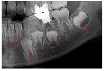
Finally, the 7 numbers obtained were added up to calculate the total maturity score, which ranges between 0 and 100. This score was converted into DA using the relevant tables and then compared with chronological age. To determine the error rate of Demirjian’s age estimation method, the obtained DA was subtracted from chronological age. If DA was greater than chronological age, the values were reported with a positive mark. Otherwise, they were recorded with a negative mark.To estimate DA by Cameriere’s method, images were opened in Adobe Photoshop-2018 to match them in terms of size and resolution. All images were observed and evaluated in a 13.3-inch screen MacBook Air 2017 (1440 × 900) and Intel HD Graphics MB 1536, 6000 graphics. Then the images were measured by two observers. In case of significant disagreement, the measurement was repeated by a radiologist. In Cameriere’s method, the 7 permanent teeth on the left side of the mandible were measured as explained in the following formula:
Age = 8.971 + 0.375 g + 1.631 X5 + 0.674 N – 1.034 S – 0.176 N.S
In single-rooted teeth (Ai, i=1…5), the distance between the inner wall of open-apex teeth was measured. In multi-rooted teeth, the mean distance between the inner walls of both roots (Ai, i=6,7) was separately calculated, and added up and then the length of the teeth was measured.
To neutralize the effects of magnification and X-ray angle, the dimensions were normalized by dividing them by the length of the tooth (Li, i=1…7) (Xi = Ai/Li = 1…7). Moreover, the number of fully developed closed-apex teeth in each participant was counted and recorded as N0.
Finally, the normalized sum of the number of open-apex teeth (Xi), the number of open-apex teeth (S), the number of closed-apex teeth (N0), and gender (0 for girls and 1 for boys) were inserted into SPSS.To estimate age by Cameriere’s method, all variables (N0, SN0, g, X1, X2… X9) were defined and put in formulas proposed by Cameriere for Italian society. The obtained figure was subtracted from chronological age to calculate the estimation error.Based on the estimation error of Cameriere’s method, a formula was developed for Iranian society. To this end, a regression equation was considered with age as the dependent variable and N0, SN0, S, X1… X7, and gender as independent variables. According to stepwise regression, variables N0, X1, X3, X7, SN0, and gender remained in the model, and the following regression line equation was obtained: Age = 9.309 + 0.636 g – 3.852 X1 – 2.505 X3 – 1.007 X7 + 0.664 N – 0.265 SN0
To reduce estimation error, the observers were unaware of the participants’ age in both methods. In addition, the chronological age of participants was obtained by subtracting the radiography date from the date of birth.
Data analysis :Statistical analyses were performed in SPSS 24. Repeated measures ANOVA and the independent t-test were employed to compare the two studied methods in terms of the absolute value of estimation error and compare the absolute value of estimation error in girls and boys, respectively. Also, the DA obtained from Cameriere’s and Demirjian’s methods was compared with chronological age (overestimation or underestimation) by the paired sample t-test. *Demirjian. error: Demirjian’s method error / ** Demirjian.error.abs: absolute value of Demirjian’s method error,*Cameriere. error: Cameriere’s method error / ** Cameriere.error.abs: absolute value of Cameriere’s method error.
RESULTS AND DISCUSSION
The research involved the analysis of 306 panoramic images, comprising 153 girls and 153 boys aged between 6 and 14 years. The participants were divided into different age groups, each spanning a one-year interval, with each age group consisting of 17 boys and 17 girls.
The results of repeated measures ANOVA for investigating the accuracy of Cameriere’s and Demirjian’s methods in the estimation of participants’ chronological age are shown in Table 2. As shown in Table 2, Demirjian’s method overestimated chronological age by 0.89 years, on average (0.86 in boys and 0.92 in girls). The absolute value of the mean estimation error for this method was 1.12 years (1.15 in boys and 1.09 in girls). Furthermore, Table 2 indicates that Cameriere’s method underestimated chronological age by 0.20 years, on average (0.29 in boys and 0.10 in girls). The absolute value of the mean estimation error for this method was 0.75 years (0.79 in boys and 0.71 in girls).
Table 3 presents the results of the paired sample t-test to investigate the degree of overestimation or underestimation of age by Cameriere’s and Demirjian’s methods. The results showed that the DA estimated by both Cameriere’s and Demirjian’s methods was significantly different from chronological age (p>0.05). The results obtained from the formula proposed in this study are shown in Table 4. Based on Table 4, our formula underestimated chronological age by 0.0008 years, on average (0.0009 in boys and 0.0006 in girls). The absolute value of the mean estimation error for this formula was 0.71 years (0.76 in boys and 0.67 in girls).
Table 5 shows the results of comparing Cameriere’s and Demirjian’s methods and our formula in terms of mean error and their absolute values for both boys and girls.
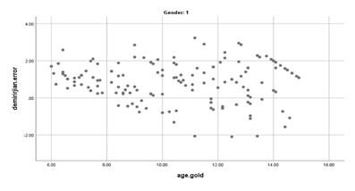
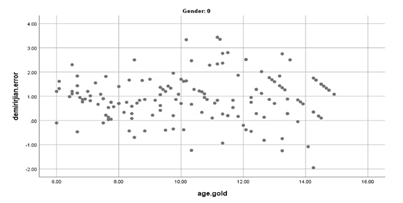
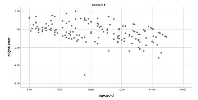
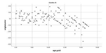
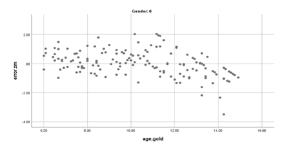
Based on this table, the highest and the lowest estimate errors were related to Demirjian’s method in boys and the formula developed in this study in girls, respectively. The highest accuracy of Demirjian’s method and Cameriere’s method was observed in the age group 8-9 years and age group 7-8 years in both sexes, respectively. Moreover, the highest accuracy of the formula developed in this study was related to the age group 6-7 years in boys and 7-8 years in girls. The three above-mentioned methods have been compared with each other in the estimation error in scatter plots 1 through 6.
When the absolute values of the estimation error of the three methods are compared, it can be concluded that there is a significant difference between Cameriere’s method and Demirjian’s method (p<0.05) and also between the formula developed in this study and Demirjian’s method (p<0.05). By contrast, there was no significant difference between the formula developed in this study and Cameriere’s method in this regard (p>0.05). The results of the independent t-test indicated no significant difference between boys and girls in terms of the absolute value of the estimation error of all methods (p>0.05).
Figure 1: Stages of dental development by Demirjian’s method
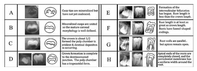
Figure 2: Measurement of dental length and width in Cameriere’s metho
Scatter plot 1: Estimation error of Demirjian’s method compared
to chronological age in boys
Scatter plot 2: Estimation error of Demirjian’s method compared
to chronological age in girls
Scatter plot 3: Estimation error of Cameriere’s method compared
to chronological age in boys
Scatter plot 4: Estimation error of Cameriere’s method compared
to chronological age in girls
Scatter plot 5: Estimation error of Mohammadi’s( proposed formula) method
compared to chronological age in boys
Scatter plot 6: Estimation error of Mohammadi’s( proposed formula)
method compared to chronological age in girls
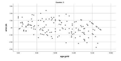
Table 1. Dental indices in each stage for girls and boys
| Tooth | A | B | C | D | E | F | G | H | |
| Boys | 2nd molar | 0.18 | 0.48 | 0.71 | 0.8 | 1.31 | 2 | 2.48 | 4.17 |
| 1st molar | – | – | – | 0.69 | 1.14 | 1.6 | 1.95 | 2.15 | |
| 2nd premolar | 0.08 | 0.05 | 0.12 | 0.27 | 0.33 | 0.45 | 0.4 | 1.15 | |
| 1st premolar | 0.15 | 0.56 | 0.75 | 1.11 | 1.48 | 2.03 | 2.43 | 2.83 | |
| Canine | – | – | – | 0.04 | 0.31 | 0.47 | 1.09 | 1.9 | |
| Lateral incisor | – | – | 0.55 | 0.63 | 0.74 | 1.08 | 1.32 | 1.64 | |
| Central incisor | – | – | 1.68 | 1.49 | 1.5 | 1.86 | 2.07 | 2.19 | |
| Girls | 2nd molar | 0.14 | 0.11 | 0.21 | 0.32 | 0.66 | 1.28 | 2.09 | 4.04 |
| 1st molar | – | – | – | 0.62 | 0.9 | 1.56 | 1.82 | 2.21 | |
| 2nd premolar | -0.19 | 0.01 | 0.27 | 0.17 | 0.35 | 0.35 | 0.55 | 1.51 | |
| 1st premolar | -0.95 | -0.15 | 0.16 | 0.41 | 0.6 | 1.27 | 1.58 | 2.19 | |
| Canine | – | – | 0.6 | 0.54 | 0.62 | 1.08 | 1.72 | 2 | |
| Lateral incisor | – | – | – | 0.29 | 0.32 | 0.49 | 0.79 | 0.9 | |
| Central incisor | – | – | 1.83 | 2.19 | 2.34 | 2.82 | 3.19 | 3.14 |
Table 2. Mean, standard deviation, maximum, and minimum estimation error and
their absolute values for Cameriere’s and Demirjian’s methods
| Number | Minimum | Maximum | Mean | Std. Deviation | |
| Demirjian.error* | 306 | -2.10 | 3.43 | 0.8971 | 0.99261 |
| Demirjian.error.abs** | 306 | 0.03 | 3.43 | 1.1235 | 0.72537 |
| Cameriere.error* | 306 | -5.08 | 1.99 | -0.2017 | 0.96282 |
| Cameriere.error.abs* | 306 | 0.00 | 5.08 | 0.7554 | 0.62869 |
Table 3. Results of the paired sample t-test for determining the overestimation or underestimation
of age by Cameriere’s and Demirjian’s methods and their p-values
| Mean | Std. Deviation | Std. Error Mean | 95% Confidence Interval of the Difference | df | Sig. (2-tailed) | ||
| Lower | Upper | ||||||
| camereiere.age – chronological.age | -0.20165 | 0.96282 | 0.05504 | -0.30996 | -0.09334 | 305 | 0.000 |
| demirjian.age – chronological.age | 0.89706 | 0.99261 | 0.05674 | 0.78540 | 1.00872 | 305 | 0.000 |
Table 4. Mean, standard deviation, maximum, and minimum
estimation error of the proposed formula
| Number | Minimum | Maximum | Mean | Std.
Deviation |
|
| Our Method.error* | 306 | -3.50 | 2.39 | 0.0008 | 0.91146 |
| ABS.error.Our Method** | 306 | 0.00 | 3.50 | 0.7193 | 0.55829 |
* Our Method.error: estimation error of the formula proposed in this study **ABS.error.
Our Method absolute value of estimation error of the formula proposed in this study
Table 5. Mean, standard deviation, maximum, and minimum estimation error of Cameriere’s and Demirjian’s
methods and the formula proposed in this study and their absolute values for both boys and girls for a number of 153
| Girls | Boys | |||||||
| Minimum | Maximum | Mean | Std. Deviation | Minimum | Maximum | Mean | Std. Deviation | |
| demirjian.error | -1.95 | 3.43 | 0.9291 | 0.93254 | -2.10 | 3.23 | 0.8650 | 1.05135 |
| demirjian.error.abs | 0.03 | 3.43 | 1.0920 | 0.73355 | 0.03 | 3.23 | 1.1550 | 0.71813 |
| error.Our Method | -3.50 | 2.04 | 0.0006 | 0.87000 | -2.75 | 2.39 | 0.0009 | 0.95398 |
| ABS.error.Our Method | 0.00 | 3.50 | 0.6744 | 0.54690 | 0.03 | 2.75 | 0.7642 | 0.56769 |
| Cameriere.error | -3.58 | 1.88 | -0.1080 | 0.89360 | -5.08 | 1.99 | -0.2953 | 1.02174 |
| Cameriere.error.abs | 0.00 | 3.58 | 0.7170 | 0.54110 | 0.01 | 5.08 | 0.7939 | 0.70522 |
Table 6. A summary of previous studies conducted about the accuracy of
Cameriere’s and Demirjian’s methods
| Author(s)
|
Year of publication | Country | Accuracy of Demirjian’s method in girls | Accuracy of Demirjian’s method in boys | Accuracy of Cameriere’s method in girls | Accuracy of Cameriere’s method in boys |
| Rozylo et al. 60 | 2008 | Poland | -1.03 | -0.89 | ||
| Qudeimat et al. | 2009 | Kuwait | -0.67 | -0.71 | ||
| Bagherpoor et al. | 2010 | Iran( Mashahad) | +0.25 | +0.34 | ||
| El Bakary e al. | 2010 | Egypt | -0.26 | -0.49 | ||
| Sheikhi et al. | 2011 | Iran(Babol) | +0.04 | +0.02 | ||
| Bagherian et al.61 | 2011 | Iran(Rafsanjan) | +0.21 | +0.15 | ||
| Lee et al. | 2011 | South Korea | +0.86 | +0.64 | ||
| Ogodecu et al. | 2011 | Romania | +0.36 | -0.04 | ||
| Javadinejad et al. | 2012 | Iran(Isfahan) | +0.47 | +0.94 | ||
| Pinchi et al. | 2012 | Italy | +0.41 | +0.68 | -0.96 | -1.07 |
| Grover et al. | 2012 | India | +0.56 | +0.66 | ||
| De luca et al.62 | 2012 | Mexico | 0.63 | 0.52 | ||
| Abbesi et al.63 | 2012 | Iran (Babol) | 0.05 | 0.72 | ||
| Sheikhi et al. | 2013 | Iran(Rasht) | -0.1 | +0.28 | ||
| Kumaresan et. al. | 2013 | Malaysia | +0.97 | +0.97 | -0.39 | -0.44 |
| Javadinejad et al. | 2015 | Iran(Isfahan) | +0.85 | +0.90 | -0.11 | -0.27 |
| Ginzelova et al. | 2015 | Czech Republic | +0.13 | +0.09 | ||
| Wolf et al. | 2016 | Germany | +0.17 | +0.16 | -0.08 | -0.07 |
| Melo et al. | 2017 | Spain | +0.85 | +0.85 | ||
| Apaydin et al. | 2018 | Turkey | +0.30 | +0.31 | -0.55 | -0.60 |
| Halilah et al. | 2018 | Germany | -0.38 | -0.64 | ||
| Wang et al. | 2018 | China | -0.62 | -0.66 | ||
| Sobieska et al. | 2018 | Poland | -0.31 | -0.31 | ||
| Kermani et al.64 | 2019 | Iran(shiraz) | 1.47 | 0.85 | ||
| Mohanty et al. | 2019 | India | +0.56 | +0.66 | ||
| Alqadi et al. | 2019 | Yemen | -0.40 | -0.73 | ||
| Moness Ali et al. | 2019 | Egypt | +0.32 | +0.46 | ||
| Ranasinghe et al. | 2019 | Sri Lanka | +0.19 | +0.19 | ||
| Da luz et al. | 2019 | Brazil | 1.05 | 1.08 | ||
| Croatia | 1.19 | 1.2 | ||||
| Lan et al. | 2019 | China | -0.15 | -0.11 | -0.72 | -0.83 |
| Pan et al.65 | 2020 | China | |0.79| | |0.73| | ||
| Karimi et al.66 | 2021 | Kuwait | +0.33 | -0.14 |
Various methods with different accuracies have been proposed to evaluate the evolution of dental structure, such as Demirjian, Cameriere, Smith, Willems, and Haavikko, (Demirjan et al 1973, Cameriere, et al 2008, Haayiikko 1974, Smith 1991).
This study aimed to investigate the accuracy of Cameriere’s and Demirjian’s methods in DA estimation from panoramic radiographic images in the Iranian population and to propose a formula suitable for Iranian society. The results showed that the DA estimated by both Cameriere’s and Demirjian’s methods was significantly different from chronological age in the studied Iranian population (p<0.05). The results also indicated that the estimation error of Demirjian’s method was higher than that of Cameriere’s method. The mean absolute value of estimation error was 1.1 years in Demirjian’s method and 0.7 in Cameriere’s method. The estimation error of both methods was observed in both genders and all age groups (p<0.05).
Demirjian’s method overestimated DA in both genders and all age groups, whereas Cameriere’s method underestimated DA in all age groups of girls but overestimated in the age group 6-10 years. Cameriere’s method followed no specific pattern, as it overestimated DA in age groups 6, 7, and 10 years but underestimated DA in other age groups.
Previous studies have reported that the estimation error of Demirjian’s method compared to chronological age ranged between 0.13 and 0.97 years in girls and between 0.09 and 0.98 years in boys, ( Grover et al 2011, Sakhdari, et al 2015, Wolf et al 2016, Pinchi et al 2016, Apaydeen et al 2018, Ginzelová et al. 2015, Mohanty et al 2019, Ali et al 2019).
However, some studies have shown that Demirjian’s method underestimates DA, (Alqadi et al 2019, Lan et al 2019), as it ranges between 0.15 and 1.03 years in girls and between 0.04 and 0.89 years in boys (Table 6). It seems that the results of DA estimation by Demirjian’s method do not follow a clear pattern in different populations.Most studies conducted on Demirjian’s method in Iran reported that this method overestimates DA compared to chronological age, Javadinejad, 2015, Shaeikhi et al 2019), the latter conducted two similar studies on children aged 5-16 years in Babol and Rasht. Their results demonstrated that Demirjian’s method underestimated DA by 0.04 years in Babol but overestimated DA by 0.02 years in Rasht compared to chronological age, (Shaeikhi et al 2012, 2013). This discrepancy can be attributed to ethnic, environmental, nutritional, and socioeconomic differences as well as differences in sample size and statistical analysis.
After Cameriere carried out a study on a large European population and developed a formula for DA estimation in 2006, different studies reported different results of age estimation by this method. 15, 22-24, 39, 42 Cameriere (2006) stated that the accuracy of his method was -0.11 years 12. In the present study, the mean accuracy of Cameriere’s method in age estimation was obtained at -0.20 years (-0.11 years in girls and -0.29 in boys). Various studies have shown that the estimation error of Cameriere’s method is less than that of chronological age. These studies have reported that the estimation error of Cameriere’s method ranges between 0.08 and 0.96 years in girls and between 0.07 and 1.07 years in boys, ( El Bakery 2010, Kumaresan 2014), (Table 6).
It can be stated that previous studies have reported different accuracies for Cameriere’s and Demirjian’s methods; some studies have shown that Demirjian’s method is more accurate than Cameriere’s method, (Wolf et al 2016, Pinchi et al 2012, Timmins et al 2011), whereas other studies, like the present study, concluded that Cameriere’s method is more accurate, (Timmins et al (2011), Javadidenaj 2015).
Some studies have stated that the accuracy of Cameriere’s or Demirjian’s methods is higher in a certain age group. For example, Bagherpour et al. 2010, and Javadinejad et al. 2015, and Timmins et al (2011) showed that the accuracy of Demirjian’s method was higher in age groups 9-13, 6-11, and 16 years, respectively. Timmins et al. (2011) also reported that Demirjian’s method was more accurate for older ages. This discrepancy may be attributed to racial, socioeconomic, and nutritional differences as well as differences in sample size.A comparison of the absolute values of the mean estimation error in this study showed that the highest accuracy of Demirjian’s method was observed in age groups 8-9 and then 7-8 years in both genders and the highest accuracy of Cameriere’s method was related to the age group 7-8 years in both genders.
The accuracy of Cameriere’s method in different age groups has been measured in different studies, ( Galic et al 2010, Javadinejad et al. 2015, da Luz et al (2019). Consistent with the findings of the present study, Javadinejad et al. (2015) reported that Cameriere’s method was more accurate in girls aged 6-11 years and boys aged 6-12 years.
In addition, Da Luz et al. (2019), Golsahi et al. (2015) and Rivera et al. ( 2017), showed that the highest accuracy of Cameriere’s method was observed in people aged 8, 9, and 13 years, respectively. Galic et al. (2010) stated that the highest accuracy of Cameriere’s method was related to the age of 15 years in boys and 12 years in girls, (Galic et al 2010). Available studies have reported that DA of boys is higher than girls in children aged under 8.5 years, whereas dental development is faster in girls in children aged 9-12 years. This can be attributed to the fact that girls reach puberty at this age, (Feijoo 2012, Halilah 2018).
In this study, the DA of boys aged under 9 years was about 0.2 years more than DA of girls of the same age. By contrast, the DA of girls was more than boys by 0.45 and 0.53 years in children aged 10 and 11 years, respectively. However, these differences were not statistically significant.
Previous studies have generally reported that both Cameriere’s and Demirjian’s methods underestimate DA at older ages and overestimate DA at younger ages, (Liversidge 2010, Guo, et al (2014). There are various reasons for DA underestimation at older ages. One of the main reasons is that all teeth have developed and there are a few teeth with non-developed roots or lately-developed roots in children of this age group. The number of developing teeth decreases as people grow older, and only the third molar remains non-developed by the age of 14 years, Liversidge 2010).
One of the reasons for the different results in different studies is the choice of different age ranges. According to available radiology references, since surgical and orthodontic treatments are not usually recommended in patients aged under 6 years (except for emergency cases), it is preferable to perform radiology and treatment at the same time, (White and Pharaoh 2014). When the apex of the distal root of the second molar is closed in children, the course of dental development is considered to be accomplished. Since the third molar is not investigated in most methods of DA estimation for children, these methods select the age of 14 years as the end of the studied age range. In the present study, the age range of participants was 6-14 years.
However, the findings of previous studies suggest that Demirjian’s method can be used for age estimation before puberty. Accordingly, if the second molar, canine, and the first premolar teeth are at F, F, and E stages, respectively, it can be stated that one is in the pre-pubertal phase (equivalent to phases CS1 and CS2 in cervical vertebrae cervical maturation). When vertebral and dental development phases are compared, it can be also concluded that girls reach puberty at the age of 12 years, which is equivalent to Stage G in the second premolar, (Tafakhori et al 2016).
Various methods of DA estimation are used to predict the beginning and completion of orthodontic treatments. In cases where there is a delay in tooth eruption, we can use the patient’s radiograph and compare the conditions of each tooth with the standard conditions to predict the time of tooth eruption. Demirjian and Levesque stated that a tooth is about to erupt if it is at Stage G but it takes about one year to erupt if it is at Stage F. They also suggest that if a permanent tooth is at Stage F, the deciduous tooth covering it should be extracted., (Levesque et al (1980).
Chronological age estimation based on Cameriere’s formula will have the least error in Iranian society when the obtained figure is added to 0.2. Moreover, to maximize the accuracy of Demirjian’s method, 0.87 should be subtracted from the figure obtained from this formula.
Considering the estimation error obtained for Cameriere’s formula in this study, a domestic formula was designed in this study based on Cameriere’s formula to suit the Iranian population. The designed formula was as follows:
Age = 9.309 + 0.636 g – 3.852 X1 – 2.505 X3 – 1.007 X7 + 00.664 N – 0.265 SN0
Compare the developed formula with Cameriere’s formula:
Age = 8.971 + 0.375 g + 1.631 XS + 0.674 N – 1.034 S – 0.176 N.S
Where, the dental index was considered to be 5 (X5), and teeth 1, 3, and 7 (X1, X3, and X7) were replaced.
Some previous studies have developed population-specific formulas for age estimation. However, the accuracy of the developed formulas is not always more than that of Cameriere’s method. Hallilah (2018) argues that this is due to the unequal number of samples in different age groups. This discrepancy was also observed in the present study despite the acceptable number and uniform distribution of samples in different age groups.When the formula developed in this study is compared with Cameriere’s and Demirjian’s methods, it can be concluded that the accuracy of the formula developed in this study was higher than that of the other two methods (p<0.05). Hence, researchers are recommended to use this formula in studies conducted on Iranian populations.
Some of the limitations of these methods that can cause differences in results are as follows: Those who have a missing tooth cannot be included in the study. Cameriere’s method requires the measurement of open-apex teeth. This is difficult to do in teeth where the apex is closed. Conduction of studies on people of different age groups or genders can lead to a discrepancy in results. Nutritional, social, and economic issues can affect the course of dental development, (Timmins et al 2011). There is a difference between races and ethnicities in terms of the rate of dental development, ( Shaikhi et al 2013).
Although the panoramic technique has many advantages, slight changes in the X-ray tube angle or patient position (the object is places a bit backward or forward in the focal trough) can cause dimensional changes in the resulting images, (Tafakhori et al 2016). Considering technological advantages, age estimation methods are recommended to be based on the use of cone beam computed tomography (CBCT), as this method has been used in recent studies, ( Kazmi et al 2019, Molina et al 2020).
CONCLUSION
Both Cameriere’s and Demirjian’s methods are not highly accurate and the DA estimated by them is significantly different from chronological age. However, the study findings revealed that Cameriere’s method was more accurate than Demirjian’s method. The formula designed in this study for Iranian society was more accurate than both of the above-mentioned methods. All three methods had almost the same accuracy in both genders. However, Cameriere’s method underestimated the age, and Demirjian’s method overestimated the age compared to the chronological age.
Conflict of Interest: Authors declare no conflict of interest.
REFERENCES
Abesi, F., Haghanifar, S., Sajadi, P., et al. (2013). Assessment of dental maturity of children aged 7-15 years using Demirjian method in a selected Iranian population. Journal of Dentistry, 14(4), p.165.
Ali, A.M.M., Ahmed, W.H. and Khattab, N.M. (2019) ‘Applicability of Demirjian’s method for dental age estimation in a group of Egyptian children,’ BDJ Open, 5(1). https://doi.org/10.1038/s41405-019-0015-y.
Alqadi, M.A. and Abuaffan, A.H. (2019). Validity of the Demirjian and Fishman Methods for Predicting Chronological Age Amongst Yemeni Children. Sultan Qaboos University Medical Journal [SQUMJ], 19(1), p.26. doi:https://doi.org/10.18295/squmj.2019.19.01.006.
Andrade, V. M., Fontenele, R.C.,Souza, A. C. et al. (2019) ‘Age and sex estimation based on pulp cavity volume using cone beam computed tomography: development and validation of formulas in a Brazilian sample,’ Dento-maxillo-facial Radiology/Dentomaxillofacial Radiology, 48(7), p. 20190053. https://doi.org/10.1259/dmfr.20190053.
Apaydin BK, Yasar F. (2018). Accuracy of the demirjian, willems and cameriere methods of estimating dental age on turkish children. Nigerian Journal of Clinical Practice, 21(3), pp.257–257. doi:https://doi.org/10.4103/1119-3077.226966.
Baccetti, T., Franchi, L. and McNamara, J.A. (2005) ‘The Cervical Vertebral Maturation (CVM) method for the assessment of optimal treatment timing in dentofacial orthopedics,’ Seminars in Orthodontics, 11(3), pp. 119–129. https://doi.org/10.1053/j.sodo.2005.04.005.
Bagherian, A. and Sadeghi, M. (2011) ‘Assessment of dental maturity of children aged 3.5 to 13.5 years using the Demirjian method in an Iranian population,’ Journal of Oral Science, 53(1), pp. 37–42. https://doi.org/10.2334/josnusd.53.37.
Bagherpour, A., Imanimoghaddam, M., Bagherpour, M.R. and Einolghozati, M. (2010). Dental age assessment among Iranian children aged 6–13 years using the Demirjian method. Forensic Science International, 197(1-3), pp.121.e1–121.e4. doi:https://doi.org/10.1016/j.forsciint.2009.12.051.
Butti, A.C. et al. (2009) ‘Haavikko’s method to assess dental age in Italian children,’ European Journal of Orthodontics, 31(2), pp. 150–155. https://doi.org/10.1093/ejo/cj. Hauk, M. J., Moss, M. E.,
Cameriere, R. et al. (2008) ‘The measurement of open apices of teeth to test chronological age of over 14-year olds in living subjects,’ Forensic Science International, 174(2–3), pp. 217–221. https://doi.org/10.1016/j.forsciint.2007.04.220.
Cameriere, R., De Angelis, D., Ferrante, L., Scarpino, F. and Cingolani, M. (2007). Age estimation in children by measurement of open apices in teeth: a European formula. International Journal of Legal Medicine, 121(6), pp.449–453. doi:https://doi.org/10.1007/s00414-007-0179-1.
Cameriere, R., Ferrante, L. and Cingolani, M. (2005). Age estimation in children by measurement of open apices in teeth. International Journal of Legal Medicine, 120(1), pp.49–52. doi:https://doi.org/10.1007/s00414-005-0047-9.
da Luz, L.C.P., Anzulović, D., Benedicto, E.N., Galić, I., Brkić, H. and Biazevic, M.G.H. (2019). Accuracy of four dental age estimation methodologies in Brazilian and Croatian children. Science & Justice, 59(4), pp.442–447. doi:https://doi.org/10.1016/j.scijus.2019.02.005.
De Luca, S., De Giorgio, S., Butti, A.C., et al. (2012). Age estimation in children by measurement of open apices in tooth roots: Study of a Mexican sample. Forensic Science International, 221(1-3), pp.155.e1–155.e7. doi:https://doi.org/10.1016/j.forsciint.2012.04.026.
Demirjian, A. and Goldstein, H. (1976) ‘New systems for dental maturity based on seven and four teeth,’ Annals of Human Biology, 3(5), pp. 411–421. https://doi.org/10.1080/03014467600001671.
Demirjian, A., Goldstein, H. and Tanner, J.M. (1973). A new system of dental age assessment. PubMed, 45(2), pp.211–27.
El-Bakary, A.A., Hammad, S.M. and Mohammed, F. (2010). Dental age estimation in Egyptian children, comparison between two methods. Journal of Forensic and Legal Medicine, 17(7), pp.363–367. doi:https://doi.org/10.1016/j.jflm.2010.05.008.
Feijóo, G., Barbería, E., De Nova, J., Prieto, J. L. (2012) ‘Dental age estimation in Spanish children,’ Forensic Science International, 223(1–3), p. 371.e1-371.e5. https://doi.org/10.1016/j.forsciint.2012.08.021.
Feijóo, G., Barbería, E., Joaquín De Nova and Jose Luis Prieto (2012). Permanent teeth development in a Spanish sample. Application to dental age estimation. Forensic science international, 214(1-3), pp.213.e1–213.e6. doi:https://doi.org/10.1016/j.forsciint.2011.08.024.
Galić, I., Vodanović, M., Cameriere, R., et al. (2010). Accuracy of Cameriere, Haavikko, and Willems radiographic methods on age estimation on Bosnian–Herzegovian children age groups 6–13. International Journal of Legal Medicine, 125(2), pp.315–321. doi:https://doi.org/10.1007/s00414-010-0515-8.
Ginzelová, K., Dostálová, T., Eliášová, H., et al. (2015). Using Dental Age to Estimate Chronological Age in Czech Children Aged 3–18 Years. Prague Medical Report, 116(2), pp.139–154. doi:https://doi.org/10.14712/23362936.2015.52.
Graber, L.W., Vanarsdall, R.L. and Vig, K.W.L. (2012) Orthodontics: Current Principles & Techniques. Chapter 14: P 482,483.
Grover, S., Marya, C.M., Avinash, J. and Pruthi, N. (2011). Estimation of dental age and its comparison with chronological age: accuracy of two radiographic methods. Medicine, Science and the Law, 52(1), pp.32–35. doi:https://doi.org/10.1258/msl.2011.011021.
Gulsahi, A., Tirali, R.E., Cehreli, S.B., De Luca, S., Ferrante, L. and Cameriere, R. (2015). The reliability of Cameriere’s method in Turkish children: A preliminary report. Forensic Science International, 249, pp.319.e1–319.e5. doi:https://doi.org/10.1016/j.forsciint.2015.01.031.
Guo, Y., Yan, C., Lin, X., Zhou, H., Li, J., Pan, F., Zhang, Z., Wei Kuang Lai, Tang, Z. and Chen, T. (2014). Age estimation in northern Chinese children by measurement of open apices in tooth roots. 129(1), pp.179–186. doi:https://doi.org/10.1007/s00414-014-1035-8.
Haavikko H., (1974) ‘Tooth formation age estimated on a few selected teeth. A simple method for clinical use,’ PubMed, 70(1), pp. 15–9. https://pubmed.ncbi.nlm.nih.gov/4821943.
Halilah, T., Khdairi, N., Jost-Brinkmann, P.-G. et al. (2018). Age estimation in 5–16-year-old children by measurement of open apices: North German formula. Forensic Science International, 293, pp.103.e1–103.e8. doi:https://doi.org/10.1016/j.forsciint.2018.09.022.
Javadinejad, S., Sekhavati, H. and Ghafari, R. (2015). A Comparison of the Accuracy of Four Age Estimation Methods Based on Panoramic Radiography of Developing Teeth. Journal of Dental Research, Dental Clinics, Dental Prospects, 9(2), pp.72–78. doi:https://doi.org/10.15171/joddd.2015.015.
Karimi A, Qudeimat MA, Lucas VS, Roberts G. (2021) ‘Dental age estimation: Development and validation of a reference data set for Kuwaiti children, adolescents, and young adults,’ Archives of Oral Biology, 127, p. 105130. https://doi.org/10.1016/j.archoralbio.2021.105130.
Kazmi, S., Mânica, S., Revie, G., Shepherd, S. and Hector, M. (2019). Age estimation using canine pulp volumes in adults: a CBCT image analysis. International Journal of Legal Medicine, 133(6), pp.1967–1976. doi:https://doi.org/10.1007/s00414-019-02147-5.
Kermani, M., Tabatabaei Yazdi, F. and Abed Haghighi, M. (2019). Evaluation of the accuracy of Demirjian’s method for estimating chronological age from dental age in Shiraz, Iran: Using geometric morphometrics method. Clinical and Experimental Dental Research, 5(3), pp.191–198. doi:https://doi.org/10.1002/cre2.169.
Koshy, S. and Tandon, S. (1998) ‘Dental age assessment: The applicability of Demirjian’s method in South Indian children,’ Forensic Science International, 94(1–2), pp. 73-85. https://doi.org/10.1016/s0379-0738(98)00034-6.
Kumaresan, R., Cugati, N., Chandrasekaran, B. and Karthikeyan, P. (2014). Reliability and validity of five radiographic dental-age estimation methods in a population of Malaysian children. Journal of Investigative and Clinical Dentistry, 7(1), pp.102–109. doi:https://doi.org/10.1111/jicd.12116.
Lan LM, Yang ZD, Sun SL. et al. (2019) ‘Application of Demirjian’s and Cameriere’s Method in Dental Age Estimation of 8-16 Year Old Adolescents from Hunan Han Nationality.,’ PubMed, 35(4), pp. 406–410. https://doi.org/10.12116/j.issn.1004-5619.2019.04.005.
Lee, S.-S., Kim, D., Lee, S., Lee, U-Young., Seo, J.S., Ahn, Y.W. and Han, S.-H. (2011). Validity of Demirjian’s and modified Demirjian’s methods in age estimation for Korean juveniles and adolescents. Forensic Science International, 211(1-3), pp.41–46. doi:https://doi.org/10.1016/j.forsciint.2011.04.011.
Lehtinen, A., Oksa, T., Helenius, H., & Rönning, O. (2000) ‘Advanced dental maturity in children with juvenile rheumatoid arthritis,’ European Journal of Oral Sciences, 108(3), pp. 184–188. https://doi.org/10.1034/j.1600-0722.2000.108003184.x.
Leurs, I E. Wattel, Irene, E.J. Etty and Birte Prahl-Andersen (2005). Dental age in Dutch children. European Journal of Orthodontics, 27(3), pp.309–314. doi:https://doi.org/10.1093/ejo/cji010.
Levesque, Gilles-Y. and Demirjian, A. (1980). The Inter-examiner Variation in Rating Dental Formation from Radiographs. Journal of Dental Research, 59(7), pp.1123–1126. doi:https://doi.org/10.1177/00220345800590070401.
Liversidge, H.M., Smith, B.H. and Maber, M. (2010). Bias and accuracy of age estimation using developing teeth in 946 children. American Journal of Physical Anthropology, 143(4), pp.545–554. doi:https://doi.org/10.1002/ajpa.21349.
Mani SA, Naing L, John J , Samsudin AR. (2008) ‘Comparison of two methods of dental age estimation in 7–15‐year‐old Malays,’ International Journal of Paediatric Dentistry, 18(5), pp. 380–388. https://doi.org/10.1111/j.1365-263x.2007.00890.x.
Marjatta Nyström, Ranta, R., Matti Kataja and Hilkka Silvola (1988). Comparisons of dental maturity between the rural community of Kuhmo in northeastern Finland and the city of Helsinki. Community Dentistry and Oral Epidemiology, 16(4), pp.215–217. doi:https://doi.org/10.1111/j.1600-0528.1988.tb01757.x.
Melo, M. and Ata-Ali, J. (2017). Accuracy of the estimation of dental age in comparison with chronological age in a Spanish sample of 2641 living subjects using the Demirjian and Nolla methods. Forensic Science International, 270, pp.276.e1–276.e7. doi:https://doi.org/10.1016/j.forsciint.2016.10.001.
Mohanty, I., Panda, S., Dalai, R. P., & Mohanty, N. (2019). Predictive accuracy of Demirjian’s, Modified Demirjian’s and India specific dental age estimation methods in Odisha (Eastern Indian) population. The Journal of forensic odonto-stomatology, 37(1), pp. 32–39.
Molina, A., Bravo, M., Fonseca, G.M., Márquez-Grant, N. and Martín-de-las-Heras, S. (2020). Dental age estimation based on pulp chamber/crown volume ratio measured on CBCT images in a Spanish population. International Journal of Legal Medicine, 135(1), pp.359–364. doi:https://doi.org/10.1007/s00414-020-02377-y.
Moshfeghi, M., Rahimi, H., Rahimi, H., et al. (2013). Predicting mandibular growth increment on the basis of cervical vertebral dimensions in Iranian girls. Progress in Orthodontics, 14(1), p.3. doi:https://doi.org/10.1186/2196-1042-14-3.
Ogodescu, A.E., Bratu, E., Tudor, A. and Ogodescu, A. (2011). Estimation of child’s biological age based on tooth development. Romanian Journal of Legal Medicine, 19(2), pp.115–124. doi:https://doi.org/10.4323/rjlm.2011.115.
Pan, J., Shen, C., Yang, Z., Fan, L., Wang, M., Shen, S., Tao, J. and Ji, F. (2021). A modified dental age assessment method for 5- to 16-year-old eastern Chinese children. 25(6), pp.3463–3474. doi:https://doi.org/10.1007/s00784-020-03668-9.
Pinchi, V., Norelli, G.-A., Pradella, F., et al,. (2012). Comparison of the applicability of four odontological methods for age estimation of the 14 years legal threshold in a sample of Italian adolescents. PubMed.
Qudeimat, M.A. and Behbehani, F. (2009). Dental age assessment for Kuwaiti children using Demirjian’s method. Annals of Human Biology, 36(6), pp.695–704. doi:https://doi.org/10.3109/03014460902988702.
Rai, B. and Bhagwat Dayal Sharma (2006). Tooth Developments: An Accuracy of Age Estimation of Radiographic Methods.
Ranasinghe, S., Perera, J., Taylor, J.A. et al. (2019). Dental age estimation using radiographs: Towards the best method for Sri Lankan children. Forensic Science International, 298, pp.64–70. doi:https://doi.org/10.1016/j.forsciint.2019.02.053.
Rivera, M., Stefano de Luca, Aguilar, L., Luz Amparo Palacio, Ivo Galić and Cameriere, R. (2017). Measurement of open apices in tooth roots in Colombian children as a tool for human identification in asylum and criminal proceedings. 48, pp.9–14. doi:https://doi.org/10.1016/j.jflm.2017.03.005.
Różyło-Kalinowska, I., Kiworkowa-Rączkowska, E. and Kalinowski, P. (2008). Dental age in Central Poland. Forensic Science International, 174(2-3), pp.207–216. doi:https://doi.org/10.1016/j.forsciint.2007.04.219.
Sakhdari, S., Mehralizadeh, S., Zolfaghari, M. and Madadi, M. (2015). Age Estimation from Pulp/Tooth Area Ratio Using Digital Panoramic Radiography. [online] Semantic Scholar. Available at: https://api.semanticscholar.org/CorpusID:35482431 [Accessed 23 May 2024].
Sheikhi M, Dakhilalian M, Jamshidi M, Nouri Sh, Babaei M. Estimation of chronologic ages of 5-16 year-old children and adolescents in Rasht by Demirjian method. J Shahid Sadoughi Univ Med Sci 2013; 21(1): 85-93.
Sheikhi, M., Dakhilalian, M., Madani, M. and Ghorbanizadeh, S., 2012. Evaluation of the accuracy of Demirjian method in estimating chronologic ages of 5-17 year-old children and adolescents in Babol. Journal of Isfahan Dental School, Special Issue 7(5), pp.488-492. [in Persian].
Smith, B.H., (1991). Standards of human tooth formation and dental age assessment. In: M.A. Kelley and C.S. Larsen, eds. Advances in Dental Anthropology. St. Louis: Wiley-Liss, pp. 143-168.
Sobieska, E., Fester, A., Nieborak, M. and Zadurska, M. (2018). Assessment of the Dental Age of Children in the Polish Population with Comparison of the Demirjian and the Willems Methods. Medical Science Monitor, 24, pp.8315–8321. doi:https://doi.org/10.12659/msm.910657.
Timmins, K., Liversidge, H., Farella, M., Herbison, P. and Kieser, J. (2011). The usefulness of dental and cervical maturation stages in New Zealand children for Disaster Victim Identification. Forensic Science, Medicine, and Pathology, 8(2), pp.101–108. doi:https://doi.org/10.1007/s12024-011-9251-8.
Wang, J., Bai, X., Wang, M., et al. (2018). Applicability and accuracy of Demirjian and Willems methods in a population of Eastern Chinese subadults. Forensic Science International, [online] 292, pp.90–96. doi:https://doi.org/10.1016/j.forsciint.2018.09.006.
Weinberg, G. A., & Berkowitz, R. J. (2001). Delayed tooth eruption: association with severity of HIV infection. Pediatric dentistry, 23(3), 260–262.
White, S.C. and Pharoah, M.J. (2014). Oral Radiology – E-Book : Principles and Interpretation. Mosby, Chapter 16, p. 261.
White, S.C. and Pharoah, M.J. (2014). Oral Radiology – E-Book : Principles and Interpretation. Mosby,Chapter 11, P. 170.
Willems, G., Van Olmen, A., Spiessens, B. and Carels, C. (2001). Dental Age Estimation in Belgian Children: Demirjian’s Technique Revisited. Journal of Forensic Sciences, 46(4), p.15064J. doi:https://doi.org/10.1520/jfs15064j.
Wolf, T.G., Briseño-Marroquín, B., Callaway, A. et al., (2016). Dental age assessment in 6- to 14-year old German children: comparison of Cameriere and Demirjian methods. BMC Oral Health, 16(1). doi:https://doi.org/10.1186/s12903-016-0315-8.
Zahra Tafakhori, Mahboubeh Shokrizadeh and Mahmood Sheikh Fathollahi (2016). Relationship between Dental Development and Cervical Vertebrae Development Assessed Using Radiography in an Iranian Population. Dentomaxillofacial radiology, pathology and surgery, 5(2), pp.17–23. doi:https://doi.org/10.18869/acadpub.3dj.5.2.17.


