1Doctor of Biological Sciences, ASRIVEA – Branch of Tyumen Scientific Centre SB RAS, FSBEI HE Northern Trans-Ural SAU Tyumen Russia
2 FSBEI HE Northern Trans-Ural SAU, Yuri Valerievich Glazunov, Doctor of Veterinary Sciences, ASRIVEA – Branch of Tyumen Scientific
Centre SB RAS, FSBEI HE Northern Trans-Ural SAU Tyumen Russia
3Candidate of Veterinary Sciences, ASRIVEA – Branch of Tyumen Scientific Centre SB RAS, FSBEI HE Northern Trans-Ural SAU Tyumen Russia
4Doctor of Veterinary Sciences, ASRIVEA- Branch of Tyumen Scientific Center SB RAS, FSBEI HE Northern Trans- Ural SAU Tymen Russia
Corresponding author email: ilmedv1@yandex.ru
Article Publishing History
Received: 27/10/2020
Accepted After Revision: 19/12/2020
The objective of the research was to study the spread of toxoplasmosis in people and animals in the Tyumen region. Studies on the diagnosis of toxoplasmosis in humans were carried out at the Federal Budgetary Institution of Science “Tyumen Scientific and Research Institute of Territorial Infectious Pathology” of Rospotrebnadzor (Tyumen) in 2000-2015. Diagnosis of toxoplasmosis in animals was carried out in the Tyumen Regional Veterinary Laboratory in 2011-2016. The disease incidence among people in the region was analyzed using the results of immunological studies of various age groups of the population. To diagnose toxoplasmosis in animals, parasitological and immunological methods of diagnosis were used. It was established that toxoplasmosis is a widely common disease among people living in the Tyumen region. The maximum number of positive reactions to toxoplasmosis was recorded in 2002, 2003, 2008, and from 2012 to 2016, when the level of seropositive reactions was 26.2; 20.2; 12.26; 17.08; 11.36; and 10.59% of people examined for toxoplasmosis. In 2014, the highest seropositivity rates for T.gondiiwere 250.35 and 151.09 per 100 people, respectively, in Abatsky and Omutinsky districts of the Tyumen region. Among children, the invasion was recorded at the age of over one year with the maximum seropositive level – 4.36% in 2012. Among dogs and cats examined for the presence of antibodies to T.gondii, the maximum level of seropositivity was found in 2013 and 2016. These indicators were at the level of 15.4 and 15.0%, respectively. During the examination of seropositive cats, the release of oocysts of T.gondii was found in only 0.42%.
Toxoplasmosis, Seropositivity, Tyumen Region, Dogs, Cats
Domatsky V. N, Antimirova A. A, Glazunova L. A, Glazunov Y. V. Spread of Toxoplasmosis in Humans and Animals in the Tyumen Region. Biosc.Biotech.Res.Comm. 2020;13(4).
Domatsky V. N, Antimirova A. A, Glazunova L. A Glazunov Y. V. Spread of Toxoplasmosis in Humans and Animals in the Tyumen Region. Biosc.Biotech.Res.Comm. 2020;13(4). Available from: <a href=”https://bit.ly/39FZ27y”>https://bit.ly/39FZ27y</a>
Copyright © This is an Open Access Article distributed under the Terms of the Creative Commons Attribution License (CC-BY). https://creativecommons.org/licenses/by/4.0/, which permits unrestricted use distribution and reproduction in any medium, provided the original author and sources are credited.
INTRODUCTION
One of the important problems of medicine and veterinary medicine that has serious socio-economic importance is toxoplasmosis. Special attention to this disease is justified by its consequences, which are most important for a person, and especially to a developing fetus when a pregnant woman is infected during pregnancy. The widespread occurrence of toxoplasmosis among animals, especially free-living and wild, does not allow for its control and affects more and more susceptible organisms(Gao, et al., 2020).
For the first time, the causative agent of toxoplasmosis – Toxoplasma gondii, was mentioned in 1908 in Tunisia and Brazil in Gundi (a species of rodents) and rabbits, respectively (Jitender Prakask, 2016; Jones and Dubey, 2012; Pappas et al., 2009; Lehmann et al., 2006; Nasiru Wana et al., 2020). The first case of congenital toxoplasmosis was diagnosed in 1923 (Chandramathi and Nissapatorn, 2019; Petersen et al., 2010). To date, the number of persons infected with toxoplasma exceeds 1.5 billion people, and the prevalence of toxoplasma in different areas varies from 14 to 90%, averaging at least 35% (Fiedler et al., 1999; Hofhuis et al., 2011; Ben-Harari and Connolly, 2019; Kijlstra and Jongert, 2008 ). The minimal prevalence of the population is noted in the Nordic countries – 14%, low, in New Zealand, Great Britain and Australia – 25%, the average level – 35-50% in many countries of Asia, Africa, America, and Europe (Fiedler et al., 1999; Glazunova et al., 2018; Ben-Harari and Connolly, 2019; Jones et al., 2009; Lehmann et al., 2006).
This is evidenced, in particular, by the fact that a high percentage (60–90%) of individuals with antibodies to toxoplasma (Robert-Gangneux and Dardé, 2012; Lehmann et al., 2006) was detected among the population of several countries in Asia and Western Europe. Annual rates of seroconversion in countries with a high prevalence of the population are more than 3%, in the “toxoplasmosis-safe” countries of Northern Europe and in the “relatively safe” UK and USA, this indicator is less than 1% (Chandramathi and Nissapatorn, 2019). But even if looking at the minimum indicator, 0.6% of the US population who annually suffer the acute phase of invasion amount to about 1.5 million cases of the disease, and approximately 15% of them are clinically significant (Mead et al., 2000). In different territories of Russia, invasion of the population (according to quite incomplete data) is on average 30-35% (Aliyu et al., 2020; Jitender Prakask, 2016).
Due to the lack of awareness of the population each year, hundreds of cats are euthanized or thrown into the streets, aggravating the problem of neglected animals. Home-living animals who have never encountered T. gondii, risk being infected, as a result of which they become subjects of the excretion of Toxoplasma cysts, infecting children’s playgrounds, especially sandboxes, municipal water bodies, and lawns. A vicious circle occurs. The spread of toxoplasmosis in populated areas is due to the lack of a systematic approach to the regulation of the number of homeless animals (Gao, et al., 2020).
In 2012-2018 in Tyumen, 9623 dogs were taken from the streets of the city, most of which were returned back without prior examination for toxoplasmosis and no action taken depending on the situation. Thus, a systematic approach to the problem of toxoplasmosis is required, where human doctors, veterinarians, breeders, and owners of animal nurseries, epidemiological control services, and every person would work to educate the population, early diagnose, prevent the disease, interrupt the development cycle of the pathogen and eliminate it from the environment.
Given the significance of the problem and the lack of current knowledge about the distribution of toxoplasmosis in the Tyumen region, we set a goal to study the spread of toxoplasmosis in humans and domestic animals in the Tyumen region. In other words, in the current study, it was tried to study and analyze the spread of toxoplasmosis in people and animals in the Tyumen region. Studies on the diagnosis of toxoplasmosis in humans were carried out at the Federal Budgetary Institution of Science “Tyumen Scientific and Research Institute of Territorial Infectious Pathology” of Rospotrebnadzor (Tyumen) in 2000-2015.
MATERIAL AND METHODS
Studies on the diagnosis of toxoplasmosis in humans were carried out at the Federal Budgetary Institution of Science “Tyumen Scientific and Research Institute of Territorial Infectious Pathology” of Rospotrebnadzor (Tyumen) in 2000-2015. Diagnosis of toxoplasmosis in animals was carried out in the Tyumen Regional Veterinary Laboratory in 2011-2016. The disease incidence among people in the region was analyzed using the results of immunological studies of various age groups of the population. To diagnose toxoplasmosis in animals, parasitological and immunological methods of diagnosis were used.
The parasitological methods were the microscopy of smears of the affected organs and stool tests. 723 stool tests were conducted. The immune-chromatographic analysis was performed using the rapid test system (Quicking Biotech Co., Ltd.); the immunological methods were enzyme immunoassay used to detect IgG and IgM (Hellmann et al., 2018; Sierra et al., 2020).
Immunology: The immune system is a complex system of structures and processes that have evolved to protect us from the disease. The function of these components is divided into two parts. The first part of which responds non-specifically to all microbes in the same way, which is called the innate immune response. The second part is the responses that specifically respond to a specific microbe known as acquired immunity. Immunology is the study of the immune system and is a very important branch of medical science and biology. Our immune system protects us from various infections. If the immune system does not work this way, it can lead to diseases such as autoimmune diseases, allergies and cancer.
Toxoplasmosis: Toxoplasmosis is one of the parasitic diseases of humans and animals is toxoplasmosis, the initial infection of which is asymptomatic. The severe and acute type of disease is accompanied by fever and enlargement of the lymph nodes. Symptoms of a rare form of the disease that is rarely seen include brain symptoms, pneumonia, generalized muscle disease, and death. The disease is caused by Toxoplasma gondii, an intracellular protozoan that is transmitted through fecal-oral feces from cat feces that contain infected oocytes or parasite eggs. The causative agent may occur through eating uncooked meat, transfusing blood or organ transplants, or through the placenta into the fetus despite an acute infection in a pregnant woman.
Chromatography: Chromatography is an analytical method commonly used to separate the components of a chemical mixture. As a result, with the “chromatography” method, it is possible to analyze the components of the mixtures well. There are several methods of chromatography, including liquid chromatography, gas chromatography, ion-exchange chromatography, and affinity chromatography, all of which have the same basis.
RESULTS AND DISCUSSION
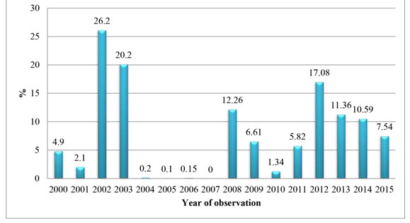
We established that the epizootological situation of toxoplasmosis among people is constantly changing and the number of seropositive reactions varies from 0 to 26.2% of those examined. Thus, no cases of toxoplasmosis among people were recorded in 2007; this period was preceded by an intense decline in positive results (Figure 1). The maximum number of positive reactions to toxoplasmosis was recorded in 2002, 2003, 2008, and from 2012 to 2016, when the level of seropositive reactions was 26.2; 20.2; 12.26; 17.08; 11.36; and 10.59% of people examined for toxoplasmosis.
Figure 1: The level of the infected population of the Tyumen region, share, % (2000-2015)
The frequency of detection of antibodies to T.gondii varied depending on the residential area of the population. The population of Abatsky and Omutinsky districts turned out to be the most infected. In 2014, the rate of infection of the population with toxoplasmosis per 100 thousand people reached its maximum of 250.35 and 151.09, respectively. In 2015, the situation remained virtually unchanged, and the level of invasion in the same areas was 229.4 and 137.5. At the same time, in the neighbouring areas, the infection rate was significantly lower and ranged from 0 to 27.38.
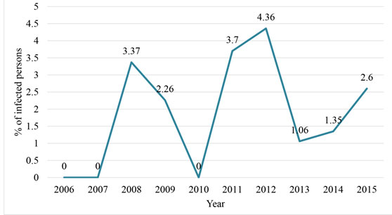
The situation of T.gondii invasion among urban residents of the Tyumen region has also been clarified. The results of research in the cities of Tyumen, Ishim, and Tobolsk were analyzed. Residents of the regional centre were found to be the most infected population, while the maximum number of seropositive people in 2012 was 32.34 per 100 thousand population. In other cities, the level of the seropositive population did not exceed 4.18. Moreover, in 2006-2015, the T.gondii invasion rate was studied among children aged 0 to 17 years. It was revealed that only one child under one year had antibodies to the specified pathogen.
Figure 2: The T.gondii invasion rate among children aged 0-17 years in the Tyumen region (2006-2015).
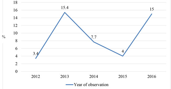
The peaks of the invasion were in 2008, 2011, and 2012, when the detection rate of antibodies to T.gondii was 3.37; 3.7; and 4.36%. We should note that the level of seropositive children under 14 is higher. In 2008 it was 4.31, in 2011 – 3.96, and the maximum indicator among children was found in 2012 – 5.13. Considering that animals are the main spreaders of invasion, we found out the prevalence of toxoplasmosis in domestic animals. It was found that in five studies (2012-2016) the level of seropositive animals varied (Figure 3). Thus, in 2012 and 2015, the level of seropositivity among dogs and cats was 3.4 and 4.0%, respectively. Whereas in 2013 and 2016, these indicators were 15.4 and 15.0%, respectively.
Figure 3: The share of T.gondii seropositive dogs and cats in the Tyumen region (2012-2016).
In parallel with the immunological method of diagnosis, 723 stool test of cats were conducted to detect oocysts of toxoplasma (Figure 4). We found that T.gondii oocysts are extremely rare in seropositive cats. In 2013-2015, only three samples contained T.gondii oocysts.
Figure 4: Oocysts in T.gondii seropositive cats (2011-2016)
It is considered that 25-30% of the world’s population is infected with T.gondii (Robert-Gangneux and Dardé, 2012). The level of the seropositive population in the Tyumen region is within the average statistical indicators for Russia. Thus, in the Russian Federation, 25% of the population surveyed in Moscow and 32% of the population surveyed in the Oryol region are seropositive to T.gondii (Aliyu et al., 2020 Prakask, 2016).
In Krasnoyarsk Krai, 28.6% were seropositive the level of seropositivity among the surveyed pregnant women in the Belgorod region was 23.6±1.4% ( Prakask, 2016). The results of immunological studies among people from the neighbouring Omsk region, where the growth of toxoplasmosis was 2.0 times higher in 1992-2006, coincide. The Tyumen region also had the highest rates in 2002 and 2003 – 26.2 and 20.2%, respectively, while in 2004-2007, there was the lowest seropositivity recorded – 0-0.2% (Prakask, 2016 Gao, et al., 2020).
There is evidence that the level of seropositivity to T.gondii depends on the socio-economic situation. Thus, the study of the epidemiological situation in Brazil found that antibodies against toxoplasma are found in 84, 62, and 23% of the population of the low, medium, and high class, respectively (Aliyu et al., 2020; Robert-Gangneux and Dardé, 2012; Schlüter et al., 2014). In the Russian Federation, 83.5% of the children surveyed in the Omsk region, who were seropositive for toxoplasmosis, were from single-parent large families with poor sanitary and living conditions (Jones et al., 2018 Ben-Harari and Connolly, 2019). The highest prevalence of toxoplasmosis (41.0%) was observed in children from rural areas on the background of tuberculosis infection.
Our results also confirm the assumption of the significance of social status and culture on the risk of T.gondii invasion. In the Tyumen region, the population living in rural areas is more seropositive, which may be due to the sanitary and hygiene offences, as well as a large number of homeless and free-living cats in the private sector. Considering the low level of release of T.gondii oocysts from seropositive domestic animals (0.42%), the probability of invasion of people eating semi-raw meat is also high (Glazunova et al., 2018; Jones and Dubey, 2012; Lindsay et al., 2002; Kijlstra and Jongert, 2008; Zulpo et al., 2018, Wana et al. 2020).
CONCLUSION
Analysis of the results allows us to conclude that toxoplasmosis is widespread among people living in the Tyumen region. The maximum number of positive reactions to toxoplasmosis was recorded in 2002, 2003, 2008, and from 2012 to 2016, when the level of seropositive reactions was 26.2; 20.2; 12.26; 17.08; 11.36; and 10.59% of people examined for toxoplasmosis. In 2014, the highest seropositivity rates for T.gondiiwere 250.35 and 151.09 per 100 people, respectively, in Abatsky and Omutinsky districts of the Tyumen region. As for urban residents of the region, toxoplasmosis is common among residents of the regional centre, with 32.34 seropositive people per 100 thousand population found in 2012. Among children, the invasion was recorded at the age of over one year with the maximum seropositive level – 4.36% in 2012. Among dogs and cats examined for the presence of antibodies to T.gondii, the maximum level of seropositivity was found in 2013 and 2016. These indicators were at the level of 15.4 and 15.0%, respectively. During the examination of seropositive cats, the release of oocysts of T.gondii was found in only 0.42%.
ACKNOWLEDGMENTS
The article was prepared with the financial support of the Program of Fundamental Research of the Russian Academy of Sciences, registration number AAAA-A18-118020690240-3 “Monitoring of the epizootic situation and forecasts of the development of possible outbreaks of parasitic animal diseases”.
Conflict of Interest: Authors declares no conflicts of interests to disclose.
Ethical Clearance Statement: The Current Research Work Was Ethically Approved by the Institutional Review Board (IRB) of Scientific Centre SB RAS, FSBEI HE Northern Trans-Ural SAU Tyumen Russia.
REFERENCES
Aliyu MB, Maikai BV, & Magaji AA (2020). Toxoplasma gondii infection and risk factors associated with its spread at live bird markets in Katsina Metropolis, Nigeria. Sokoto Journal of Veterinary Sciences, 18(1), 39-46.
Ben-Harari RR, Connolly MP (2019). High burden and low awareness of Toxoplasmosis in the United States. Postgraduate Medicine. 17;131(2):103-8.
Chandramathi SR, Nissapatorn V. Prevalence and risk factors of Toxoplasma infection–an update in Malaysian pregnant women. Tropical Biomedicine. 2019;36(3):694-702.
Fiedler K, Hülsse C, Straube W, Briese V(1999). Toxoplasmosis-antibody seroprevalence in Mecklenburg-Western Pomerania. Zentralblatt fur Gynakologie.121(5):239-43.
Gao JM, Yi SQ, Geng GQ, Xu ZS, Hide G, Lun Z. R, & Lai DH (2020). Infection With Trypanosoma Lewisi or Trypanosoma Musculi May Promote The Spread of Toxoplasma gondii
Glazunova LA, Glazunov YV, Ergashev AA (2018).Ecological-Epizootical Situation On Telasiosis Among Large Cattle In Northern Ural Region. Research Journal of Pharmaceutical, Biological and Chemical Sciences.9(4):1687-93.
Hellmann MD, Mangarin LM, Abu-Akeel M, Liu C (2018).Genomic features of response to combination immunotherapy in patients with advanced non-small-cell lung cancer. Cancer cell. 33(5):843-52.
Hofhuis A, Havelaar AH, Kortbeek LM (2011). Decreased prevalence and age-specific risk factors for Toxoplasma gondii IgG antibodies in The Netherlands between 1995/1996 and 2006/2007. Epidemiology & Infection. 139(4):530-8.
Jones JL, Dargelas V, Roberts J, Press C, Remington JS, Montoya JG (2009).Risk factors for Toxoplasma gondii infection in the United States. Clinical Infectious Diseases.49(6):878-84.
Jones JL, Dubey JP (2012). Foodborne toxoplasmosis. Clinical infectious diseases. 55(6):845-51.
Jones JL, Kruszon-Moran D, Elder S, Rivera HN, Press C, Montoya JG, McQuillan GM (2018). Toxoplasma gondii infection in the United States, 2011–2014. The American Journal of Tropical Medicine and Hygiene. 98(2):551-7.
Kijlstra A, Jongert E (2008). Control of the risk of human toxoplasmosis transmitted by meat. International journal for parasitology. 38(12):1359-70.
Lehmann T, Marcet PL, Graham DH, Dahl ER, Dubey JP (2006). Globalization and the population structure of Toxoplasma gondii. Proceedings of the National Academy of Sciences. 103(30):11423-8.
Lindsay DS, Blagburn BL, Dubey JP. (2002). Survival of nonsporulated Toxoplasma gondii oocysts under refrigerator conditions. Veterinary parasitology. 103(4):309-13.
Mead PS, Slutsker L, Dietz V, McCaig LF (2000). Food-related illness and death in the United States. Journal of Environmental Health.62(7):9.
Pappas G, Roussos N, Falagas ME (2009). Toxoplasmosis snapshots: global status of Toxoplasma gondii seroprevalence and implications for pregnancy and congenital toxoplasmosis. International journal for parasitology. 39(12):1385-94.
Prakask J D (2016). Toxoplasmosis of animals and humans. CRC press.
Petersen E, Vesco G, Villari S, Buffolano W (2010). What do we know about risk factors for infection in humans with Toxoplasma gondii and how can we prevent infections?. Zoonoses and public health. 57(1):8-17.
Robert-Gangneux F, Dardé ML (2012). Epidemiology of and diagnostic strategies for toxoplasmosis. Clinical microbiology reviews. 25(2):264-96.
Schlüter D, Däubener W, Schares G, Groß U, Pleyer U, Lüder C (2014). Animals are key to human toxoplasmosis. International Journal of Medical Microbiology. 304(7):917-29.
Sierra MF, Ricoy G, Sosa S, Colavecchia SB, Santillán G, López CM, Mundo SL, Sommerfelt IE (2020). Humoral immune response of pigs infected with Toxocara cati. Experimental Parasitology. 107997.
Zulpo DL, Sammi AS, dos Santos JR, Sasse JP, Martins TA, Minutti AF, Cardim ST, de Barros LD, Navarro IT, Garcia JL (2018). Toxoplasma gondii: a study of oocyst re-shedding in domestic cats. Veterinary parasitology. 249:17-20.


