Department of Electronics and Communication Engineering, UIT RGPV, Bhopal, India
Corresponding author email: virendrasingh1180@gmail.com
Article Publishing History
Received: 21/10/2020
Accepted After Revision: 11/12/2020
With the time, as there is advancement in medical technology, there is also a huge increment in number of physical disabilities and diseases. Among them some are curable and some aren’t. Low back pain & sub-acute pain growing simultaneously. Ignorance of such pain becomes chronic disease which not only affect our spine, but also affect our work. Cervical traction is most preferred treatment for cervical spine disabilities in intervertebral disc, the vertebrae, the ligaments, and the facet joints but its effectiveness as a part of physical therapy is a matter of discussion. First step for spinal column diagnosis is vertebra detection. Due to presence of skull bone in C-spine region this task becomes more difficult as X-ray images considered are noisy low contrast images. In this paper an automatic approach is proposed for calculating intervertebral posterior height and based on the distance between different vertebrae traction force at C-spine region is calculated.
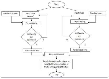
The Proposed method consists of following steps: 1) Vertebra detection 2) Intervertebral posterior height calculation 3) calculating traction weight. Data of 17 volunteers and X-ray Image data of 12 cases are taken to validate our approach by using a total of 60 cervical vertebrae. The objective is to find ROI and localize cervical spine centers. Thus, an automatic approach is proposed which gives generalization to traction therapy. Image analysis of C-spine is achieved with an accuracy of 98 %. Image characterized as normal or abnormal cervical image; also, exact traction weight is calculated using physics behind traction. 8% of the body weight will be the ideal weight to start traction with minimal side effect and highest therapeutic efficacy. Therefore, clinicians could adopt this weight as minimum weight in managing neck disorder requiring traction and goes up to the weight as calculated by proposed algorithm.
Hough Transforms, Spinal Traction, Spectral Transformation, Intervertebral Posterior Height
Singh V, Nigam v. S. Spinal Column Diagnosis in Cervical Vertebrae Interface Using Image Spectral Techniques. Biosc.Biotech.Res.Comm. 2020;13(4).
Singh V, Nigam v. S. Spinal Column Diagnosis in Cervical Vertebrae Interface Using Image Spectral Techniques. Biosc.Biotech.Res.Comm. 2020;13(4). Available from: https://bit.ly/35K3pfp
Copyright © This is an Open Access Article distributed under the Terms of the Creative Commons Attribution License (CC-BY). https://creativecommons.org/licenses/by/4.0/, which permits unrestricted use distribution and reproduction in any medium, provided the original author and sources are credited.
INTRODUCTION
In this paper, a smart approach is discussed to detect and localize changes in x-ray images of the cervical spine. The method discussed here for the treatment of cervical pain is a combination of image Processing and Physics theory, this analysis in future bears a major aid to doctors and will save considerable time. The goal of this research is to evaluate biomechanical efficacy and to reduce the range of the traction force (25lbs-4lbs) used for the treatment of cervical spinal traction by analyzing spinal structure. The study will help to improve outcomes, quality of care, and provide some standardization of treatment. For spinal column diagnosis the first step is vertebra detection. This task becomes more problematic in case of the cervical X-ray images as these images are usually characterized by their low contrasts and noise due to skull bones.
Preprocessing is done to locate and enhance the spine region of interest in x- ray images; this stage is usually followed by the processing stage which includes the boundary representation and segmentation algorithms based on feature vector extraction. Image processing has been used in all areas of research. It has become an important tool for visualization and analysis of data. In recent years, the field of medical imaging has required that the role of image processing to expand for better results (Huihao et al., 2019).
References till 2019-2020: In current scenario, back pain is a leading cause of work absenteeism. Ignorance of such pain becomes chronic disease which not only affect our spine, but also affect our work, afterwards it become major symptoms of disabilities. (Ehrlich et al., 1999; George E 2003). Three-dimensional imaging tests like MRI and CT scan, is ideal for analyzing& visualizing pathology of the IVD (intervertebral disc), neural structures such as the spinal cord. Imaging methods provide superior structural information and a better resolution (Schmitz et al., 2011); (Runshengwang et al., 2014); proposed a new method in which semiautomatic image analysis routine is used to analyze dendrite and synapse characteristics in immune-fluorescence images. Another method effective in reducing pain or improving intervertebral motions is HVLA-SM (High velocity low amplitude spinal manipulation) (Reed et al., 2015).
Another area of concerns is the cervical spine trauma which causes majority of spinal lesions. Many of these problems are the consequence of an abnormal spinal motion. A model of the human cervical spine was developed with a focus on accurate representation of the cervical spine using image processing algorithms. Models of each segment were constructed from the basic building blocks of the cervical spine: the intervertebral disc, the vertebrae, the ligaments, and the facet joints. The capsule ligament stiffness made an impact on segmental mobility and vertebral spatial position, and the sagittal angle of articular facet joint exerted an influence on disc pressure distribution. To predict effectiveness possibility of cervical traction the least absolute shrinkage and selection operator regression model was used to select potentially relevant features (Huihao et al., 2019; Yang et al., 2020).
The results of the study indicate that the no other algorithm at present using smart image processing algorithm for calculating exact traction weight in case of cervical spine. This research contribution is generalized traction weight calculation. The objective is to find ROI and localize cervical spine centers. The results of the study indicate that the no other algorithm is present using smart image processing algorithm for calculating exact traction weight in case of cervical spine. Individually in image processing algorithm the model was in reasonable agreement with the experimental data, and compared better than current models (Reed and Pickar, 2015).
MATERIAL AND METHODS
To find the accuracy of traction treatment a small survey was conducted under which data of 17 volunteer data and 12 X-ray image data were collected from 3 hospitals. In which 16 wereales and 13 were females. Following observations are mention as: Traction force is not only dependent to the fractional part of body weight but also on several other factors which is not included in the main parameters for applying the traction force. For cervical traction the force applied i.e. 1/7th part of body weight and for lumbar traction the force applied i.e. 1/3rd of body weight is not practically implemented, but it varies according to age, gender, bone density, blood pressure, etc which is not included at present in the treatment. On the basis of survey conducted, the following data were recorded. The following data were taken from Narmada Trauma Centre (Bhopal), Rajiv Gandhi Hospital (Trilanga, Bhopal) and Peoples hospital (Bhanpur, Bhopal) (Yang et al., 2020).
Table 1. Survey Report Of Some Patients Who Were Going Under The Treatment of Traction
| Volunteer data base & the amount of traction weight applied | ||||
| Patient number (M/F) | Age of patient (in years) | Body Weight (in kg) | Type of traction | Applied Weight (in kg) |
| Patient 1 (M) | 40 | 65 | Cervical | 7 |
| Patient 2 (F) | 38 | 55 | Lumbar | 20 |
| Patient 3
(M) |
33 | 55 | Cervical | 6 |
| Patient 4 (M) | 55 | 68 | Cervical | 8 |
| Patient 5 (F) | 22 | 58 | Cervical | 6 |
| Patient 6 (F) | 21 | 62 | Cervical | 6 |
| Patient 7 (M) | 38 | 52 | Cervical | 6 |
| Patient 8 (F) | 55 | 70 | Cervical | 8 |
| Patient 9 (F) | 49 | 75 | Lumbar | 25 |
| Patient 10 (M) | 39 | 90 | Cervical | 10 |
| Patient 11 (F) | 59 | 70 | Cervical | 5 |
| Patient 12 (M) | 50 | 75 | Cervical | 8 |
| Patient 13 (M) | 23 | 59 | Cervical +
Shoulder pain |
8 |
| Patient 14 (M) | 40 | 60 | Cervical | 2 |
| Patient 15 (M) | 55 | 60 | Lumbar | 20 |
| Patient 16 (M) | 55 | 75 | Lumbar | 25 |
| Patient 17 (M) | 30 | 54 | Cervical | 7 |
[source: narmada trauma centre (bhopal), rajiv gandhi hospital (trilanga, bhopal)]
Figure 1: ORIGINAL X-ray image of C-spinE
[Source: Peoples HOSPITAL, BHOPAL]
![[SOURCE: PEOPLES HOSPITAL, BHOPAL]](https://bbrc.in/bbrc/wp-content/uploads/2020/11/Vol_13No_4_Spi_Do_Fig1.jpg)
This data leads to us to do work for the improvement of traction treatment. The proposed research is a smart approach in which cervical vertebra detection method using a statistical algorithm and template matching approach based on the Spectral Transformation technique. In this method an edge-based recognition process used to extract vertebra shapes and its invariance to scale change, rotation and translation. This research is based on following steps:
A. Modelling –modelling is aimed to make template of the vertebra, edge detection and to apply Hough transform.
B. Centers detection-to enhance X-ray images histogram equalization is used as pre-processing. Canny and Sobel operators are used for edge detection and gradient computation.
C. Post-processing-In the last step adaptive filtering is used to find intervertebral distance.D. Mathematical calculation-after determining the intervertebral distance, mathematics is applied to calculate traction weight.
Figure 2: Flow chart for developing an algorithm for Spinal Traction.
Physics behind traction
Figure: free body diagram of traction machine setup
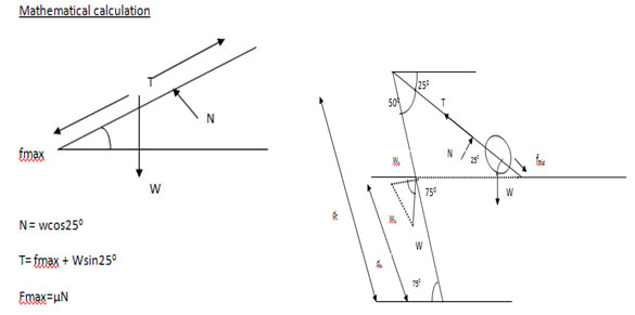
Below calculations are based on the traction machine setup, i.e. distance from ground, machine weight and total height. The cervical traction is applied to a patient; traction force is developed in backward while maintaining the system in equilibrium. Machine weight =15kg Center of mass height from ground= 102cm (max). If the distance from bottom to the center of mass is 102cm, then the total height from where force is applied is 173cm as distance from bottom to the center of mass 59% of total height.
From the free body diagram establishing momentum equation about the bottom
Ty.dT=Wy.dw Where: the distance from the bottom to the center of mass is
dW=59% of 1.73m =1.02m and dT= 85% 0f 1.73m = 1.46mtherefore
Ty.dT=Wy.dw Tsin500.dT = Wy.dw, (fmax +wsin250)sin500x1.46 = Wcos750x1.02 As fmax = µ.N
In this paper for two body weight of a patient, calculations are done. Body weight is 70kg and 75kg, so head weight i.e. 8% of the total body weight are 5.6kg and 6kgs respectively. (µ.N + wsin250) sin500x 1.46 = Wcos750x1.02, Where N = Wcos250, here w is weight of the head of a patient
And the value of µ = 0.2, Now equating the values in the equation
(µ. wcos250 + wsin250) sin500x 1.46 = Wcos750x1.02
(0.2×5.6cos250+ 5.6sin250) sin500x 1.46 = Wcos750x1.02
The exact W i.e. traction weight can be calculated as
W= [(0 cos 750 X 1.46] W= 15kgs
The calculations have been coded and required results have been obtained.
RESULTS AND DISCUSSION
In this research results have been calculated for radicular pain which occurs when the intervertebral separation is < 3mm. It has been suggested that the present research is not for patients with radiculopathy as it describes neurological condition. As the data set used in this research consists of X-ray images. The major problem faced while using X-Ray images are, the image data base is blurry, so vertebrae localization becomes the challenging task to perform manually. In order to enhance the image and to find region of interest image processing algorithm is used (Huihao et al., 2019).Next step is to resize the image [Fig-4].Longest segment has been detected using spectral transformation technique, in this research we use Hough transform for finding longest segment [Fig-5]. Following operation is formed to get Region of Interest.
- Morphological operations are performed now for boundary extraction [Fig-5].
- Plot the contour of the spectrum.
- In this step a matrix is formed according to brightness pixel value by applying condition BW1=1
- By above step we get Region of interest (ROI)[Fig-6].
Figure 4: C-spine image for 3 cases [source: image data base collects from Peoples hospital, Bhopal]
![Fig:4 C-spine image for 3 cases [source: image data base collects from Peoples hospital, Bhopal]](https://bbrc.in/bbrc/wp-content/uploads/2020/11/Vol_13No_4_Spi_Do_Fig4.jpg)
Figure 5: Image transformations using spectral transformation [source: Processed image of the collected image database]
![Fig: 5 Image transformations using spectral transformation [source: Processed image of the collected image database]](https://bbrc.in/bbrc/wp-content/uploads/2020/11/Vol_13No_4_Spi_Do_Fig5.jpg)
Figure 6: Initial segmentation stage [source: Processed image of the collected image database]
![Fig 6: Initial segmentation stage [source: Processed image of the collected image database]](https://bbrc.in/bbrc/wp-content/uploads/2020/11/Vol_13No_4_Spi_Do_Fig6.jpg)
Figure 7: Locating ROI [source: Processed image of the collected image database]
![Fig 7: Locating ROI [source: Processed image of the collected image database]](https://bbrc.in/bbrc/wp-content/uploads/2020/11/Vol_13No_4_Spi_Do_Fig7.jpg)
Figure 8: Intervertebral Posterior height calculated for three images from the image data base collected from Peoples Hospital, Bhopal
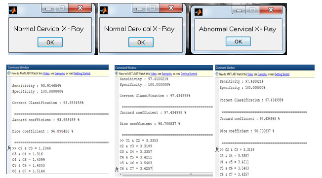
After characterizing image as normal or abnormal image, an algorithm used to detect amount of traction weight required.
Figure 9: C++ code to detect traction weight based on body weight of patient under medical supervision for normal and abnormal images from image database collected from Peoples hospital, Bhopal.
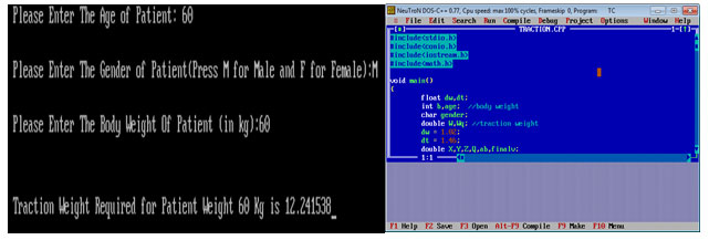
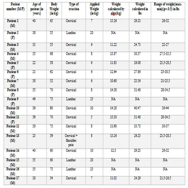
Table 3 shows the applied and proposed traction weights for the patient under medical supervision for neck disorder.
Table 3. Table shows the result of applied weight and proposed weight for C-spine traction. [source: Narmada Trauma Centre (Bhopal), Rajiv Gandhi Hospital (Trilanga, Bhopal)
The intervertebral disc (IVD) is comprised of three distinct components: (a) the annulus fibrous, (b) the nucleus pulposus, and (c) the cartilaginous endplates. The annulus fibrous is the tissue of the intervertebral disc that surrounds the nucleus pulposus and forms the outer portion of the disc. Due to compressive load the posterior section of the intervertebral disc will undergo tension loading that causes the annulus to contract toward the center of the disc (White et al., 1990; Runshengwang and Ward, 2014). The intervertebral height for cervical spine is mentioned in the table given below.
Table 4. Accuracy Recognition
| S.No | Study | IVD height | C23 | C34 | C45 | C56 |
| 1. | Gilad & Nissan, 1986 | Posterior | 3.4mm | 3.3mm | 3.0mm | 3.0mm |
| 2. | Przybylski et al,1998 | Posterior | 3.4mm | 3.4mm | 3.7mm | 3.9mm |
| 3. | Present Research | Posterior | 3.3mm | 3.3mm | 3.4mm | 3.5mm
|
| Accuracy Recognition | 97% | 97% | 98.5% | 98.5% |
Due to load, the intervertebral posterior height reduces. In this research, image processing algorithm is used to detect the intervertebral posterior height. As the study says for spinal traction relieves pressure on cervical spine, it can be done manually or mechanically. To get better results x-ray images of cervical area must be studied along with the symptomatic problems. In this research both the symptoms as well as image analysis is performed in order to get better results (Fabian et al., 2012; Runshengwang and Ward, 2014). None of the study at present will work on reducing the range of traction force (25lbs-40lbs), as the amount of force applied will depends on the experience of physiotherapist. Most of the researches are using image data base of CT scan or MRI imaging but X-ray imaging is economic among the imaging technique used. 8% of the body weight will be the ideal weight to start traction with minimal side effect and highest therapeutic efficacy. Therefore, clinicians could adopt this weight as minimum weight in managing neck disorder requiring traction and goes up to the weight as calculated by proposed algorithm (Martiel et al., 2015).
CONCLUSION
In this paper, a new smart approach is used to detect C-spine vertebrae in X-ray images. A model of the human cervical spine was developed with a focus on accurate representation of the cervical spine using image processing algorithms. Models of each segment were constructed from the basic building blocks of the cervical spine: the intervertebral disc, the vertebrae, the ligaments, and the facet joints. This research contribution is generalized traction weight calculation. The objective is to find ROI and localize cervical spine centers. The results of the study indicate that the no other algorithm is present using smart image processing algorithm for calculating exact traction weight in case of cervical spine.
Individually in image processing algorithm the model was in reasonable agreement with the experimental data, and compared better than current models. Furthermore, it was recommended that the future goal of this model would be to implement so that a Clinical prediction has been developed which will improve decision-making and reduce time & cost. Thus, an automatic approach is proposed which gives generalization to traction therapy. Considering the advantages (feasible, accurate and economic) of the goal, if achieved, this may be used to develop traction machines in future which will be algorithm based and would work automatically on the basis of the factors to be considered while applying the therapy. Angle of head and table selected is 250, in future different angle settings will be done for C1-C2 & C6-C7.
ACKNOWLEDGEMENTS
We show our gratitude to Narmada Trauma Centre (Bhopal), Rajiv Gandhi Hospital (Trilanga, Bhopal) and Peoples hospital (Bhanpur, Bhopal). We thank Principal Rajiv Gandhi Physiotherapy College (Trilanga, Bhopal) for giving letter of recommendation for above research.
Conflict of Interest
The authors and planners have disclosed no potential conflicts of interest, financial or otherwise.
REFERENCES
Adams MA, Freeman BJ and Morrison HP (2000). Mechanical initiation of intervertebral disc degeneration. Hopkins clinic report on spinal decompression therapy, Spine 25(13): 1625-36. Available at: http://www.hopkinsclinic.com/decompression-therapy-dts.html.
Bagchi K, Mohaideen A, Thomson JD and Foley LC (2002). Hardware complications in scoliosis surgery. PediatrRadiol; 32:465–475.
Ehrlich GE and Khaltaev NG (1999). Low back pain initiative Geneva: World Health Organization.
Lecron F, Benjelloun M and Mahmoudi S (2012). Cervical Spine Mobility Analysis on Radiographs: A Fully Automatic Approach. Computerized Medical Imaging and Graphics.
Ehrlich GE. (2003). Low Back Pain, Bulletin of the World Health Organization, 81 (9).
Heaven Stubblefield (2014) Published on January 14. Available at: http://www.healthline.com/health/spinal-traction#Overview1
Huihao, Kuan, Zhen, Xiaofei, Yi-Xian, Hongsheng and Wenxin (2019). Effects of facet joint degeneration on stress alterations in cervical spine C5–C6: A finite element analysis. Mathematical Biosciences and Engineering 16(6):7447-7457. DOI: 10.3934/mbe.2019373
Martiel L, Lea A and Kurzawa L (2015). Measurement of cell traction forces with Image J. in Methods in Cell Biology, Ed., vol. 125, chapter 15, pp. 269–287, Academic Press.
Raney NH, Petersen EJ and Smith TA (2009). Development of a clinical prediction rule to identify patients with neck pain likely to benefit from cervical traction and exercise. European Spine Journal; 18:382–391.
Reed WR and Pickar JG (2015). Paraspinal Muscle Spindle Response to Intervertebral Fixation and Segmental Thrust Level during Spinal Manipulation in an Animal Model. 40(13) E752-E759.
Runshengwang and Ward MM (2014). Arthritis of the Spine ChapterSpinal Imaging and Image Analysis Volume 18 of the series Lecture Notes in Computational Vision and Biomechanics pp 31-66.
Schmitz S. K., Hjorth J. J. J. and Joemai R.M. S. (2011). Automated analysis of neuronal morphology, synapse number and synaptic recruitment. Journal of Neuroscience Methods, vol. 195, no. 2, pp. 185–193.
White, A.A. and Panjabi, M.M. (1990) Clinical Biomechanics of the Spine. ISBN 0-397-50720-8 Second Edition.
Wong and L.K.F.(2013). Cervical Spine Simulation model for Traction Therapy Analysis. System Integration (SII), IEEE/SICE International Symposium, pg. no.516 – 520.
Yang, Qisen, Shiping, Taotao, Linquan, Shengxiong, Bin, Qin, Zhengru, Wenge and Zhenyu (2020). Is Cervical Traction Effective in Chronic Nonspecific Neck Pain Patients with Unsatisfactory NSAID Control? A Nomogram to Predict Effectiveness. World neurosurgery. Volume 139, July 2020, Pages e245-e254. Available at: https://doi.org/10.1016/j.wneu.2020.03.195.

