Department of Biotechnology, G B Pant Engineering College, Pauri Garhwal, Uttarakhand, India
Corresponding author Email: vermajyoti983@gmail.com
Article Publishing History
Received: 19/03/2018
Accepted After Revision: 16/04/2018
The present study was conducted to evaluate the in vitro inhibitory effect of ethanolic extract of Quisqualis indica leaves on the digestive enzyme á-amylase and to characterize the compounds responsible for it as a means of managing hyperglycaemia. The presence of various phytochemicals such as flavonoids, phenols, alkaloids, tannins may be responsible for the plant biological activities. The ethanolic leaf extract of the plant was further subjected to á-amylase inhibitory assay where it showed the dose dependent inhibitory effect on the á-amylase enzyme when compared with the positive control acarbose. GC-MS analysis of the ethanolic plant leaf extract revealed the presence of phytol in the highest concentration and other compounds like linolenic acid, pentadecanoic acid and 9, 12-linoleic acid in moderate concentrations which were further evaluated for their biological activities using PASS and compared with the biological activity profile of the anti-diabetic drug acarbose. The various modes of action of these compounds include á-amylase inhibition, á-glucosidase inhibition, insulin inhibition, etc. In silico studies were also done using AutoDock to study the compound’s minimum free energy of stabilization in complex with the á-amylase enzyme. Further Ligplot+ was used to study the presence of hydrogen bonding in the complex. Drug parameters of the identified compounds in the extract were evaluated and compared with the acarbose. The results obtained were successfully compared to the pharmacological and toxicological activity information available for the studied compounds.
Autodock Vina, Diabetes Mellitus, Gc-Ms Analysis, Pass, Quisqualis Indica, Ligplot+ V.1.4.3.
Verma J, Arora D, Singh A. Evaluation of Anti-Hyperglycaemic Potential of the Ethanolic Leaf Extract of Quisqualis Indica. Biosc.Biotech.Res.Comm. 2018;11(2).
Verma J, Arora D, Singh A. Evaluation of Anti-Hyperglycaemic Potential of the Ethanolic Leaf Extract of Quisqualis Indica. Biosc.Biotech.Res.Comm. 2018;11(2). Available from: https://bit.ly/2TIYrYK
Introduction
Diabetes mellitus is a chronic heterogeneous endocrine disorder characterized by elevated blood glucose level resulting in serious metabolic disturbances in carbohydrate, protein and fat metabolism causing premature fatality (Patel et al. 2011; Warjeet 2011). After cancer, cardiovascular and cerebrovascular disease, it is the third most life-threatening disease posed to the health of mankind (Chauhan et al. 2010). Annually, a rise of 4-5 % in number of diabetic patients is observed (Wagman and Nuss 2001). It is caused due to insufficient insulin production or psychological unresponsiveness to insulin. Pancreatic á-amylase enzyme is an endoglucanase that catalyzes the internal á-1, 4 glycosidic bond hydrolysis in starch and other polysaccharides to yield maltose and maltotriose polysaccharides. Inhibition of this enzyme can help to manage the disorder by lowering the level of glucose released in the blood. There is considerable evidence that lipid peroxidation owing to free radical activity causes induction of oxidative stress that plays a crucial role in the onset of the abnormal condition. Alteration in anti-oxidant enzymes, impaired glutathione metabolism and decreased ascorbic acid levels are the main causes of disturbance of anti-oxidant defence system (Patel et al. 2011). This leads to accumulation of advanced glycation products (AGEs) and sorbitol concentration that cause complications such as retinopathy, neuropathy and renal dysfunction (Safi et al. 2014). The physical symptoms include cycle of heavy thirst and frequent sugar loaded urination along with presence of sugar in mucus, sweat and breath. In the past recent years, natural pharmacologically bioactive compounds derived from terrestrial and marine organisms had received considerable attention to cure potentially vulnerable diseases due to their lesser or virtually no side effects as compared to synthetic drugs (Yuan
et al. 2016).
Recently many R&D based pharmaceutical company are employing more time and money on development of herbal therapies rather than formulating synthetic drugs due to their unpredictable adverse effects. Ayurveda, which is the renowned traditional system of medicines native to India, had always promoted the use of plant biodiversity for several therapeutic uses due to their ease of availability and low cost of production (Patwardhan and Hopper 1992). Plant-derived molecules (PDMs) could be chemically elaborated to generate novel leads and to screen molecules from drug-like libraries and hence can be proved effective to systematically extract unique molecular scaffolds. Quisqualis indica, commonly known as ‘rangoon creeper’ is an important medicinal plant of the Indian subcontinent, Africa and Indo Malaysian region (Joshi 2002). It is a vine with pink and white flowers. It is also used as anthelmenic by the inhabitants of North Annan to expel parasitic worms (helminths) and other internal parasites from the body (Kirtikar and Basu 2006). It has also been reported that the extract of flowers of Q. indica exhibit hypoglycaemic and hypocholesterolemic activity against animal models (Bairagi et al. 2012). Various parts of the plant are used individually or mixed with other ingredients as a cure for ailments like antiflatulence, coughs, diarrhea (Khare 2007), body pains, toothache (Padua et al. 1999).
With the available therapeutic knowledge, scientists have been thriving hard to explore the safe and effective treatment of the disease which is yet to be achieved. Clinically various oral anti-diabetic drugs are used such as biguanides that increase glucose uptake, sulfonylureas that increase insulin secretion and digestive enzyme inhibitors that delay complex carbohydrate digestion and absorption. Large scale research is being done worldwide, exploiting the known ethno-botanical knowledge and phytochemical interpretations that could be an effective approach for the diabetes treatment. In the designing of drug, the in silico analysis using bioinformatics tools is a boon for the researchers as they minimize the time and labour employed during the work. PASS is one such tool that uses algorithms based on the physico-chemical methods, that predicts the possible activity of the drug against a target using the intrinsic property of the compounds (Filimonov et al. 1995; Filimonov and Poroikov 1996). In 1972, the National Registration System of New Chemical Compounds organized in the USSR formulated the computer program PASS which was suggested by researcher V. Avidon (Burov et al. 1990; Poroikov et al. 2003).
Computational based approaches, such as molecular docking, hydrogen bonding analysis, evaluation of drug parameters have been widely used in the modern drug discovery to explore drug-receptor interactions. For designing of novel inhibitors, molecules from a database of organic compounds should be screened based on steric and electrostatic complementarity with the binding pocket of protein. Molecular docking simulation studies were performed using AutoDock Vina. Docking of the individual compounds with the á-amylase enzyme which is the key enzyme involved in the regulation of the metabolic pathway was done using Autodock Tools 1.5.4 package. Ligplot+ v.1.4.3 software can help in refined assessment of docked complexes and in obtaining detailed information on protein-ligand interaction (Laskowski and Swindells 2011).
Chemicalize.orgbeta software tool by ChemAxon was used to study the drug like activities of the compounds identified through GC-MS analysis. Therefore, combination of these methods can be used to study mechanisms of drug-receptor interactions, and provide structural insights by which molecules interact within binding pocket of the receptor. Further experimental evaluation and validation are required to establish the clinical significance of leads obtained that are promising candidates.
Materials and Methods
Plant material and preparation of extract
The plant leaves of Quisqualis indica Linn. were obtained from Forest Research Institute (FRI), Dehradun, India. The plant was botanically identified at FRI. Leaves were thoroughly cleaned with tap water to remove any dust particles and further shade dried for 3 weeks to make them crisp and completely devoid of moisture. The dried leaves were finely grinded and 10 g of the powdered material was extracted with 250 mL of ethanol procured from Merck, India using Soxhlet apparatus for 48 hrs. The extract was concentrated using vacuum rotary evaporator at 78 °C. The extract was filtered through Whatmann filter paper No.1 and stored at 4 °C until use. Rest of the chemicals were of analytical grade. Preliminary screening: Phytochemical screening of the leaf extract was done using the methods described by Trease and Evans (1996), and Sofowora (2006).
In vitro á-amylase inhibitory assay
The assay was carried out according to the standard protocol (Hansawasdi et al. 2000), with slight modifications. 2 mg of starch azure was suspended in 0.2 mL substrate solution containing 0.5 M Tris-HCl buffer (pH 6.9) and 0.01 M CaCl2. The substrate solution in tubes were boiled for 5 min and then preincubated at 37 °C for 5 min. Ethanol extract of Q. indica was dissolved in DMSO to obtain concentrations of 10, 20, 40, 60, 80, and 100 µg/mL. Then, 0.2 mL of plant extract of particular concentration was added to the substrate solution in respective tubes. Further, 0.1 mL of porcine pancreatic á-amylase procured from Hi-Media in Tris-HCl buffer (2 units/mL) was added to the tube containing the plant extract and substrate solution. The reaction was carried out at 37 °C for 10 min. The reaction was terminated by addition of 0.5 mL of 50 % acetic acid in each tube. The reaction mixture was centrifuged at 3000 rpm for 5 min at 4 °C. The absorbance of resulting supernatant was measured at 595 nm using spectrophotometer. Acarbose, a known á-amylase inhibitor was used as a standard drug control. The experiments were done in triplicates. The á-amylase inhibitory activity was calculated by using following formula:
The á-amylase inhibitory activity = [{(Ac+) – (Ac–)}
– {(As – Ab)}/ {(Ac+) – (Ac–)}] × 100,
where Ac+, Ac-, As, and Ab are defined as the absorbance of 100 % enzyme activity (only solvent with enzyme), 0 % enzyme activity (only solvent without enzyme), a test sample (with enzyme), and a blank (a test sample without enzyme), respectively. The percentage inhibitory effects of acarbose and plant extract on á-amylase activity were determined. Statistical analysis was performed using Microsoft Excel. All values were expressed as mean ± standard deviation.
GC-MS was carried out at Advanced Instrumentation Research Facility (AIRF) at Jawaharlal Nehru University (JNU), New Delhi, India. The analysis was done on GCMS-QP2010 Ultra under the following conditions to study the phytochemical components present in the extract. Column-Rtx-5 MS (30 m X 0.25 mm i.d. X 0.25µm film thickness) was used. 2 µL of the plant sample was injected in split mode at a constant column flow rate of 1.21 mL/ min with linear velocity 40.9 cm/sec flow control mode and purge flow of 3.0 mL/ min. The column temperature was programmed to 100 °C, ion source temperature at 220 °C and the injection temperature was 260 °C. Total GC-MS running time was 45 min. Inbuilt libraries WILEY8.lib and NIST11.lib were used for the identification and comparison of the organic compounds. The name, molecular weight and structure of the phytochemical components of the extract were ascertained.
The activity list comprises of names of pharmaco-therapeutic effects as well as names of mechanism of action. The compounds present in high concentration in the plant leaf extract were studied using PASS for their biological activity prediction, whose structures were drawn using MarvinSketch v5.10.0 and compared with acarbose. The mean accuracy of prediction is about 85 % in leave-one-out cross-validation (LOOCV), hence it is reasonable using this tool to find and optimize new lead compounds which is a crucial step in pharmaceutical research and development process. The chemical structures of molecules were drawn and edited using MarvinSketch v.5.10.0 software (https://www.chemaxon.com), an advanced chemical structure editor. The structures were saved in 3D MOL2 format. Individual MOL2 files were converted into PDBQT format (acceptable format for AutoDock Vina package (Trott and Olson 2010)), using the python script ‘prepare_ligand4.py’ available in Autodock Tools 1.5.4 package (Morris et al. 2008). During this conversion, appropriate charges were added to ligands. The commercially available drug acarbose as well as the compounds present in high concentration in the extract were docked with enzyme á-amylase using software AutoDock Vina to analyze their free energy. Further presence of hydrogen bonding were analyzed using software Ligplot+ v.1.4.3 software and molecular interactions between protein and ligands were predicted. Drug parameters were studied using chemicalize.orgbeta by ChemAxon.
| Table 1: Phytochemical screening of ethanolic leaf extract of Q. indica | |||||
| S. No. | Phytochemicals | + = present;
– = absent |
S. No. | Phytochemicals | + = present;
– = absent |
| 1. | Anthraquinones | – | 6. | Tannins | + |
| 2. | Flavonoids | + | 7. | Terpenoids | – |
| 3. | Reducing sugar | – | 8. | Phenols | + |
| 4. | Saponins | + | 9. | Alkaloids | + |
| 5. | Steroids | – | 10. | Quinones | + |
| Table 2: [á-amylase inhibitory effects of Q. indica leaf extract in comparison with standard drug acarbose | |||
| S. No. | Concentration (µg/mL) | % inhibition by acarbose | % inhibition by ethanolic leaf extract of Q. indica |
| 1.
2. 3. 4. 5. 6. |
10
20 40 60 80 100 |
20.19 ± 0.96
27.04 ± 0.98 37.17 ± 1.47 43.91 ± 1.46 57.68 ± 1.92 71.79 ± 0.55 |
14.86 ± 1.93
24.35 ± 0.88 26.15 ± 3.35 38.97 ± 1.17 53.07 ± 2.03 56.40 ± 2.35 |
Results and Discussion
The plant leaf extract was found to contain the phytoconstituents like flavonoids, saponins, tannins, phenols, alkaloids and quinones upon preliminary screening among which flavonoids, tannins and phenols are reported to play a crucial role in the management of diabetes mellitus (Table 1, S. No. 1-10). The percentage inhibitory activity exhibited by the extract and comparison with acarbose is shown in Table 2. (S. No. 1-6). Acarbose at concentration 100 µg/mL showed 71.79 ± 0.55 % inhibitory effects on the á-amylase activity. The ethanolic leaf extract of Q. indica at a concentration 100 µg/mL exhibited 56.40 ± 2.35 % of á-amylase inhibitory activity. The plant extract showed potent á-amylase inhibitory activity in a dose dependent manner as compared with acarbose (Fig. 1). Acarbose was used as a positive control which is a secondary metabolite which belongs to the class of drugs called digestive enzyme inhibitors. It is obtained via a multistep batch fermentation process from bacterium Actinoplanes species SE50. It is approved for treating type II diabetic patients. This reveals that the extract exhibited a comparable inhibitory effect on the á-amylase enzyme as compared to the standard drug acarbose.
| Table 3: Phytochemical components identified as major percentage in the ethanolic extract of leaves of Q. indicaby GC-MS | |||||
| R.Time | Mol. Wt. | Formula | Name | Area% | Activity already reported |
| 16.509 | 242 | C15H30O2 | Pentadecanoic acid | 8.73 | – |
| 17.959 | 296 | C20H40O | (E)-Phytol | 18.07 | anti-microbial, anti-cancer, anti-inflammatory, anti-diuretic, immunostimulatory, anti-diabetica |
| 18.173 | 280 | C18H32O2 | 9,12-Linoleic acid | 3.48 | anti-inflammatory, nematicide, insectifuge, hypocholesterolemic, cancer preventive, hepatoprotective, anti-histaminic, anti-acne, anti-arthritic, anti-eczemic, 5-á reductase inhibitor, anti-androgenic, anti-coronaryb |
| 18.259 | 278 | C18H30O2 | Linolenic acid | 9.52 | prevent heart attacks, lowers high blood pressure, lowers cholesterol, reverse “hardening of the blood vessels (atherosclerosis)c, treatment of rheumatoid arthritis (RA), multiple sclerosis (MS), lupus, anti-diabetic, treatment of chronic obstructive pulmonary disease (COPD), migraine headache, skin cancer, depression, allergic and inflammatory conditions such as psoriasis and eczemad |
| aVenkata et al. 2012; bSermakkani and Thangapandian 2012; cBrouwer et al. 2004; dChristensen et al. 2000 | |||||
 |
Figure 1: Percentage of á-amylase inhibitory effects of acarbose (standard drug) and ethanolic leaf extract of Q. indica |
Although the presence of flavonoids and phenols contribute to the anti-diabetic activity, the specific bioactive components of the plant extract were studied through GC-MS analysis (Fig. 2). The peaks clearly show that compounds like phytol, linolenic acid, pentadecanoic acid and 9, 12-linoleic acid were present in subsequently higher concentrations. GC-MS study of the Q. indica leaf extract has shown the presence of a number of phytochemical constituents which confer the medicinal property to the plant (Table No 3).
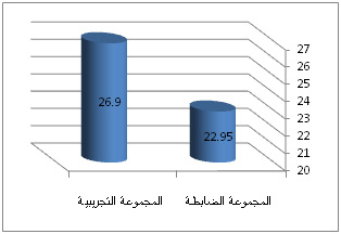 |
Figure 2: Chromatogram of the ethanolic leaf extract of Q. indica by GC-MS |
This study depicted the presence of 55 compounds. Among these, 21 compounds with their biological activities have been reported earlier. Compounds like lauric acid, 9, 12-linoleic acid, linolenic acid, vitamin E, stigmasterol, á-sitosterol are reported to posses hypocholesterolemic activity. Compounds like neophytadiene, squalene, heptacosanol, solanesol, á-tocopherol, vitamin E, stigmasterol, á-sitosterol, fucosterol are reported to have anti-oxidant activity. Both of these activities play a direct role in the management of diabetes mellitus by blocking the sugar metabolism. Phytol also known as phytanic acid is the test compound to be studied which was present in the highest concentration in the plant Q. indica leaves. Phytol is an acyclic diterpene alcohol molecule which is a precursor of synthetic vitamin E and K1 (Thomas and Netscher 2007; Daines et al. 2003). It was first obtained by chlorophyll hydrolysis and now obtained in the process of chlorophyll separation from alfalfa. It is reported as transcription factors peroxisome proliferator activated receptor (PPAR-á) and retinoid X receptor (RXR) activator. It is already reported to be a cholesterol lowering agent in patients with type II diabetes, obesity and cardiovascular diseases. It is also reported to possess anti-inflammatory as well as metabolic properties.
The predicted biological activity spectrum of the compounds by PASS contributing to their anti-hyperglycaemic potential is shown in Table 4 (S. No. 1-5). The PASS result is obtained in the form of names of biological activity whose probability value ranges from 0.000 to 1.000. Only activity types for which Pa>Pi, are considered possible. Pa and Pi are the probability measures for the compound to be active and inactive respectively for the respective activities in the biological activity spectrum. The various predicted metabolic pathways of phytol involved in control of diabetes include dextranase inhibition, glycerol-3-phosphate dehydrogenase inhibition, á-glucuronidase inhibition, diabetic neuropathy treatment, etc. It shows 7 possible pathways known to play a role in diabetes management that can be further studied using wet lab experiments to explore its potential to be used as drug for treatment of diabetes. Other compounds like stigmasta-5, 23-dien-3â-ol, squalene, stearic acid, tetracontane, heptacosanol, stigmasterol were also predicted for the presence of their biological activities based on their chemical structures that could play a role in the management of diabetes.
| Table 4: Biological activity spectrum of standard drug acarbose and peak compounds identified by GC-MS | ||||
| S. No. | Compound | Pa | Pi | Biological activity |
| 1. | Acarbose | 0,972
0,962 0,958 0,943 0,779 0,752 0,691 0,681 0,636 0,611 0,512 0,508 0,499 0,447 0,333 |
0,000
0,000 0,000 0,000 0,002 0,005 0,005 0,002 0,009 0,001 0,002 0,003 0,003 0,016 0,002 |
Sucrose á-glucosidase inhibitor
4-á-glucanotransferase inhibitor á-glucosidase inhibitor á-amylase inhibitor â-amylase inhibitor Anti-diabetic Fructan â-fructosidase inhibitor â-glucosidase inhibitor â-glucuronidase inhibitor â-galactosidase inhibitor Amylo-á-1,6-glucosidase inhibitor Isoamylase inhibitor Oligo-1,6-glucosidase inhibitor Galactose oxidase inhibitor á-L-fucosidase inhibitor |
| 2. | Phytol | 0,541
0,491 0,465 0,401 0,369 0,373 0,349 |
0,018
0,013 0,020 0,030 0,006 0,112 0,010 |
Dextranase inhibitor
Sorbitol-6-phosphate 2-dehydrogenase inhibitor Glycerol-3-phosphate dehydrogenase inhibitor Fructan â-fructosidase inhibitor á-N-acetylgalactosaminidase inhibitor Diabetic neuropathy treatment á-glucuronidase inhibitor |
| 3. | Linolenic acid | 0,902
0,852 0,826 0,597 0,574 0,515 0,490 0,491 0,467 0,466 0,447 0,464 0,430 |
0,003
0,004 0,004 0,015 0,011 0,004 0,006 0,013 0,006 0,012 0,016 0,051 0,021 |
Dextranase inhibitor
Cholesterol antagonist Antihypercholesterolemic Insulin promoter Fructan â-fructosidase inhibitor á-glucuronidase inhibitor á-amylase inhibitor Sorbitol-6-phosphate 2-dehydrogenase inhibitor 1,2- á-L-fucosidase inhibitor Anti-diabetic (type II) Galactose oxidase inhibitor â-glucuronidase inhibitor Diabetic neuropathy treatment |
Binding simulation studies revealed stable complexes of enzyme and the respective compounds with their energy minimization values shown in Table 5 (S. No. 1-5). AutoDock studies have revealed the minimum free energy needed to stabilize the complex of the á-amylase enzyme with the studied compounds. Interestingly, in our docking studies, we found that phytol could be a potent á-amylase inhibitor with quite a strong binding affinity and made remarkable inhibitory interactions with critical residues. It was observed that the chemical interactions established between phytol and á-amylase binding pocket residues were the most stable as compared with other studied compounds like linolenic acid, pentadecanoic acid and 9, 12-linoleic acid. Hydrogen bondings were visualized using Ligplot+ v.1.4.3 software to study the stability of the complex of the enzyme and the target compounds. Drug parameters of the target compounds were analyzed and compared with acarbose, as shown in table 6 (S. No. 1-6). The molecular mass, polar surface area and molar refractivity of phytol, linolenic acid, pentadecanoic acid and 9, 12-linoleic acid were found to be in range in compliance with Lipinski rule. The results of the conducted study clearly indicate the anti-hyperglycaemic property of the plant Q. indica. All the test compounds studied above may contribute to the anti-diabetic potential of the plant.
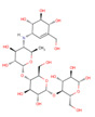 |
Table 5a |
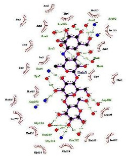 |
Table 5b |
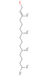 |
Table 5c |
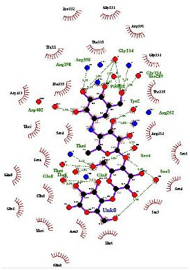 |
Table 5d |
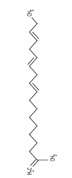 |
Table 5e |
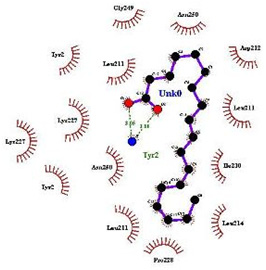 |
Table 5f |
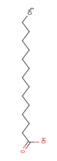 |
Table 5g |
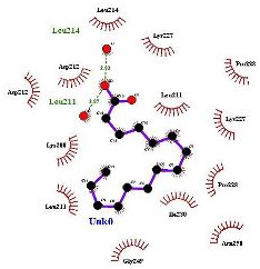 |
Table 5h |
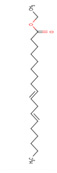 |
Table 5i |
 |
Table 5j |
| Table 6: Evaluation of drug parameters of peak compound identified by GC-MS analysis and comparison with the anti-diabetic drug acarbose | ||||||
| S. No. | Drug parameters | Acarbose | Phytol | Linolenic acid | Pentadecanoic acid | 9,12 linoleic acid |
| 1. | Mass | 646 | 296 | 267 | 227 | 267 |
| 2. | Formula | C25H43NO18 | C20H40O | C18H30O2 | C15H30O2 | C18H32O2 |
| 3. | Log P | -7.61 | -7.04 | -6.06 | -5.81 | -6.42 |
| 4. | Polar Surface Area | 321.17 | 20.23 | 37.30 | 37.30 | 37.30 |
| 5. | Molar Refractivity | 136.524780 | 95.561760 | 79.009491 | 69.104492 | 80.161491 |
| 6. | Lipinski Rule of 5 | No | No | No | No | No |
Conclusion
In the context of already known mechanisms central to diabetes and its complications, we proposed that bioactive compounds of Q. indica could be prospected for, in search of potential á-amylase inhibitors. To the best of our knowledge, this is the first report of screening á-amylase inhibitor from Q. indica leaves for alleviation of diabetes and related complications. The molecular interaction patterns observed in the conducted study may enable the designing of novel drug structures with considerable inhibitory action. We believe that the study conducted could provide leads for designing novel drug inhibitors of á-amylase with better efficacy and comparatively lesser side-effects. Such a formulation could be exploited for the development of an effective dosage form for drugs that inhibit the digestive enzymes, helping to attain maximum therapeutic efficacy at reduced dose and minimum toxicity.
Acknowledgments
This study was conducted in the Department of Biotechnology, G. B. Pant Engineering College (GBPEC), Pauri Garhwal (Uttarakhand), India. Authors gratefully acknowledge the assistance provided by Dr. Ajai Kumar (system analyst) at AIRF-JNU and Dr. Ritu Chowdhary. Jyoti Verma is thankful to TEQIP-II (Technical Education Quality Improvement Programme, Government of India) for financial assistance.
Conflict of Interest
Authors have no conflict of interest regarding the publication of paper.
References
Ariful HM, Azmal IH, Tridib KP, Mariz S, Himel NK (2010) A survey of medicinal plant usage by folk medicinal practitioners in two villages by the Rupsha river in Bagerhat District, Bangladesh. American-Eurasian J Sustainable Agri 4(3): Pages 349-356.
Bairagi VA, Sadul N, Senthilkumar KL, Y Ahire (2012) Anti-diabetic potential of Quisqualis indica Linn in rats. Int J Pharm Phytopharmacol Res 1(4): Pages 166-171.
Brouwer IA, Katan MB, Zock PL (2004) Dietary alpha-linolenic acid is associated with reduced risk of fatal coronary heart disease, but increased prostate cancer risk: a meta-analysis. J Nutr 134(4): Pages 919-922.
Burov YV, Poroikov VV, Korolchenko LV (1990) National system for registration and biological testing of chemical compounds: facilities for new drugs search. Bull Natl Center for Biologically Active Compounds (Rus.) 1: Pages 4-25.
Chakraborty AK, Shrivastava A, Garg S, Shrivastava TP (2015) Pharmacognostic and phytochemical study of whole plant of Quisqualis indica Linn. International Journal of Medicine and Pharmaceutical Research 3(2): Pages 970-977.
Chauhan A, Sharma PK, Srivastava P, Kumar N, Dudhe R (2010) Plants having potential anti-diabetic activity: A review. Der Pharmacia Lettre 2(3): Pages 369-387.
Christensen JH, Christensen MS, Toft E, Dyerberg J, Schmidt EB (2000) Alpha-linolenic acid and heart rate variability. Nutr Metab Cardiovasc Dis 10(2): Pages 57-61.
Colditz GA (2000) Changing dietary patterns and cancer prevention: alpha-linolenic acid health risks and benefits. Cancer Causes Control 11(8): Pages 677-678.
Connor WE (1999) Alpha-linolenic acid in health and disease. Am J Clin Nutr 69(5): Pages 827-828.
Daines A, Payne R, Humphries M, Abell A (2003) The synthesis of naturally occurring vitamin K and vitamin K analogues. Current Organic Chemistry 7(16): Pages 1625-1634.
Ding Z, Lu Y, Lu Z, Lv F, Wang Y, Bie X, Wang F, Zhang K (2010). Hypoglycaemic effect of comatin, an anti-diabetic substance separated from Coprinus comatus broth, on alloxan-induced-diabetic rats. Food Chem 121(1): Pages 39-43.
Djousse L, Arnett DK, Pankow JS, Hopkins PN, Province MA, Ellison RC (2005) Dietary linolenic acid is associated with a lower prevalence of hypertension in the NHLBI Family Heart Study. Hypertension 45: Pages 368-373.
Filimonov DA, Poroikov VV (1996) PASS: Computerized prediction of biological activity spectra for chemical substances. In: Bioactive compound design: Possibilities for industrial use, Oxford: BIOS Scientific Publishers 47-56 http://www.ibmh.msk.su/PASS
Filimonov DA, Poroikov VV, Borodina Y, Gloriozova T (1999) Chemical similarity assessment through multilevel neighborhoods of atoms: definition and comparison with the other descriptors. J Chem Inf Comput Sci 39: Pages 666-670.
Filimonov DA, Poroikov VV, Karaicheva EI, Kazaryan RK, Boudunova AP, Mikhailovsky EM, Rudnitskih AV, Goncharenko LV, Burov YV (1995) Computer-aided prediction of biological activity spectra of chemical substances on the basis of their structural formulae: computerized system PASS. Experimental and Clinical Pharmacology (Rus) 8(2): Pages 56-62.
Guidelines on registration of traditional medicines in the WHO African Region (2004). Brazzaville: World Health Organization Regional Office for Africa, Background and Purpose: 40.
Hansawasdi C, Kawabata J, Takanori K (2000) á-amylase inhibitors from Roselle (Hibiscus sabdariffa Linn.) Tea. Biosci Biotechnol Biochem 64: Pages 1041-1043.
Joshi SG (2002) Medicinal plants, 1st edn. Mohan Primlani for Oxford and IBH publishing Co pvt Ltd, Delhi, Page 141.
Khare CP (2007) Indian Medicinal Plants. An Illustrated Dictionary Berlin/Heidelberg: Springer-Verlag. Pages 649-650.
Kirtikar KR, Basu BD (2006) Indian medicinal plant, 2nd edn. Prashant Gahlot at valley offset publishers, New delhi, Page 1037.
Laskowski R, Swindells M (2011) LigPlot+: multiple ligand-protein interaction diagrams for drug discovery. Journal of Chemical Information and Modeling 51: Pages 2778-2786.
Morris GM, Huey R, Olson AJ (2008) Using AutoDock for ligand-receptor docking. Curr Protoc Bioinformatics Chapter 8:8.14.
Padua LS, Bunyapraphatsara N, Lemmens RM (1999) Plant resources of South-East Asia in medicinal and poisonous plants. Source Backhuys Publications Leiden In: the Netherlands 12(1): Pages 255-259.
Patel DK, Kumar R, Laloo D, Hemalatha S (2011) Evaluation of phytochemical and antioxidant activities of the different fractions of Hybanthus enneaspermus (Linn.) F. Muell (Violaceae). Asian Pac J Trop Med 4(5): Pages 391-396.
Patel DK, Kumar R, Prasad SK, Sairam K, Hemalatha S (2011) Anti-diabetic and in vitro antioxidant potential of Hybanthus enneaspermus (Linn.) F. Muell in streptozotocin-induced diabetic rats. Asian Pac J Trop Biomed 1(4): Pages 316-322.
Patwardhan B, Hopper M (1992). Ayurveda and future drug development. J Altern Complement Med 3: Pages 9-11.
Poroikov VV, Filimonov DA, Ihlenfeldt WD, Gloriozova TA, Lagunin AA, Borodina YV, Stepanchikova AV, Nicklaus MC (2003) J Chem Inf Comput Sci 43(1): Pages 228-236.
Safi SZ, Qvist R, Kumar S, Batumalaie K, Ismail ISB (2014) Molecular Mechanisms of Diabetic Retinopathy, General Preventive Strategies, and Novel Therapeutic Targets. BioMed Research International PMC4106080
Sermakkani M, Thangapandian V (2012) GC-MS analysis of Cassia italica leaf methanol extract. Asian J Pharm Clin Res 5(2): Pages 90-94.
Sofowora A (2006) Medical plants and traditional medicine in Africa. Spectrum Books, Ibadan, Nigeria.
Thomas, Netscher (2007) Synthesis of vitamin E. Vitamins and hormones 76: Pages 155-202.
Trease GE, Evans WC (1996) Pharmacognosy. WB Saunders, Philadelphia, Pa, USA
Trott O, Olson A (2010) AutoDock Vina: Improving the speed and accuracy of docking with a new scoring function, efficient optimization, and multithreading. Journal of Computational Chemistry 31: Pages 455-461.
Venkata RB, Samuel LA, Pardha SM, Narashimha RB, Naga VKA, Sudhakar M, Radhakrishnan TM (2012). Antibacterial, antioxidant activity and GC-MS analysis of Eupatorium odoratum. Asian J Pharm Clin Res 5(2): Pages 99-106.
Wagman AS, Nuss JM (2001) Current therapies and emerging targets for the treatment of diabetes. Curr Pharm Des 7(6): Pages 417-450.
Warjeet SL (2011) Traditional medicinal plants of Manipur as anti-diabetics. J Med Plants Res 5(5): Pages 677-687.
Wetwitayaklung P, Phaechamud T, Keokitichai S (2007) The study of antioxidant activities of edible flower. In: Proceeding of International Workshop on Medicinal and Aromatic Plants, Chiang Mai: Thailand 75.
Yuan H, Ma Q, Ye L, Piao G (2016) Traditional Medicine and Modern Medicine from Natural Products. MDPI Molecules 21(559): Pages 1-18.

