Division of Orthodontics, Department of Pediatrics Dentistry and Orthodontics,
College of Dentistry, King Saud University, Riyadh, Saudi Arabia.
Corresponding author email: dr_aljazi@hotmail.com
Article Publishing History
Received: 23/03/2021
Accepted After Revision: 28/06/2021
Digital intraoral scanners (IOS) have a variety of applications in the orthodontic clinic. Without recommending a particular scanner, the aim of this article is to provide an overview of the currently available IOSs for orthodontic use, their advantages and disadvantages over conventional approaches, features of available scanners, and clinical software. In the next decades, this technology will become ordinary in orthodontic practice. Digital dentistry is the future since it provides efficiency and effectiveness for both the clinician and the patient. A number of different intraoral scanners are available on the market. The clinician should be aware of the available device features, maintenance, prices, and advantages. Digital scanning of the dental arch is expected to become routine in the dental clinic, and more progress in the IOS is expected in coming years. The aim of this article is to provide an overview of currently available IOSs for orthodontic use, their advantages and disadvantages over conventional procedures, features of available scanners, and clinical software.
Intraoral Scanners, Orthodontics, Digital Imaging, Digital Models.
Aljabaa A. Applications of Digital Orthodontic Scanners: Unleashing A New Era. Biosc.Biotech.Res.Comm. 2021;14(3).
Aljabaa A. Applications of Digital Orthodontic Scanners: Unleashing A New Era. Biosc.Biotech.Res.Comm. 2021;14(3). Available from: <a href=”https://bit.ly/2U8EBeg“>https://bit.ly/2U8EBeg</a>
Copyright © Aljabaa This is an open access article distributed under the terms of the Creative Commons Attribution License (CC-BY) https://creativecommns.org/licenses/by/4.0/, which permits unrestricted use distribution and reproduction in any medium, provide the original author and source are credited.
INTRODUCTION
Digital orthodontics is in a new era that is progressing rapidly. The availability of this technology has simplified orthodontic practice by reducing inconsistencies when fabricating dental casts, reducing their storage, and reducing the inconvenience of using impressions. Digital intraoral scanners (IOS) have a variety of applications in the orthodontic clinic (Kravitz et al., 2014). The introduction of 3D scanning in dentistry began with the use of computer-aided design and computer-aided manufacturing (CAD/CAM) by Dr. Francois Duret in 1973 (Duret, 1973; Logozzo et al., 2008). Afterwards, Sirona Dental Systems manufactured a chairside scanning device (CEREC) utilizing CAD/CAM technology (Brandestini and Moermann, 1989; Mormann, 2006).
Although CEREC had its own limitations and seemed imperfect at that time, it was without competitors until the Cadent iTero digital impression system was launched in 2006, introducing full-arch intraoral scanning (Kravitz et al., 2014; Hwang et al., 2020). In 2011, Align Technology purchased Cadent, allowing clinicians to submit 3D scans instead of physical impressions for Invisalign fabrication. Since then, dental companies around the world are focused on producing superior IOS. More than 14 scanners were demonstrated at the 2017 International Dental Show in Cologne (Hwang et al., 2020).
Digital scanning of the dental arch is expected to become routine in the dental clinic, and more progress in the IOS is expected in coming years. The aim of this article is to provide an overview of currently available IOSs for orthodontic use, their advantages and disadvantages over conventional procedures, features of available scanners, and clinical software.
Advantages and applications of 3D scanners in orthodontics: Impression-taking might be an unpleasant experience for patients, which might cause them discomfort and anxiety, especially with a sensitive gag reflex (Kravitz et al., 2014). Alginate and Polyvinyl siloxane (PVS) impressions have been associated with many drawbacks, such as voids, bubbles and improper tray size, temperature sensitivity, limited working time, poor pouring, and improper trimming, as well as model breakages. Studies have shown that full-arch digital scans are as accurate as conventional impressions (Ender and Mehl, 2011).
In orthodontics, the use of digital scanning has reduced the chair time, expedited submission of the records to the labs, expanded accessibility from different locations, reduced storage problems, eliminated model breakages, improved accuracy of the appliances, enhanced the workflow, and facilitated the fabrication of customized appliances, aligners, and retainers (Kravitz et al., 2014; Martin et al., 2015; De Luca et al., 2015; Hwang et al., 2020).
For patients, it has resulted in a better demonstration of their case and the expected results, while reducing chair time, anxiety, and discomfort (Kravitz et al., 2014). Depending on the device, software, and the clinician, IOS can be used for a variety of applications in orthodontics. Usage includes treatment planning (Rheude et al., 2005), indirect bonding (Wiechmann et al., 2003), customized lingual brackets (Wiechmann et al., 2003), clear aligner fabrication (Hilliard, 2006), orthognathic surgery simulation (Gateno et al., 2007; Cousley and Turner, 2014), as well as scoring surgical outcomes in cleft lip and palate patients (Asquith and McIntyre, 2012).
The accuracy of IOS has been investigated in several studies. Although the early version of IOS was not adequate for workflow (Ender and Mehl, 2013), scanning time and accuracy were improved by improving optical technology (Grunheidet al., 2014). Patzel et al. (2014a; 2014b; 2014c) did several studies to verify the accuracies of three to four types of IOS. They found that results in accuracy were comparable and that digital workflow was more time efficient. Other investigators compared the digital with conventional methods, and they found that, even with some local deviation in complete-arch test results, higher precision (range 42.9–82.8 µm; average 50 µm) was achieved with digital scanners than with conventional alginate impressions (162.2 µm) (Mehl et al., 2009; Ender and Mehl, 2013; Ender and Mehl, 2015; Ender et al., 2016 Hwang et al., 2020).
The technical methods of scanners: Scanning performance depends on the technology used in their imaging processors. There are different types of available technologies, but the most common image technologies follow are as:
Confocal laser scanning: The three-dimensional structure is produced by repossessing two-dimensional images at different confocal planes. A filter with a tiny pinhole produces the laser. Only the area within the conofocal plane is captured. This imaging technology is used by TRIOS and iTero (van der Meer et al., 2012; Wong et al., 2017).
Triangulation technique: The Pythagorean theorem is used in this technique to calculate distance and angulation. It is composed of three points: laser emitter, object surface, and sensor. This technique is used by the CEREC system. To produce more details, this technique requires using a radiopaque powder that contains titanium oxide (e.g., Optispray by CEREC) (Logozzo et al., 2008).
3D in-motion video recording: This technology uses high-definition video with three small cameras (trinocular imaging) to capture three precise images of the tooth. A unique characteristic of this technique is the high data redundancy, in addition to high accuracy. This method requires a thin layer of powder to serve as a connector for location references. This technique is used by 3M’s Lava C.O.S. and True Definition (Syrek et al., 2010; Kravitz et al., 2014).
Most popular IOS products: Below, I will discuss different types of commercially available scanners, their clinical performance, and their popularity.
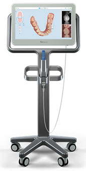
iTero scanner by Align Technology: In 2007, Cadent LTD introduced the iTero digital scanner. Later, in 2013, Align Technology restructured and introduced its iTero scanner to the market. The technical method used in this scanner is confocal imaging technology with a red-light laser beam (Babayoff and Glaser-Inbari, 2004). Since the iTero scanner was associated with Invisalign, it became more popular, especially among Invisalign practitioners. The advantage of the iTero scanner is that it is a powder-free scanner, so it provides operators with a real simulation and progress of Invisalign patients. The scan tip can directly contact the tooth surface. The iTero software uses an open-source standard triangulation language (STL), which makes it compatible with different systems, i.e., Invisalign, Incognito, Sure Smile, etc.
Although the scanner wand is considered bulky compared with other types of scanners, the manufacturer argues that this was done to take a wider view, which in turn will obtain a shorter scanning time and high accuracy. The scan wand has a reflective mirror design, which, according to the company, will make it easier to scan the most distal tooth. Further, to prevent infection, the wand has a disposable sleeve (figure 1).
Figure 1: iTero Scanner
True definition scanner by 3M ESPE: In 2008, 3M produced Lava C.O.S. using an active wavefront sampling technology. This classic scanner provided good performance at that time. Later, the company improved its specifications and produced the True Definition scanner. In 2016, it launched the Mobile True Definition, which has the same software/hardware as the original. The advantages of this scanner are that it provides a high accuracy and the tip is the size of the hand piece. This type of scanner requires a thin coat of powder for high accuracy. The scanner doesn’t offer real-time full-color scans. The software is an open STL format. The data files are compatible with Incognito, Invisalign customized lingual brackets, Sure Smile, and Clear Correct. Also, the software is designed to work with UnitekTM Treatment Management Portal Digital Model software for treatment planning, such as Bolton and space analysis (figure 2).
Figure 2: True definition Scanner

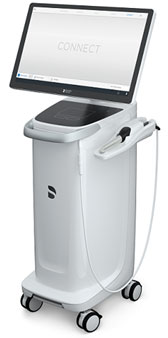
CEREC Omnicam by Sirona Dentsply: The CEREC system gained its popularity by manufacturing CAD/CAM. It has a strong name in restorative dentistry. Its scanning technology is the triangulation imaging method. The scanner provides video streaming instead of static images, and powdering is not required in the scanning procedure. In orthodontics, the system has been advanced by the possibility of producing a full-arch scan (figure 3).
Figure 3: CEREC Scanner
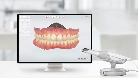
TRIOS 3/TRIOS 3 wireless by 3Shape: In 2010, 3Shape launched the first edition of TRIOS 3. In the beginning, there were a lot of complaints that there was fogging in the wand, but later, the company solved this problem. The system has the advantage that the operator can perform the scanning as soon as it is turned on, since the new scanner tip is preheated. Moreover, the scanner wand is downsized, and the wand sleeve can be sterilized by autoclave.
The scanning technology is the Ultrafast Optical Sectioning, which is based on the conofocal laser principal. The wireless version of this scanner was launched in 2017, offering the operator flexibility and freedom in scanning. The system also has an advanced treatment simulator and a built-in recording feature of dynamic occlusion (figure 4).
Figure 4: TRIOS 3 Scanner
Which one to use?
Suggestion of a specific scanner is not easy, and that is not an aim of this article. A comparison of different features of the popular scanners will be discussed below. One of the most important features in any scanner is the accuracy of the scanning (Goracci et al., 2016). Two factors affect scanner accuracy: the trueness and precision. Trueness means how truly the scanner can replicate the real dimensions, while precision means how reproducible the scanner is (Huang et al., 2020).
Though many previous studies suggested that iTero and True Definition have higher accuracy than TRIOS and Omnicam (Ryakhovskiy and Kostyukova, 2016; Amin et al., 2017; Renne et al., 2017), accuracy comparison between iTero and True Definition was not studied. Scanning accuracy is affected by the material being scanned (Nedelcu and Persson, 2014). Powdering improves the scan accuracy. In vitro study revealed that lingual brackets are less accurate than buccal ones, though TRIOS and iTero produced accurate scans under these circumstances (Park et al., 2016 Marghalani et al. 2018; Nedelcu et al., 2018).
Scanning time is also important. Kim et al. (2016) found that iTero may have a longer scanning time than the traditional impression method, but as the clinician’s experience increases, the time decreases. Moreover, they reported that TRIOS takes a shorter time than the traditional impression and that it is suitable for clinicians with less experience with scanners (Kim et al., 2016). Finally, the machine prices: the IOS prices range from $36,000 (USD) to $90,000 (USD). The price depends on the company, the machine, the software, and annual fees. Investing in IOS is an important step that needs critical thinking. It’s a good idea to ask a colleague who has had experience with scanners before buying. Also, ask about the company you are buying from—its reputation, as well as the after-sales service and maintenance.
CONCLUSION
Digital dentistry is the future since it provides efficiency and effectiveness for both the clinician and the patient. A number of different intraoral scanners are available on the market. The clinician should be aware of the available device features, maintenance, prices, and advantages.
Conflict of Interest: Author has no conflict of interest
REFERENCES
Amin, S., Weber, H.P., Finkelman, M., El Rafie, K., Kudara, Y. and Papaspyridakos, P. (2017). Digital vs. conventional full-arch implant impressions: a comparative study. Clin Oral Implants Res, 28: 1360–1367.
Asquith, J. and Mcintyre, G. (2012). Dental arch relationships on three-dimensional digital study models and conventional plaster study models for patients with unilateral cleft lip and palate. Cleft Palate Craniofac J, 49: 530–534.
Babayoff, N. and Glaser-Inbari, I. (2004). Imaging a three-dimensional structure by confocal focussing an array of light beams. US Patent, International Patent.
Brandestini, M. and Moermann, W.H. (1989). Method and apparatus for the three-dimensional registration and display of prepared teeth. Google Patents.
Cousley, R.R. and Turner, M.J. (2014). Digital model planning and computerized fabrication of orthognathic surgery wafers. J Orthod, 41: 38–45.
De Luca Canto, G., Pacheco-Pereira, C., Lagravere, M.O., Flores-Mir, C. and Major, P.W. (2015). Intra-arch dimensional measurement validity of laser-scanned digital dental models compared with the original plaster models: a systematic review. Orthod Craniofac Res, 18: 65–76.
Duret, F. (1973). The Optical Impression (dissertation), Universite Claude Bernard.
Ender, A., Attin, T. and Mehl, A. (2016). In vivo precision of conventional and digital methods of obtaining complete-arch dental impressions. J Prosthet Dent, 115: 313–320.
Ender, A. and Mehl, A. (2013). Accuracy of complete-arch dental impressions: a new method of measuring trueness and precision. J Prosthet Dent, 109: 121–128.
Ender, A. and Mehl, A. (2015). In-vitro evaluation of the accuracy of conventional and digital methods of obtaining full-arch dental impressions. Quintessence Int, 46: 9–17.
Ender, A. and Mehl, A. (2011). Full arch scans: conventional versus digital impressions—an in vitro study. Int J Comput Dent, 14: 11–21.
Gateno, J., Xia, J.J., Teichgraeber, J.F., Christensen, A.M., Lemoine, J.J., Liebschner, M.A. et al. (2007). Clinical feasibility of computer-aided surgical simulation (CASS) in the treatment of complex cranio-maxillofacial deformities. J Oral Maxillofac Surg, 65: 728–734.
Goracci, C., Franchi, L., Vichi, A. and Ferrari, M. (2016). Accuracy, reliability, and efficiency of intraoral scanners for full-arch impressions: a systematic review of the clinical evidence. Eur J Orthod, 38: 422–428.
Grunheid, T., McCarthy, S.D. and Larson, B.E. (2014). Clinical use of a direct chairside oral scanner: an assessment of accuracy, time, and patient acceptance. Am J Orthod Dentofacial Orthop, 146: 673–682.
Hilliard, J.K. (2006.) U.S. Patent No. 7,077,646. U.S. Patent and Trademark Office, Washington, DC.
Hwang, H.H.M., Chou, C.W., Chen, Y.J. and Yao, C.C.J. (2020). An overview of digital intraoral scanners: past, present and future—from an orthodontic perspective. Taiwanese Journal of Orthodontics, 30(3): 3.
Kim J., Park, J.M., Kim, M., Heo, S.J., Shin, I.H. and Kim, M. (2016). Comparison of experience curves between two 3-dimensional intraoral scanners. J Prosthet Dent, 116: 221–230.
Kravitz, N.D., Growth, C., Jones, P.E., Graham, J.W. and Redmond, W.R. (2014). Intraoral digital scanners—overview. JCO, 48: 337–347.
Logozzo, S., Franceschini, G., Kilpelä, A., Caponi, M., Governi, L. and Blois, L. (2008). A comparative analysis of intraoral 3d digital scanners for restorative dentistry. The Internet Journal of Medical Technology, 5: 1–18.
Marghalani, A., Weber, H.P., Finkelman, M., Kudara, Y., El Rafie, K. and Papaspyridakos, P. (2018). Digital versus conventional implant impressions for partially edentulous arches: an evaluation of accuracy. J Prosthet Dent, 119: 574–579.
Martin, C.B., Chalmers, E.V., McIntyre, G.T., Cochrane, H. and Mossey, P.A. (2015). Orthodontic scanners: what’s available? J Orthod, 42: 136–143.
Mehl, A., Ender, A., Mormann, W. and Attin, T. (2009). Accuracy testing of a new intraoral 3D camera. Int J Comput Dent, 12: 11–28.
Mormann, W.H. (2006). The evolution of the CEREC system. J Am Dent Assoc, 137: 7S–13S.
Nedelcu, R., Olsson, P., Nystrom, I., Ryden, J. and Thor, A. (2018). Accuracy and precision of 3 intraoral scanners and accuracy of conventional impressions: a novel in vivo analysis method. J Dent, 69: 110–118.
Nedelcu, R.G. and Persson, A.S. (2014). Scanning accuracy and precision in 4 intraoral scanners: an in vitro comparison based on 3-dimensional analysis. J Prosthet Dent, 112: 1461–1471.
Park, J.M., Choi, S.A., Myung, J.Y., Chun, Y.S. and Kim, M. (2016). Impact of orthodontic brackets on the intraoral scan data accuracy. Biomed Res Int, 2016: 5075182.
Patzelt, S.B., Bishti, S., Stampf, S. and Att, W. (2014a). Accuracy of computer-aided design/computer-aided manufacturing-generated dental casts based on intraoral scanner data. J Am Dent Assoc, 145: 1133–1140.
Patzelt, S.B., Emmanouilidi, A., Stampf, S., Strub, J.R. and Att, W. (2014b). Accuracy of full-arch scans using intraoral scanners. Clin Oral Invest, 18: 1687–1694.
Patzelt, S.B., Lamprinos, C., Stampf, S. and Att, W. (2014c). The time efficiency of intraoral scanners: an in vitro comparative study. J Am Dent Assoc, 145: 542–551.
Renne, W., Ludlow, M., Fryml, J., Schurch, Z., Mennito, A., Kessler, R. and Lauer, A. (2017). Evaluation of the accuracy of 7 digital scanners: an in vitro analysis based on 3-dimensional comparisons. J Prosthet Dent, 118(1): 36–42.
Rheude, B., Lionel Sadowsky, P., Ferriera, A. and Jacobson, A. (2005). An evaluation of the use of digital study models in orthodontic diagnosis and treatment planning. Angle Orthod, 75: 300–304.
Ryakhovskiy, A.N. and Kostyukova, V. (2016). Comparative analysis of 3D data accuracy of single tooth and full dental arch captured by different intraoral and laboratory digital impression systems. Stomatologiia (Mosk), 95: 65–70.
Syrek, A., Reich, G., Ranftl, D., Klein, C., Cerny, B. and Brodesser, J. (2010). Clinical evaluation of all-ceramic crowns fabricated from intraoral digital impressions based on the principle of active wavefront sampling. J Dent, 38: 553–559.
van der Meer, W.J., Andriessen, F.S., Wismeijer, D. and Ren, Y. (2012). Application of intra-oral dental scanners in the digital workflow of implantology. PLoS One, 7: e43312.
Wiechmann, D., Rummel, V., Thalheim, A., Simon, J.S. and Wiechmann, L. (2003). Customized brackets and archwires for lingual orthodontic treatment. Am J Orthod Dentofac, 124: 593–599.
Wong, K.Y., Esguerra, R.J., Chia, V.A.P., Tan, Y.H. and Tan, K.B.C. (2017). Three-dimensional accuracy of digital static interocclusal registration by three intraoral scanner systems. J Prosthodont, 27(2): 120-128.

