1Department of Pediatric Dentistry and Orthodontics, College of Dentistry, King Saud University, Riyadh, Saudi Arabia.
2Dental Intern, College of Dentistry, King Saud University, Riyadh, Saudi Arabia.
Corresponding author email: maldosari@outlook.com
Article Publishing History
Received: 26/10/2020
Accepted After Revision: 14/12/2020
The present study investigated patterns of root resorption in mandibular primary molars of Saudi children. The dental records of a randomly selected 460 Saudi children matched by age and gender who were treated in pediatric dentistry clinics at College of Dentistry, King Saud University in Riyadh, Saudi Arabia, were reviewed. Primary mandibular molar teeth were identified on the digital panoramic radiographs that were taken as part of the children’s routine examinations. A trained and calibrated single examiner carried out all the examination procedures. The parameters such as symmetry and the level of mesial or the distal root resorption were investigated. The collected data were recorded on a special form and statistically computed. There were statistically significant differences in the distribution of the level of root resorption in the primary first and second molars; both the mesial and distal roots showed significantly less than a quarter root resorption followed by one-fourth to three-fourth levels of root resorption (p<0.001). The majority of the root resorption occurred symmetrically for the primary first (63.7%) and second (71.1%) molars. The resorption in the distal roots were comparatively more than the mesial roots in the primary first molars and vice versa in case of the primary second molars. A relatively high incidence of uneven level of root resorption was evident, more in the mandibular second primary molars with majority of the primary molars exhibiting less than a quarter root resorption. This requires close monitoring to ensure prevention of over-retention of primary molars. Though symmetrical resorption of mesial and distal roots of primary molars was observed in this study, variations does occur.
Primary Dentition, Root Resorption, Mandibular Molars
Al-Sehaibany F. S, Alomran W, Almutairi A, Al-Homaidhi M. A, Aldosari M. A, Albarakati S. F. Patterns of Root Resorption in Primary Mandibular Molars of Saudi Children. Biosc.Biotech.Res.Comm. 2020;13(4).
Al-Sehaibany F. S, Alomran W, Almutairi A, Al-Homaidhi M. A, Aldosari M. A, Albarakati S. F. Patterns of Root Resorption in Primary Mandibular Molars of Saudi Children. Biosc.Biotech.Res.Comm. 2020;13(4). Available from: <a href=”https://bit.ly/388BxBT”>https://bit.ly/388BxBT</a>
Copyright © Al-Sehaibany et al., This is an Open Access Article distributed under the Terms of the Creative Commons Attribution License (CC-BY) https://creativecommons.org/licenses/by/4.0/, which permits unrestricted use distribution and reproduction in any medium, provide the original author and source are credited.
INTRODUCTION
Eruption of the primary dentition, their exfoliation as a result of root resorption, followed by eruption of permanent dentition is a complex, age specific event and occurs in an orderly sequence. (Pahkala et al., 1991) It is a critical milestone in the overall development of a child and is influenced by racial, ethnic, sexual, and individual factors. (Proffit et al., 2014) Initial resorption begins between one and three years after closure of the apices, and exfoliation follows by approximately three years. (McDonald et al., 2011).The roots of primary teeth resorb when the succeeding permanent tooth begins active eruption and the follicle comes into juxtaposition with the root surface. Though the pressure of the erupting permanent tooth is believed to be a contributing factor initiating resorption, (Sahara, 2001) the presence of a permanent successor, however, is not a prerequisite for this process to occur. In the absence of a permanent successor, primary teeth do eventually resorb, although their exfoliation may be delayed than usual timings. (Cate, 1998, Obersztyn, 1963). There are several factors which affect the resorption of primary molar roots such as dental caries, congenital absence of permanent tooth bud and a number of pathological causes, (Sharma et al., 2016).
The development of the premolars commences between the diverging roots of the primary molars and the pattern of root resorption is affected by the position and size of the premolar follicles,(Fulton and Liversidge, 2016). Resorption of primary molar roots can be analogous, or perhaps irregular with one root considerably more resorbed than the other. Anatomically, the primary lower second molars’ roots are very curved and divergent, and the inter-root distance is often greater than the size of the follicle of its successor. Unequal influences may be applied to the roots depending on the successor’s position. This can explain the irregular root resorption detected in more than one third of all lower second molars at any given time after its initiation. On the other hand, the incidence of irregular root resorption is comparatively lower for the primary first molars and this is claimed to be due to the smaller difference between its inter-root distance and the size of the crown of its successor. (Prove et al., 1992a).
Longitudinal studies utilizing radiographs have been used to determine the extent and timing of resorption of the roots of primary teeth by comparing the resorbed root and the total root length, (Knott and O’Meara, 1967, Moorrees et al., 1963). However, the pattern of root resorption on radiographs is variable and can appear as a rounded or sharp apical margin, distinct or additional lateral thinning at initial stages of resorption and a horizontal or diagonal edge at advanced stages. (Fulton and Liversidge, 2016) Regional information regarding the patterns of root resorption of primary teeth are sparse. Hence, the present study investigated patterns of root resorption in mandibular primary molars of Saudi children.
MATERIAL AND METHODS
The study protocol and consent form were approved by the Research and Ethical Committee of Human Studies at King Saud University, College of Dentistry Research Center (IR 0259). The dental records of a randomly selected 460 Saudi children matched by age and gender who were treated in pediatric dentistry clinics at College of Dentistry, King Saud University in Riyadh, Saudi Arabia, were reviewed. Children were selected for the study, if they were not diagnosed with any systemic diseases or taking any medications for chronic diseases.
Primary mandibular molar teeth were identified on the digital panoramic radiographs that were taken as part of the children’s routine examinations and on which the teeth were present and fully visible. All radiographs were taken with Planmeca ProMax (Version 3.6.0, Helsinki, Finland), operating at 5 mA, 62 kVp. The radiographs were reviewed in identical conditions using Planmeca Romexis software (Version 4.0, Helsinki, Finland). A single examiner (WA) who was trained and calibrated (kappa=0.84) with a senior faculty in recording features of root resorption patterns carried out all the examination procedures. The intra-examiner reliability was established by re-examination of 40 radiographs on 2 different occasions with one week apart (Kappa=0.88).Radiographs were selected for the present study based on the following criteria :Clear image of the roots of the primary mandibular molars, clear image of the cementoenamel junction (CEJ), and no pulp treatment and no stainless-steel crowns.
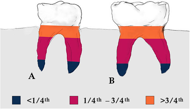
The following parameters were investigated:1. The resorption of the mesial and distal roots (i.e. symmetrical or if either the mesial or the distal root were more resorbed); symmetry was defined if the line connecting the apices was parallel to a line connecting the CEJ at the mesial and distal surfaces.2. The level of resorption in the mesial and distal roots, according to the defined stages suggested by Moorrees et al.(1963) (less than a quarter, one-quarter to three-fourths, and greater than three-fourths of the root) as presented in Figure 1.
Figure 1: Index of root resorption (Source: Moorrees et al (Moorrees et al., 1963)). A. Mandibular first primary molar, B. Mandibular second primary molar.
The collected data were recorded on a special form and the Statistical Package for Social Science (SPSS V. 20, Chicago, IL, USA) was utilized for statistical computation. Data analysis included frequency distribution and chi-square was used to determine the significance of differences in level of root resorption with the level of significance set at P<0.05.
Table 1. Level of root resorption for the mandibular first primary molars.
| Level of Root Resorption | Primary left first molar* | Primary right first molar* |
| Mesial Distal
n (%) n (%) |
Mesial Distal
n (%) n (%) |
|
| < 1/4th | 297(64.1) 249 (54.4) | 306 (66.5) 247 (53.7) |
| 1/4th – 3/4th | 128(28.0) 167 (36.3) | 115 (24.8) 173 (37.6) |
| > 3/4th | 35 (7.9) 44 (9.3) | 39 (8.7) 40 (8.7) |
| Total | 460 (100) 460 (100) | 460 (100) 460 (100) |
* P<0.001, significantly different using Chi-square test
Table 2. Level of root resorption for the mandibular second primary molars.
| Level of Root Resorption | Primary left second molar* | Primary right second molar* |
| Mesial Distal
n (%) n (%) |
Mesial Distal
n (%) n (%) |
|
| < 1/4th | 307 (66.7) 334 (72.6) | 319 (69.4) 247 (53.7) |
| 1/4th – 3/4th | 146 (31.7) 121 (26.1) | 136 (29.3) 173 (37.6) |
| > 3/4th | 7 (1.6) 5 (1.3) | 5 (1.3) 40 (8.7) |
| Total | 460 (100) 460 (100) | 460 (100) 460 (100) |
* P<0.001, significantly different using Chi-square test
RESULT AND DISCUSSION
The age of the children ranged between 6-10 years, with a mean (±SD) age of 7.61 ± 1.24 years. Males and females were equally distributed (230 males, 230 females). There were statistically significant differences in the distribution of the level of root resorption in the primary first molars (Table 1) and primary second molars (Table 2). For both the molars, the mesial and distal roots showed significantly less than a quarter root resorption followed by one-fourth to three-fourth levels of root resorption (p<0.001). For the primary first molars, the more number of mesial roots, irrespective of the side of the jaw, showed less than a quarter root resorption, when compared to the distal roots. For the primary right second molars, more number of the mesial roots showed less than a quarter root resorption than the distal roots on the right side. On the other hand, for the primary left second molars, more number of distal roots showed less than a quarter root resorption than the distal roots on the left side.
Table 3 shows the distributions of symmetry of root resorption of the mandibular primary molars which was found to be significantly different (p<0.001). The majority of the root resorption occurred symmetrically for the primary first molars (63.7%) and primary second molars (71.1%). The root resorption in the distal roots were comparatively more than the mesial roots in the primary first molars and vice versa in case of the primary second molars.This study investigated the patterns of root resorption of primary mandibular molars in a sample of 6-10-year-old Saudi children. Root resorption is a three-dimensional process and viewing this in two-dimensions certainly has some limitations. Resorption patterns in the bucco-lingual plane is less likely to appear well defined than in the mesio-distal plane, (Fulton and Liversidge, 2016).
Table 3. Symmetry of root resorption for the mandibular primary molars.
| Root Resorption | Primary First Molar* n (%) | Primary Second Molar* n (%) |
| > Distal Root | 133 (28.9) | 51 (11.1) |
| Symmetrical | 293 (63.7) | 327 (71.1) |
| > Mesial Root | 34 (7.4) | 82 (17.8) |
| Total | 460 (100) | 460 (100) |
*P<0.001, significantly different using Chi-square test
There was symmetrical root resorption of the mesial and the distal roots in approximately 64 percent of the first primary molars and 71 percent of the second primary molars. This finding is higher in comparison to 5 to 12-year-old Israeli children where symmetrical root resorption of the mesial and the distal roots were found in approximately 41 percent of the cases.(Peretz et al., 2013) This finding is in contrast to previous studies where the distal roots(Peretz et al., 2013, Sharma et al., 2016) or the mesial roots(Moorrees et al., 1963) showed more resorption. The different study populations and variations in the study methodologies might have attributed to the contrasting findings. While Moorrees et al.(Moorrees et al., 1963) used lateral or oblique radiographs, Peretz et al.(2013) used peri-apical or bitewing radiographs and Sharma et al.( 2016) used radiovisiographs. The present study utilized digital panoramic radiographs.
The position and size of the permanent tooth bud plays an imperative role in influencing asymmetrical pattern of resorption of primary tooth roots. This may be further justified when upper primary molars are taken into consideration, whereby the palatal root is often spared from resorption due to its highly divergent nature. Likewise, in anterior tooth, the completed crown of the permanent successor is found lingual to the primary tooth apex with its eruption in labial and incisal direction causing the resorption of the lingual surfaces of the apical third of the primary tooth root,(Prove et al., 1992b). In this study, both the mesial and distal roots of all the mandibular primary molars showed significantly less than a quarter root resorption. This finding was more evident in the mandibular second primary molars as compared to the first primary molars. The mandibular primary second molars have roots that are curved and divergent and the crown width of the permanent successor is smaller than its inter root distance,(Nelson, 2014). Consequently, root portions of these teeth escape resorption and root fragments tend to remain which may affect the successors adversely, (Teague et al., 1999). Such uneven distribution of the level of root resorption warrants close monitoring and timely management to ensure no complications occurs due to over-retained primary teeth.
In this study, the mandibular primary second molars showed greater propensity for symmetrical root resorption than the roots of the mandibular primary first molars. This finding is in contrast to the study involving 84 children between the age group of 7 to 10 years recruited from the University of Queensland dental school, (Prove et al., 1992b). A high incidence of ankylosis has been documented in both primary molars and this has contributed to the disturbances in the eruption and development of the premolars, (Tieu et al., 2013). However, the ankylosed molar often exfoliates spontaneously within six months; causing complications due to delayed exfoliation such as arch-length loss, occlusal disturbance, hooked roots or impaction of permanent successors,(Tieu et al., 2013).
In this study, certain limitations merits mention in order to improve on future studies. The cross-sectional design and the exclusion of various other parameters such as the position of erupting permanent successors, clinical status of the resorbing primary teeth and assessment of canine root resorption may be considered as major drawbacks. Further studies with a longitudinal design inclusive of the above-mentioned parameters may thus be conducted to gain a better understanding of the root resorption patterns of primary dentition.
CONCLUSION
Within the limitations of this study, a relatively high incidence of uneven level of root resorption was evident, more in the mandibular second primary molars with majority of the primary molars exhibiting less than a quarter root resorption. This requires close monitoring to ensure prevention of over-retention of primary molars. Though symmetrical resorption of mesial and distal roots of primary molars was observed in this study, variations does occur.
ACKNOWLEDGEMENTS
This research project was supported by a grant from the “Research Center of the Center for Female Scientific and Medical Colleges”, Deanship of Scientific Research, King Saud University.
REFERENCES
Cate, A. T. 1998. Oral Histology: Development, Structure, And Function. 5th Edition.
Fulton, A. J. & Liversidge, H. M. 2016. A Radiographic Study Of Estimating Age By Deciduous Mandibular Canine And Molar Root Resorption. Annals Of Anatomy-Anatomischer Anzeiger, 203, 33-37.
Knott, V. B. & O’meara, W. F. 1967. Serial Data On Primary Incisor Root Resorption And Gingival Emergence Of Permanent Successors. The Angle Orthodontist, 37, 212-222.
Mcdonald, R. E., Avery, D. R. & Dean, J. A. 2011. Eruption Of The Teeth: Local, Systemic, And Congenital Factors That Influence The Process. Mcdonald And Avery Dentistry For The Child And Adolescent (Ninth Edition). Elsevier.
Moorrees, C. F., Fanning, E. A. & Hunt, E. E., Jr. 1963. Formation And Resorption Of Three Deciduous Teeth In Children. Am J Phys Anthropol, 21, 205-13.
Nelson, S. J. 2014. Wheeler’s Dental Anatomy, Physiology And Occlusion-E-Book, Elsevier Health Sciences.
Obersztyn, A. 1963. Experimental Investigation Of Factors Causing Resorption Of Deciduous Teeth. Journal Of Dental Research, 42, 660-674.
Pahkala, R., Pahkala, A. & Laine, T. 1991. Eruption Pattern Of Permanent Teeth In A Rural Community In Northeastern Finland. Acta Odontologica Scandinavica, 49, 341-349.
Peretz, B., Nisan, S., Herteanu, L. & Blumer, S. 2013. Root Resorption Patterns Of Primary Mandibular Molars And Location Of The Premolar Successors: A Radiographic Evaluation. Pediatr Dent, 35, 426-9.
Proffit, W. R., Fields, H. W. & Sarver, D. M. 2014. Contemporary Orthodontics-E-Book, Elsevier Health Sciences.
Prove, S., Symons, A. & Meyers, I. 1992a. Physiological Root Resorption Of Primary Molars. J Clin Pediatr Dent, 16, 202-6.
Prove, S. A., Symons, A. L. & Meyers, I. A. 1992b. Physiological Root Resorption Of Primary Molars. J Clin Pediatr Dent, 16, 202-6.
Sahara, N. 2001. Cellular Events At The Onset Of Physiological Root Resorption In Rabbit Deciduous Teeth. The Anatomical Record, 264, 387-396.
Sharma, A., Kulkarni, S., Swamy, N., Khandelwal, V., Nenava, D. & Agrawal, P. 2016. Co-Relation Between Root Resorption Patterns Of Deciduous Mandibular Molars And Position Of Erupting Permanent Successors: A Radiographic Evaluation. Ijar, 2, 440-444.
Teague, A. M., Barton, P. & Parry, W. J. 1999. Management Of The Submerged Deciduous Tooth: 1. Aetiology, Diagnosis And Potential Consequences. Dental Update, 26, 292-296.
Tieu, L. D., Walker, S. L., Major, M. P. & Flores-Mir, C. 2013. Management Of Ankylosed Primary Molars With Premolar Successors: A Systematic Review. The Journal Of The American Dental Association, 144, 602-611.

