Department of Prosthetic Dental Sciences, College of Dentistry,
King Saud University, Riyadh, Saudi Arabia.
Corresponding author email: Shidiibra147@gmail.com
Article Publishing History
Received: 12/10/2021
Accepted After Revision: 15/12/2021
In order to assess the influence of finishing and polishing on the surface brightness and color stability of the ceramic veneer, fifty specimens were fabricated with 10 mm diameter and 2 mm thickness using IPS E-Max Ceramic. After glazing, 10 specimens were untouched as control group, and the other 40 specimens were abraded using 125µm diamond bur to create surface roughness. Forty specimens were divided into four groups (n=10), in group 1: specimens were finished using diamond point, in group 2 specimens’ surface was polished with a polishing kit, Group 3: Each specimen surface was polished with the polishing kit as in protocol 2 and was polished a polishing past and group 4 Each specimen was glazed by heating at 621℃ for 3 minutes followed by a temperature increase of 83℃/min up to 918℃ for 30 seconds. Color measurement was performed using spectrophotometer. Color stability data were analyzed using two-way ANOVA and Tukey’s HSD test (α=0.05).
For Ra values, paired-samples t-tests were used to analyze the data and compare groups. The change in L and E showed a significant difference among the study groups; (group 1, group 2, group 3 and group 4) with respect to three variables L, a and b. A significant difference was noted when compared each group with the control; however, only group 2 showed a significant difference from group 4; the remaining groups demonstrated similar findings for all three variables. The study displayed a significant impact of the finishing and polishing technique on the surface brightness and color stability of ceramic restoration. However, it was evident that combination of two or three polishing techniques which includes polish kit and glaze enhances the surface finish and adds color stability by alternating the yellow – blue axis (increase in b) and red- green axis (decrease in a).
Color stability, Commission Internationale de l’Eclairage, Finishing, Glazing, Polishing, Veneers.
Alshiddi I. F. Effect of Different Finishing and Polishing Methods for Color Stability of the E-Max Dental Ceramics: In vitro Study. Biosc.Biotech.Res.Comm. 2021;14(4).
Alshiddi I. F. Effect of Different Finishing and Polishing Methods for Color Stability of the E-Max Dental Ceramics: In vitro Study. Biosc.Biotech.Res.Comm. 2021;14(4). Available from: <a href=”https://bit.ly/3wLhlls“>https://bit.ly/3wLhlls</a>
Copyright © This is an Open Access Article distributed under the Terms of the Creative Commons Attribution License (CC-BY) https://creativecommons.org/licenses/by/4.0/, which permits unrestricted use distribution and reproduction in any medium, sources the original author and sources are credited.
INTRODUCTION
Aesthetics is one of the pioneer aspects of a smile which adds up to the confidence and personality of the person. The appearance of the dental teeth is not only important for the patient but also for the dentist. With advanced technology, manufacturers produce wide range of range of materials to match the shade of the teeth varying across the globe (Vieira et al. 2013; Vanlıoğlu and Kulak-Özkan 2014). With advancement and greater awareness, patient demands for good functional and highly esthetic restoration. Hence, it is utmost important to select appropriate veneer materials with excellent esthetic quality. The recent developed dental ceramics presents desirable characteristics that highlights them as best choice as indirect restoration (Kilinc and Turgut 2018; Sarıkaya et al. 2018).
Dental ceramic is a dental material used extensively by the dentist for veneers, braces, inlays, onlays and crown restoration. The material is biocompatible, esthetics, insoluble and hard enough for a prefect and long-term restorations (Sarıkaya et al. 2018; AlMawash et al. 2020). Several different types of ceramics are available which includes pressed ceramics that mimics the natural tooth properties such as light transmission, color reproduction and texture. Studies have shown that the surface smoothness and color stability are interrelated. Hence after finishing and polishing procedures the optical properties of dental ceramics which enhances the surface finish to allow for proper light reflection (Sarikaya. and Güler 2011; Volpato et al. 2016; AlMawash et al. 2020).
Restorations in the oral environment deteriorate with time under constant exposure to external factors and saliva. Along with surface properties, discoloration of the restoration is equally affected in long term. Authors demonstrated long term exposure to external factors such as forceful bite, diet and habits the restoration surface which affects the color stability and brightness of the restoration (Pires-de et al. 2009; Hamza et al. 2017). These craters or surface defects produced leads to staining, plaque retention, gingival irritation, and recurrent caries causing the adherence of agents responsible for changing the color. Hence, dentist imposes great emphasis on polishing and finishing of the surface for better quality finish and restoration (Cengiz et al. 2014; Shetty et al. 2020). Nonetheless, limited literature supports the importance of types of polishing in creating a smooth finish for color stability and brightness. Thus, the study aims to evaluate the effect of the types of finishing and polishing technique on surface brightness and color stability in ceramic veneers.
MATERIAL AND METHODS
A total of 50 disc-shape specimens were fabricated with 10 mm diameter and 2 mm thickness using IPS E-Max Ceramic Material (Ivoclar Vivadent, Schaan, Liechtenstein) (Fig: 1). Each sample was fabricated initially as a wax cylindrical pattern using a split brass matrix. The wax patterns were then invested with an investment material (IPS Empress Esthetic Speed Investment; Ivoclar Vivadent AG). Wax elimination was completed in temperature of 750℃ for 6 min using oven (Meditherm 200 MP, Bego, Canada). Upon burnout, IPS e-max Press ingots (Ivoclar Vivadent, Schaan, Liechtenstein) were heat pressed using Programat® EP 3000 Oven (Ivoclar Vivadent, Schaan, Liechtenstein) at temperature of 920℃.
After bench cooling, the specimens were treated with 2% hydrofluoric acid for 10 min and with 50 µm Al2O3 at 0.2 MPa pressure for 5 sec. The specimens were then inserted in a digital ultrasonic cleaner (JP-4820, Skymen, Shenzhen, China) for 10 min, followed by stratification of the ceramic using color A2 of the Vita shade scale (Vita Zahnfabrik, Bad-Säckingen, Germany). After cleansing, 10 specimens were untouched as control group, and the other 40 specimens were abraded using 125µm diamond bur in a unidirectional motion to create surface roughness simulate. The 40 specimens were divided into four groups (n=10), and a different finishing and polishing method were completed for each group as following:
Group 1: Each specimen was finished using diamond point 862C 012 (Drendel+Zweiling, Germany) (Fig. 2), intermittently in one direction, with a cooling and high speed (Kavo do Brasil Industria, SC, Brazil), for 30 seconds. The specimen was then washed with air/water spray to remove the residues. Group 2: Each specimen surface was polished with a polishing kit (Ceramaster Finishing and Polishing Kit; Shofu Inc) (Fig. 3) using a slow-speed handpiece, as recommended by manufacturer. Group 3: Each specimen surface was polished with the polishing kit as in protocol 2 and was polished a polishing past (OptraFine polishing Paste; Ivoclar vivadent, Germany) (Fig. 4).
Group 4: Each specimen was finished as in protocol1, and then polished with polishing kit as in protocol 2. Each specimen was then auto-glazed by heating at 621℃ for 3 minutes followed by a temperature increase of 83℃/min up to 918℃ for 30 seconds. Color measurement of all specimens was completed using a laboratory spectrophotometer (HunterLab LabScan XE spectrophotometer; the sotto group, United Kingdom) (Fig. 5). This instrument takes measurements at 400nm-700nm and gives readings at intervals of 10nm. It measures the reflected colour with 0˚/45˚, and use an inclusive EasyMatch QC software for analyzing colour data. Calibration was performed before take the measurement of each group, and and it is built in calibration. The average color of each group will be compared with the color of the control group.
The Commission Internationale de l’Eclairage (CIE L*a*b*) color space is employed to determine color differences. ∆L*, ∆a*, and ∆b* are differences between two colors in the CIE-based color space. In present study, the colorimetric values of ΔL*, Δa*, and Δb* are measured from differences in the respective L*, a*, and b* values. The total color difference ΔE*ab between two colors, each given in terms of L*, a*, and b*, was calculated from the following formula: ΔE*ab = [(ΔL*) 2 +(Δa*)2 +(Δb*) 2 ]1/2Differences in color (∆E) were calculated according to the following equation:
∆E = [(L2-L1)2 + (a2-a1)2 + (b2-b1)2]1/2
Because the most important factor affecting the result of shade appearance of a new restoration is the Value “brightness”, the differences in Value (∆L) was calculated as well for each group comparing to the control group using the following equations:
∆L = [(L2-L1)2] 1/2
The formulae comprise of three variables L, a and b that represents the chromatic changes in the veneers. Each specimen was tested thrice followed by an average value for accurate measurements. This process was repeated for each specimen. In additions, after the color data was collected for each group (n=10) the spectrophotometer was recalibrated. Data collected were analyzed using a statistical program software, SPSS 13 for Windows (SPSS Inc., Chicago, IL, USA). The Normality distribution of the data was assessed using Levene’s Test. Means and standard deviations of the 4 study groups based on the finishing and polishing techniques. The outcomes of these 4 groups were compared with the control group and among the study groups to evaluate impact of the finishing and polishing on the color stability and brightness of the surface using ANOVA and Tukey’s HSD test (α=0.05).
RESULTS AND DISCUSSION
The present study demonstrated a positive effect of the type of polishing method employed on the color stability of the restoration. The Δ L and Δ E showed a significant difference among the study groups; (group 1, group 2, group 3 and group 4) with respect to three variables l, a and b.
Table 1. Mean and standard deviations (SD) for the CIE coordinates.
| Mean L | Mean a | Mean b | Δ L | Δ E | ANOVA
p value |
|
| Control | 59.66 | 0.887 | 6.67 | – | – |
0.01 |
| Group 1 | 59.66 (1.67) | 0.892 (.11) | 7.27 (.498) | 4.93 (1.07)a | 4.487 (.897)a | |
| Group 2 | 58.88 (5.90) | 0.832 (.37) | 5.77 (0.96) | 2.65 (0.77)b | 2.70 (0.03)b | |
| Group 3 | 58.68 (3.45) | 0.79 (.347) | 5.85 (1.185) | 1.93 (0.824)bc | 2.33 (0.517) bc | |
| Group 4 | 58.36 (2.20) | 0.727 (0.127) | 6.01(1.34) | 1.409c | 1.688c |
Dissimilar superscript alphabets show significant difference among the study groups (Δ E & L)
Table 2. The maximum and minimum for the CIE coordinates
| L min | L max | a min | a max | b min | b max | |
| Control | 56.13 | 62.05 | 0.69 | 1.05 | 0.81 | 8.1 |
| Group 1 | 56.1 | 62.05 | 0.69 | 1.05 | 6.72 | 8.1 |
| Group 2 | 52.01 | 68.25 | 0.51 | 1.71 | 4.91 | 8.31 |
| Group 3 | 53.81 | 64.1 | 0.49 | 1.59 | 4.63 | 8.82 |
| Group 4 | 53.9 | 61.09 | 0.51 | 0.92 | 4.07 | 9.02 |
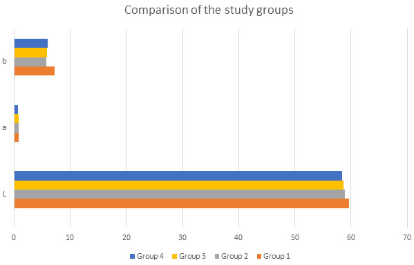
Table 1 presents mean and standard deviation of the CIE coordinates for color stability. The control group mean calculated for the three variables, L, a and b were as follows, 56.13, 0.887 and 6.67. Comparatively, the highest mean value for CIE coordinates obtained among the 4-study group was for group 1 [ 59.66 (1.670], [ 0.892 (0.11)] and [7.27 (0.498)] whereas the least value obtained was for group 4, L [ 58.36 (2.20)], a [0.727 (0.127)] and group 2, a [ 5.77 (0.96)] (Fig 1). A significant decrease in the L and a was noted; however, b increased after polishing with a paste and glazing. It was observed that using a polishing paste and glazing enhances the color components for the veneer natural appearance and esthetics. Furthermore, comparing the 4 groups, highest maximum value measured was in group 2 for L (68.25) and b (1.71) and group1 (0.69). Whereas the minimum value obtained group 2 (52.01), group 3 a (0.49) and group 4 b (4.07) (table 2).
The ANOVA for change in color, Δ L and Δ E displayed a significant difference p < 0.01. Comparatively, Δ L and Δ E mean value is suggestive of a significant difference in the effect of the polishing and finishing technique. Following the statistical calculation, the Δ L and Δ E showed that highest degree of change was observed in group 4 with minimum value of 1.688 and l (1.409) whereas group 1 demonstrated least value of Δ L (4.93) and Δ E (4.487). Moreover, a significant difference was noted when compared each group with the control; however, only group 2 showed a significant difference from group 4; the remaining groups demonstrated similar findings for all three variables. Hence, displaying bur finishing without glazing and polishing kit significantly increase the surface roughness and color instability (table 1).
Figure 1. Comparison of the CIE coordinates among the study groups
The present study assessed the effects of the 4 types of finishing and polishing methods on the color stability of the veneers. The difference of the composition and polishing methods play an important role in the final appearance and color stability of the e.max CAD ceramics. Each type of the polishing method applied has its own importance in producing a finishing look that reflects upon a natural appearance. This was evidently displayed in the present study when a significant difference was observed when compared to the control and among the study groups except few. Hence, the null hypothesis can be partially rejected.
Surface roughness is one of the main contributors for the material strength as well as the color stability of the restorations (Gönülol and Yilmazl 2012; Maciel et al. 2019). Finishing the restoration with diamond bur leaves behind great amount of surface roughness in form of surface defects and craters that alters the light perception leading to color change (Köroğlu et al. 2016; Aldahian et al. 2021). In the present study, the ANOVA showed a significant difference among the study groups; however, no significant effect was observed if the surface was only polished with polishing paste after disking.
In recent clinical practice, Sof-lex discs and shofu are popular polishing tools to produce smooth finish over the ceramic surface; however, they did not demonstrate any effect of glazing (Özarslan et al. 2016). On the contrary, few others emphasized greatly on the importance of glazing to alter the color gradient axis (Rashid et al. 2014; Sarikaya and Güleç 2019; Kim et al. 2021). Similarly, in the present study, it was observed that finishing the surface with polishing paste after using shofu did not positively influence color change; however, adding glaze to the surface protects the surface from extrinsic stains and plaque.
Hence, a significant impact was observed between group 2 and group 4 indicating combine effects of paste improves surface smoothness and glazing enhances the color stability by altering the color axis; yellow – blue axis (increase in b) and red- green axis (decrease in a). Hence, this showed that the low surface roughness reduces the chance for adhesion of colorants and plaque leading to better color stability. Nevertheless, to ensure evident color stability along with surface brightness, glazing is important (Kim et al. 2021). To understand the importance to replicate the natural color of teeth two valuable variables are required to explore greatly, the translucency and optical properties. Optical properties play a vital role in light transmission and reflection at the surface of the restoration (Bagis and Turgat 2013; Yılmaz et al. 2019; Kim et al. 2021).
According to Shahmiri et al. (2018), the appearance of the restoration greatly influences its optical property. Studies have shown that these properties are greatly influenced by the surface roughness of the restoration (Shahmiri et al. 2018). The change in the surface polish and finish causes a change in light beam reflection at different angles producing variation in color perception (Bagis and turgat 2013; Shahmiri et al. 2018). In the present study Δ L and Δ E were comparable, which showed a positive correlation. As the study moved from burs to disc followed by glazing along with polishing paste, the finer the surface became and produce a translucent and brighter surface of the restoration. Nevertheless, optical properties do not only depend upon the color but is greatly influence by the thickness and type of porcelain used as well (Alkhudhairy et al. 2020).
Previously, dentist employed using superfine diamond burs and polishing brushes to smoothen out the restorative surfaces. These polishing discs are impregnated with aluminum oxide that contributes to smoother surface preparation. However, the geometry of the restoration is a limiting factor (Jefferies et al. 1998; Saker and Özcan 2020). Moreover, studies have shown that polishing the anatomical contours is often difficult, which leaves rough areas that causes difference in color at different spots especially in the posterior region of the mouth (Ehrmann et al. 2019; Saker and Özcan 2020).
On the other, some studies support the use of polishing wheels consist of a flexible rubber-like material, a polymerized resin impregnated with an abrasive that polish effectively in contoured areas (Jung 1997). To demonstrate the impact of different finishing tools, the present study, 4 groups were prepared, the specimen in group 1 and 2 were polished using diamond burs and shofu. Among these tools dentist commonly uses rubber cups, which are compressible, and easily polishes deep areas; however, adding up polishing paste in group3 further aided the finish of surface but not at a significant rate (Ishii et al. 2020).
Some dentist believes glazing the surface of the restoration completely finishes the product and provides a good shine to the surface (Al-Wahadni et al. 1998; Saker and Özcan 2020). These creates the surface stain free and reduces the color shift of the restoration. Many dentists believe quick polishing and finishing at chairside is reasonable to save time; hence, avoids glazing (Al-Wahadni et al. 1998; Saker and Özcan 2020). Nonetheless, the studies have shown glazing the surface although adds up an extra step but reduces the number of polishing products needed in the dental operatory; hence, demonstrated a significant change that is decrease in L* and a*, and increase in b*. Hence, authors concluded change significantly after glazing process, with respect to the nature and type of composition of the ceramic (Vichi et al. 2018; Alencar et al. 2021).
Other factors have also been identified as variables influencing the color stability and brightness of the restoration. One of the main factors is type of ceramic and its composition (Turgut and Bagis 2011; Boaventura et al. 2013). Literature supports ceramic laminate veneers are often preferred for their color stability and esthetics (Vanlıoğlu and Kulak-Özkan 2014; Perroni et al. 2018). Moreover, it has been observed that the color stability of veneer restoration is greatly affected by firing temperature, number of firings, surface smoothness, ceramic thickness, manufacturer, and type of supporting structure (Vanlıoğlu and Kulak-Özkan 2014; Alencar et al. 2021). However, the present study only evaluated direct impact of the color stability and surface brightness using the effect of the varying finishing and polishing techniques.
Certain limitations were identified with regards to the clinical perspectives. Methodological limitations are also inherent in in-vitro studies. Studies have demonstrated that aging of the restoration is an important factor that influences color stability over the period. Oral environment consists of variety of fluids whose prolong exposure negatively influences the surface of the restoration and color stability. Temperature changes and pH level in the oral cavity also greatly effects the ceramics restoration surfaces.
In addition, in the present study flat specimen surfaces were used whereas clinically the surfaces are contoured such as an irregular geometric structure of convex and concave surfaces. Moreover, the present study only used one single type of ceramic to achieve a standard outcome to analyze the impact of polishing technique. Hence, in future studies comparison of the different types of material should be made to evaluate the impact of composition of ceramic influencing the surface finish (Alencar et al. 2021).
CONCLUSION
The findings of the present study displayed a significant impact of the finishing and polishing techniques on the surface brightness and color stability of ceramic restoration. However, it was evident that combination of two or three polishing techniques which includes polish kit and glaze enhances the surface finish and adds color stability by alternating the yellow – blue axis (increase in b) and red- green axis (decrease in a). Hence presenting greater finish of the surface reduces the porosity of the surface and enhances the esthetics.
ACKNOWLEDGEMENTS
This study was supported by the Dentistry Research Center at King Saud University. Authors thank the collefore for supporting in specimen testing.
Conflict of interest: Author declare no conflicts of interests to disclose.
Data Availability Statement: The database generated and /or analysed during the current study are not publicly available due to privacy, but are available from the corresponding author on reasonable request.
REFERENCES
Aldahian, N., Khan, R., Mustafa, M., et al. (2021). Influence of Conventional, CAD-CAM, and 3D Printing Fabrication Techniques on the Marginal Integrity and Surface Roughness and Wear of Interim Crowns. Applied Sciences, 11(19), 8964.
Alencar, C.M., Zaniboni, J.F., Silva, A.M., et al. (2021). Impact of Finishing Protocols on Color Stability of CAD/CAM Ceramics: A Systematic Review. The International Journal of Prosthodontics.
Alkhudhairy, F., Vohra, F., Naseem, M., et al. (2020). Color stability and degree of conversion of a novel dibenzoyl germanium derivative containing photo-polymerized resin luting cement. Journal of Applied Biomaterials & Functional Materials, 18, 2280800020917326.
AlMawash, A., Alyabis, N. and Alzaaqi, N., (2020). An in vitro evaluation of the effect of polishing on the stainability of different CAD/CAM ceramic materials. The Saudi dental journal, 32(3), pp.135-141.
Al-Wahadni, A. and Muir Martin, D., (1998). Glazing and finishing dental porcelain: a literature review. Journal-Canadian Dental Association, 64, pp.580-583.
Bagis, B. and Turgut, S., (2013). Optical properties of current ceramics systems for laminate veneers. Journal of dentistry, 41, pp.e24-e30.
Boaventura, J.M.C., Nishida, R., Elossais, A.A., et al. (2013). Effect finishing and polishing procedures on the surface roughness of IPS Empress 2 ceramic. Acta Odontologica Scandinavica, 71(3-4), pp.438-443.
Cengiz, S., Sarac, S. and Oezcan, M., (2014). Effects of simulated gastric juice on color stability, surface roughness and microhardness of laboratory-processed composites. Dental materials journal, 33(3), pp.343-348.
Alencar, C.M., Zaniboni, J.F. and Lima, D.A.N.L., (2021). Impact of Finishing Protocols on Color Stability of CAD/CAM Ceramics: A Systematic Review. The International journal of prosthodontics.
Ehrmann, E., Medioni, E. and Brulat-Bouchard, N., (2019). Finishing and polishing effects of multiblade burs on the surface texture of 5 resin composites: microhardness and roughness testing. Restorative dentistry & endodontics, 44(1).
Gönülol, N. and Yılmaz, F., (2012). The effects of finishing and polishing techniques on surface roughness and color stability of nanocomposites. Journal of Dentistry, 40, pp.e64-e70.
Hamza, T.A., Alameldin, A.A., Elkouedi, A.Y., et al. (2017). Effect of artificial accelerated aging on surface roughness and color stability of different ceramic restorations. Stomatological Disease and Science, 1, pp.8-13.
Ishii, R., Takamizawa, T., Tsujimoto, A., et al. (2020). Effects of finishing and polishing methods on the surface roughness and surface free energy of bulk-fill resin composites. Operative dentistry, 45(2), E91-E104.
Jefferies, S.R., (1998). The art and science of abrasive finishing and polishing in restorative dentistry. Dental Clinics of North America, 42(4), pp.613-627.
Jung, M., (1997). Surface roughness and cutting efficiency of composite finishing instruments. Operative dentistry, 22(3), pp.98-104.
Kilinc, H. and Turgut, S., (2018). Optical behaviors of esthetic CAD-CAM restorations after different surface finishing and polishing procedures and UV aging: An in vitro study. The Journal of prosthetic dentistry, 120(1), pp.107-113.
Kim, S. H., Choi, Y. S., Kang, K. H., et al. (2021). Effects of thermal and mechanical cycling on the mechanical strength and surface properties of dental CAD-CAM restorative materials. The Journal of Prosthetic Dentistry.
Köroğlu, A., Sahin, O., Dede, D.Ö. et al. (2016). Effect of different surface treatment methods on the surface roughness and color stability of interim prosthodontic materials. The Journal of prosthetic dentistry, 115(4), pp.447-455.
Maciel, L.C., Silva, C.F.B., de Jesus, R.H., et al. (2019). Influence of polishing systems on roughness and color change of two dental ceramics. The journal of advanced prosthodontics, 11(4), pp.215-222.
Özarslan, M.M., Büyükkaplan, U.Ş., Barutcigil, Ç., et al. (2016). Effects of different surface finishing procedures on the change in surface roughness and color of a polymer infiltrated ceramic network material. The journal of advanced prosthodontics, 8(1), p.16.
Perroni, A.P., Kaizer, M.R., Bona, A.D., et al. (2018). Influence of light-cured luting agents and associated factors on the color of ceramic laminate veneers: A systematic review of in vitro studies. Dental Materials, 34(11), pp.1610-1624.
Pires-de, F.D.C.P., Casemiro, L.A., Garcia, L.D.F.R. et al. (2009). Color stability of dental ceramics submitted to artificial accelerated aging after repeated firings. The Journal of prosthetic dentistry, 101(1), pp.13-18.
Rashid, H., (2014). The effect of surface roughness on ceramics used in dentistry: A review of literature. European journal of dentistry, 8(04), pp.571-579.
Saker, S. and Özcan, M., (2020). Effect of surface finishing and polishing procedures on color properties and translucency of monolithic zirconia restorations at varying thickness. Journal of Esthetic and Restorative Dentistry. 33(6), pp.953-963.
Sarikaya, I. and Güleç, C., (2019). The effect of polishing on surface roughness and fracture load of hybrid and resin nanoceramics. International Journal of Medical Dentistry, 23(1).
Sarikaya, I. and Güler, A.U., (2011). Effects of different surface treatments on the color stability of various dental porcelains. Journal of Dental Sciences, 6(2), pp.65-71.
Sarıkaya, I., Yerliyurt, K. and Hayran, Y., (2018). Effect of surface finishing on the colour stability and translucency of dental ceramics. BMC oral health, 18(1), pp.1-8.
Shahmiri, R., Standard, O.C., Hart, J.N. et al. (2018). Optical properties of zirconia ceramics for esthetic dental restorations: A systematic review. The Journal of prosthetic dentistry, 119(1), pp.36-46.
Shetty, S.K., Hasan, M.S., Zahid, M., et al. (2020). Evaluation of fracture resistance and color stability of crowns obtained by layering composite over zirconia and polyetheretherketone copings before and after thermocycling to simulate oral environment: An in vitro study. Journal of Pharmacy & Bioallied Sciences, 12(Suppl 1), p.S523.
Turgut, S. and Bagis, B., (2011). Colour stability of laminate veneers: an in vitro study. Journal of dentistry, 39, pp.e57-e64.
Vanlıoğlu, B.A. and Kulak-Özkan, Y., (2014). Minimally invasive veneers: current state of the art. Clinical, cosmetic and investigational dentistry, 6, p.101.
Vichi, A., Fonzar, R.F., Goracci, C., et al. (2018). Effect of finishing and polishing on roughness and gloss of lithium disilicate and lithium silicate zirconia reinforced glass ceramic for CAD/CAM systems. Operative dentistry, 43(1), pp.90-100
Vieira, A.C., Oliveira, M.C., Lima, E.M., et al. (2013). Evaluation of the surface roughness in dental ceramics submitted to different finishing and polishing methods. The Journal of Indian Prosthodontic Society, 13(3), pp.290-295.
Volpato, C.Â.M., Cesar, P.F. and Bottıno, M.A., (2016). Influence of accelerated aging on the color stability of dental zirconia. Journal of Esthetic and Restorative Dentistry, 28(5), pp.304-312.
Yılmaz, K., Gonuldas, F. and Ozturk, C., (2014). The effect of repeated firings on the color change of dental ceramics using different glazing methods. The journal of advanced prosthodontics, 6(6), pp.427-433.

