1Dr. Indu Dayal Meshri College of Science and Technology, Patan, Gujarat, India.
2Department of Chemistry, Hemchandracharya north Gujarat University, Patan, Gujarat, India.
3Government Science College, Gandhinagar, Gujarat, India.
Corresponding author email: rahulshah1691@gmail.com
Article Publishing History
Received: 25/04/2021
Accepted After Revision: 24/06/2021
The hunt for novel antimicrobials has become unavoidable due to the emergence of new infectious illnesses and the rise in treatment resistance among dangerous microorganisms. Nanoparticles are one of the most promising and new therapeutic agents currently available. The unique physiochemical features of nanoparticles, paired with their ability to impede microbe development, has sparked a boom in interest in nanoparticles and their potential as antimicrobials. Metals in the form of nanoparticles have recently come to the fore as possible antibacterial agents thanks to the convergence of nanotechnology and biology. The current invention and deployment of new technologies has ushered in a new age, the nano-revolution, which reveals the function of plants in bio and green nanoparticle synthesis, which appears to have piqued everyone’s interest in terms of synthesizing stable nanoparticles. Although nanoparticles may be made using a variety of traditional ways, biological approaches outperform physical and chemical approaches.
Plants, rather than microorganisms, are being used to synthesize nanoparticles, and the presence of a wide variety of bio-molecules in plants can function as capping and reducing agents, increasing the rate of reduction and stability of nanoparticles. Biosynthesized Palladium Nanoparticles (PdNPs) are environmentally sustainable, cost-effective, biocompatible, and expanding research areas. Because of their possible uses in the medical domain. The current research examines the possible antibacterial efficacy of biologically synthesized Palladium nanoparticles using aqueous leaf extract of Cocculus hirsutus as a reducing and capping agent. In a reduction reaction, silver (Pd+2) ions interact in leaf extract and are reduced in solution (Pd+1), resulting in the formation of stable spherical Palladium Nanoparticles. Antibacterial activity of biosynthesized palladium nanoparticles was found to be efficient and rapid against both bacterial strains (gram+ve and gram-ve). Palladium Nanoparticles were found to be highly toxic to Bacillus subtilis and Escherichia coli pathogenic bacterial strains, indicating that they could be used in biomedical research.
Antimicrobial Activity, Characterization, Cocculus Hirsutus Leaf Extract, Palladium Nanoparticles, Result.
Shah R, Parmar K, Vaghela H. Analysing Antibacterial Efficacy of Biosynthesized Palladium Nanoparticles Using Aqueous Leaf Extract of Cocculus hirsutus As The Reducing Agent. Biosc.Biotech.Res.Comm. 2021;14(2).
Shah R, Parmar K, Vaghela H. Analysing Antibacterial Efficacy of Biosynthesized Palladium Nanoparticles Using Aqueous Leaf Extract of Cocculus hirsutus As The Reducing Agent. Biosc.Biotech.Res.Comm. 2021;14(2). Available from: <ahref=”https://bit.ly/3zcDQAi“>https://bit.ly/3zcDQAi</a>
Copyright © Shah et al., This is an open access article distributed under the terms of the Creative Commons Attribution License (CC-BY) https://creativecommns.org/licenses/by/4.0/, which permits unrestricted use distribution and reproduction in any medium, provide the original author and source are credited.
INTRODUCTION
Medicinal plant mediated synthesis is a currently exploring field which gained more attention, because it can serve as an alternative option to chemical and physical methods Various types of conventional methods are available for the synthesis of PdNPs like ion exchange, Polyolmetho, chemical and electrochemical reduction and vapor deposition, thermal decomposition (Sartre et al. 1993; Son et al. 2004; Kim et al. 2003; Xiong et al. 2005; Xiong et al. 2005; Tristany et al. 2006; Iravani et al. 2011; Govindarajan et al. 2017). All of these methods are expensive and associated with toxic reducing agents (like sodium borohydride, dimethyl formamide, hydrazine etc.) stabilizers (like CTAB, dendrimers, organic ligands, etc.), high pressure, capital consumption, etc which is harmful to the environment (Govindarajan et al. 2017). So, alternative biological methods have been established which are eco-friendly and of low cost, convenient, versatile and very simple for the formation of metal nanoparticles and additionally, they can be applied for large scale production. Biological method involves mild reaction conditions, nontoxic solvents (eg. plant extract and water) and very simple one pot reaction setup and provides wide efficiency for biomedical applications (Abdelhalim et al. 2011; Fang et al. 2018).
Plant extracts have been found to be more reliable compared to microbes due to their easy scaling up procedures. Plant extracts exhibit reducing and stabilizing properties as they accumulate and deposit (capping) on the metal ion surface to form nanoparticles. There are so many natural, medicinal plant extracts containing primary and secondary metabolites, enzymes, proteins etc. biomolecules which are responsible for the reduction of metal ions to metal Nanoparticles (Mittal et al. 2013). The antimicrobial effect of PdNPs strongly is dependent on their size and shape. The ultra-small PdNPs with a difference of 1 nm in size showed that the smaller PdNPs were more toxic to E. coli than the large ones, and the ultra-small PdNPs showed a very high antimicrobial effect at even very low concentrations (10−9 M) (Adams et al. 2014). The Pd nanocrystals with two different shapes (i.e., Pd cubes and Pd octahedron) showed distinct antibacterial activity on Gram-positive and Gram-negative bacteria. The facet-dependent oxidase and peroxidase-like activities of Pd nanocrystal help them have excellent antibacterial properties by the generation of reactive oxygen species (ROS) (Fang et al. 2018).
To Gram-positive bacteria, the faceted Pd cubes have a more effective killing ability than faceted Pd octahedrons; meanwhile, the octahedrons can penetrate into Gram-negative bacterial membranes in a higher number than Pd nanocubes, thus resulting in higher antibacterial activity (Fang et al. 2018). PdNPs, which were synthesized by biogenic methods, also showed a good antibacterial property. For example, PdNPs, that were synthesized using biomass waste from petals of Moringaoleifera as a natural reducing and capping agent, showed excellent antibacterial activity against Enterococcus faecalis. PdNPs, which were synthesized by a green method using white tea extract (named Pd@W.tea NPs), also exhibited antibacterial activity (Anand et al. 2016; Azizi et al. 2017; Fang et al. 2018). Basically, nobel nanomaterials like palladium (Pd), iron (Fe), zinc (Zn), titanium (Ti), platinum (Pt), copper (Cu), silver (Ag) and gold (Au) have received formidable attention because of their inevitable unique physic-chemical characteristic compared to their macro scale counterparts (Parhi et al. 2012). These Nanomaterials have also exhibited outstanding pharmaceutical applications like imaging, drug delivery, bactericidal and cancer theranostics, cell labeling, anti-inflammatory, antioxidant and Surface Enhanced Raman Scattering (SERS) (Kharissova et al. 2013). Specifically, metal-oxide and metal Nanomaterials are considered to have strong potential in cancer therapy.
PdNPs are known as catalysts in various industrial applications. On the other hand, very few reports have shown the distinct characteristics of PdNPs for their utilization as a photochemical agent, drug delivery system and anti-microbial / anticancer therapy (Dumas et al. 2015). Tahir et al. (2016) demonstrated the use of biosynthesized PdNPs as antibacterial compounds against Pseudomonas aeruginosa. Smaller and spherical nanoparticles were observed to have more antibacterial effects compared to larger and irregular shaped Nanoparticles (Tahir et al. 2016; Hiral et al. 2020). PdNPs synthesized from Terminalia bellirica have also been tested for their antifungal properties against Aspergillus niger. At present, few reports are available which describe the biological synthesis of Novel Metal Nanoparticles using various plant extract eg. Curcuma longer tuber, Hippophaerhammoides Linn, Diopyros kaki leaf, Banana peel, Cinnamomzeylanicum bark, Oak gum, Pistaciaatlanticakurdica gum, C.comphora leaf, Rosa caninafruit, Stachyslavandulifolia, Pectin, Crateva Religiosa, Bauhinia Variegata, Moringa Pterygosperma, Cleistanthuscollinus, Morindacitrifolia, Alternantherasessilis, Ceropegiathwaitesii, Iris germanica, Aegle marmelos etc.
Metal Nanoparticles are made from a variety of medicinal plants (Sathishkumar et al. 2009; Sathishkumar et al. 2009; Yang et al. 2010; Banker et al. 2010; Song et al. 2010; Jeyaraj et al. 2013; Niraimathi et al. 2013; Khazaei et al. 2013; Kanipandian et al. 2014; Veisi et al. 2015; Nasrollahzadeh et al. 2015; Veisi et al. 2015; Bhakya et al. 2016; Veisi et al. 2016; Viswadevarayalu et al. 2016; Jayesh et al. 2016; Veisi et al. 2016; Hiral et al. 2017; Rahul et al. 2017; Hiral et al. 2018; Rahul et al. 2019; Rahul et al. 2020; Hiral et al. 2020). In this study, we report the plant-mediated synthesis of Palladium nanoparticles using the aqueous leaf extracts of Cocculus hirsutusan evergreen shrub which is found in many parts of India. We prepared metallic Palladium nanoparticles via green biogenic synthesis. The reduction of aqueous Pd+2 to Pd+1 ions and binded with Plant and its characterization and its anti-microbial activity tested against microorganisms namely Escherichia coli and Bacillus subtilis. The Palladium nanoparticles (PdNPs) synthesized was characterized by UV-Vis spectroscopy, SEM, TEM, XRD, EDAX, and FTIR. The Antibacterial/Antimicrobial Activity effects of PdNPs.
MATERIAL AND METHODS
Plant material (Cocculus Hirsutus) was obtained the Department of Botany from botanical garden at Hemchandracharya North Gujarat University, Patan (Gujarat). Analytical-grade Palladium Chloride (PdCl2) reagent was used as received from the S.d. fine chemicals (India). As a solvent, double distilled water was used in the experiment. For the Cocculus Hirsutus Leaf Extract Preparation, the newly harvested Cocculus Hirsutus plant material was carefully washed with tape water, supplemented by D.D. water, and dried for one month at room temperature in the shade. A grinder was used to process dried plant material. 10 gm dried plant powder was boiled at 500C for 30 minutes in 100 ml double distilled water. After cooling, the mixture was filtered into Whatman filter paper no.1 and the resulting aqueous filtrate was deposited at 4°C for possible PdNPs synthesis.
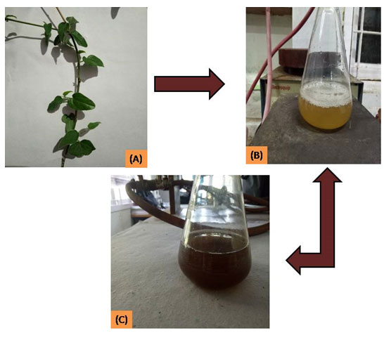
For the synthesis of PdNPs, in a conical flask, 30 ml of 1 mM PdCl2 solution was slowly applied to 20 ml of prepared plant extract for biogenic synthesis of PdNPs. After applying the salt solution, the reaction mixture was placed on a magnetic stirrer with a hot plate set to 50°C for 60 minutes, and the colour of the reaction mixture changed from light orange to dark brown (Fig.1), confirming the tentative proof of PdNPs formation by reducing the ions Pd+2 to Pd+1 condition. UV-visible spectroscopy with an SPR peak at 422 nm was used to validate the results. The reaction mixture was reduced in less than eight hours. After centrifuging the final reaction mixture for 15 minutes at 6,000 rpm at 4°C, the bottom deposited nanoparticles were extracted and dried in an oven for 1-2 hours. For further characterization and biological screening, the obtained crystalline powdered PdNPs is placed in an airtight bottle.
Figure 1: (A) Cocculus Hirsutus Leaf (B) Leaf extract and (C) Reaction mixture after 8 hr.
To analyse the characteristics of PdNPs, the UV-vis spectrometer (UV-1800, Shimadzu, Japan) was used to classify biosynthesized Palladium nanoparticles in the range of 200-800 nm. A Rigaku D/MAX 40 kV diffractometer with graphite chromatography was used for the X-ray diffraction analysis. Debye Scherer’s formula was used to calculate the average particle size.
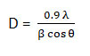
Where D is the mean crystalline dimension, FWMH (full width at half maximum), X-ray wavelength, and diffraction angle are all constants. Field electron gun scanning electron microscopy with energy dispersive spectroscopy (FEG-SEM with EDS, JEOL JSM-7600F) and high resolution transmission spectroscopy is used to investigate the structural morphology and elemental composition (HR-TEM, Tecnai G2-F30).The functional groups present in the plant extract as biomolecules were established using Fourier transform infrared spectroscopy (FTIR) (Shimadzu, range of 400-4000 cm-1) to contribute to the reduction of metal ions. Biogenic PdNPs were tested against Bacillus subtilis MTCC 121 (gram positive) and Escherichia coli MTCC 119 (gram negative) Pathogenic bacterial strains in an antibacterial assay.
Where D is the mean crystalline dimension, FWMH (full width at half maximum), X-ray wavelength, and diffraction angle are all constants. Field electron gun scanning electron microscopy with energy dispersive spectroscopy (FEG-SEM with EDS, JEOL JSM-7600F) and high resolution transmission spectroscopy is used to investigate the structural morphology and elemental composition (HR-TEM, Tecnai G2-F30).The functional groups present in the plant extract as biomolecules were established using Fourier transform infrared spectroscopy (FTIR) (Shimadzu, range of 400-4000 cm-1) to contribute to the reduction of metal ions. Biogenic PdNPs were tested against Bacillus subtilis MTCC 121 (gram positive) and Escherichia coli MTCC 119 (gram negative) Pathogenic bacterial strains in an antibacterial assay.
RESULTS AND DISCUSSION
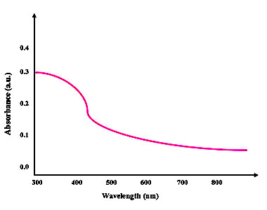
UV-Vis Spectra Analysis: UV-vis spectroscopy measurements (Shimadzu UV 1800) were carried out at room temperature in the region 800–200 nm as a function of time of the reaction. The UV–Visible absorption spectrum was used for the analysis of optical properties of biogenic synthesized Palladium nanoparticles. The mono dispersed Palladium nanoparticles are shown in synthesis figure-3. The room temperature spectra exhibited strong absorption peaks at 422 nm for samples respectively, which is in good agreement with previous work (Osonga et al. 2020).
Figure 2: UV-visible spectra of Biogenic Synthesized Palladium nanoparticles
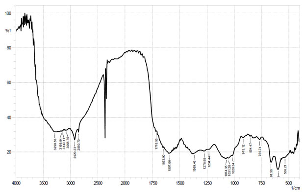
FTIR analysis of Palladium nanoparticles: FTIR spectroscopy analysis was carried out to identify the biomolecules responsible for the reduction of Pd+2 ions. FTIR spectroscopy analysis was carried out to find the biomolecules that were bound specifically on the Palladium nanoparticles surface. Fig. 4 shows the FTIR spectra of bio synthesized palladium nanoparticles. The spectrum showed sharp bands at 854 cm-1and 918 cm-1 corresponding to palladium nanoparticles. Strong bands were observed at 1234 cm-1, 1276 cm-1and 1369 cm-1 and have been referred to as C–N stretching and N=O stretching vibrations of aliphatic amines and nitro compound, respectively. The bands obtained at 1718 cm-1, 2850 cm-1, 2920 cm-1 and 3290 cm-1 have been representing to stretching vibrations of C=O stretching (Aldehyde), primary alkanes and water molecules.
Table 1. FTIR spectral data for Palladium nanoparticles.
| Functional Group | Wave number (cm-1) |
| Pd NPs | 854, 918 |
| C-N stretch
(Aliphatic amines) |
1234, 1276 |
| N=O stretch | 1369 |
| C=O stretch
(Aldehyde) |
1718 |
| C-H stretch
(alkanes) |
2850, 2920 |
| O-H stretch | 3290 |
Figure 3: FTIR spectrum of Palladium nanoparticles
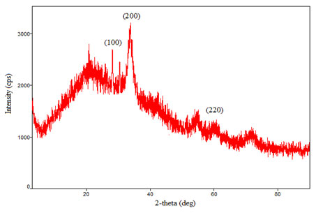
XRD analysis of Palladium Nanoparticles: X-ray diffraction (XRD) measurement of the green synthesis of palladium nanoparticles carried out on a Rigaku D/max 40 kV diffractometer equipped with the graphite mono chromator and cu target. Fig. 5 shows the XRD analysis of biogenic synthesized palladium nanoparticles. This is used for further confirmation of palladium phase of nanoparticles. The observed intense peaks are 28.220, 33.610, 60.160 and 72.100 respectively representing the (100), (200), (220) and (311) reflections indicating the face centered cubic (fcc) structure of palladium nanoparticles XRD pattern reveals the face centered cubic structure indicating the crystalline nature of palladium NPs and the particle size calculated using Debye-Scherrer equation,
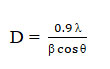
Where, D is average Particles size, λ is wavelength (1.5418 Å), θ is the Bragg’s angle and β is full width half maximum (FWHM) of corresponding peek. The Scherer’s formula was used to estimate the particles sizes and was found to around 12 nm.
Table 2. XRD spectral data of palladium nanoparticles
| 2Ɵ | Particle size (nm) | (h k l) |
| 28.22 | 35.66 | (100) |
| 33.6 | 4.68 | (200) |
| 60 | 4.36 | (220) |
| 72 | 2.28 | (311) |
| Average Particle size D = 12 nm | ||
Figure 4: XRD spectra of biogenic synthesized Palladium nanoparticles FEG-SEM and EDS analysis of Palladium Nanoparticles
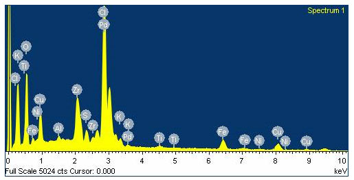
Field emission gun scanning electron microscope (FEG-SEM) and EDS images were recorded on a JSM-7600F series instrument. FEG-SEM spectra of biogenic synthesis of Palladium nanoparticles are shown in Fig.6. Palladium nanoparticles by green method show nearly mono dispersed distribution of particle sizes. The average particle size of the Pd nanoparticles is around 12 nm. The composition of Palladium nanoparticles was further probed by energy-dispersive X-ray (EDS) analysis. Fig. 7 shows the EDS pattern of Palladium NPs prepared using green synthesis, which indicates the presence of Pd and small amount of oxygen. EDS spectrum of Palladium nanoparticles shows the peaks for Palladium and respective elements indicating the formation of Palladium nanoparticles. Peak indexing of the elements is oxygen 0.5 keV and Palladium 2.8 & 3.5 keV. The compositions in the mass percentage of the elements are oxygen 41.16% and Palladium 46.21 %. The experimental composition matches with the theoretically calculated composition Rahul et al. 2019; Rahul et al. 2020; Hiral et al. 2020).
Figure 5: FEG-SEM spectra of green synthesis of Palladium nanoparticles
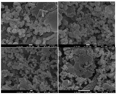
Figure 6: EDS spectra of green synthesis of Palladium nanoparticles
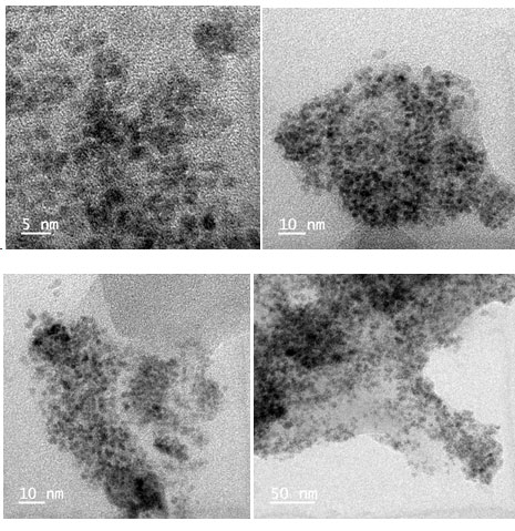
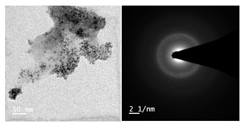
HR-TEM analysis of Palladium Nanoparticles: High-resolution Transmission electron microscope (HR-TEM) images were recorded on a Tecnai G2-F30 electron microscope. Fig. 8 shows the HR-TEM images of PdNPs prepared using Cocculus hirsutus plant. The sample preparation was carried out via the coating on carbon coated grid Cu Mesh 300 prior to the measurement. High-resolution Transmission electron microscopy (HR-TEM) has been employed to characterize the size, shape and morphology of synthesized Palladium nanoparticles.
Figure 7: -SEM spectra of Palladium nanoparticles
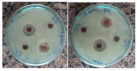
Antimicrobial Test: Using the agar well diffusion process, biosynthesized palladium nanoparticles made from Cocculus hirsutus leaf extract were tested against gram+ve (Escherichia coli MTCC 119) and gram-ve bacteria (Bacillus subtilis MTCC 121) bacteria at different concentrations. The same procedure was used to test plant extracts.
Table 3. Antimicrobial activity data for synthesized palladium nanoparticles.
|
Bacteria |
Zone of Inhibition | Average zone of
Inhibition |
||
| 1 | 2 | 3 | ||
| B. subtilis | 7 mm | 7.5 mm | 7.5 mm | 7.5 mm |
| E. Coli | 14 mm | 13 mm | 13 mm | 13 mm |
Fresh overnight cultures of each strain were swabbed uniformly by cotton on plates containing sterile Luria Bertani agar, and 4 cup borer wells (diameter size – 6 mm) were prepared. Each well was filled with 50 microlitres of sample nanoparticles, with a commercial gentamicin disc serving as a positive monitor. It was incubated for 24 hours at 37°C, during which the diameter of the inhibition region was measured in millimetres across the well (Fig. 9). (Table 3).The inhibitory compounds in the tested sample trigger the bacterial growth inhibition region. The antibacterial efficacy of the agar well diffusion process was found to be satisfactory. In comparison to Escherichia coli, Bacillus subtilis showed poor results in terms of zone of inhibition. Plant extract, on the other hand, yielded no results. The experiment was repeated three times in order to produce improved data Rahul et al. 2019; Rahul et al. 2020; Hiral et al. 2020).
Figure 8: Palladium nanoparticle antibacterial experiments (a) Escherichia coli (b) Bacillus subtilis
CONCLUSION
The Biogenic synthesis of Palladium nanoparticles performed Cocculus hirsutus leaf Extracts without involving any toxic chemicals. In this reduction reaction metal ions were reduced (Pd+2 to Pd+1) very rapidly and reaction was finally completed within 8 hours to produce Palladium nanoparticles. Different plants will take different time to complete the reaction due to have their different properties. The characterization of synthesized Palladium nanoparticles characterized by various microscopic and spectroscopic techniques which includes FEG-SEM with EDS, FTIR, HR-TEM, XRD and UV-visible confirms the formation of Palladium nanoparticles. Synthesized Palladium nanoparticles showed high stability even after few months at ordinary room temperature. XRD patterns conforms that the average particle size found to 12 nm. UV spectra shows absorption peak near 422 nm to confirm that formation of palladium nanoparticles.
ACKNOWLEDGEMENTS
Authors thank Department of Botany, Hemchandracharya North Gujarat University, Patan (Gujarat) for the plant material – Cocculus Hirsutus.
Conflict of Interests: The participating authors had no conflicts in their interests while preparing for this research.
REFERENCES
Abdel-Halim, E.S., El-Rafie, M.H. and Al-Deyab, S.S., (2011). Polyacrylamide/guar gum graft copolymer for preparation of silver nanoparticles. Carbohydrate Polymers, 85(3):692-697.
Adams, C., Walker, K.A., Obare, S.O. and Docherty, K.M., (2014). Size-dependent antimicrobial effects of novel palladium nanoparticles. PloS one, 9(1): e85981.
Anand, K., Tiloke, C., Phulukdaree, A., Ranjan, B., Chuturgoon, A., Singh, S. and Gengan, R.M., (2016). Biosynthesis of palladium nanoparticles by using Moringa oleifera flower extract and their catalytic and biological properties. Journal of Photochemistry and Photobiology B: Biology, 165:87-95.
Azizi, S., Shahri, M.M., Rahman, H.S., Rahim, R.A., Rasedee, A. and Mohamad, R., (2017). Green synthesis palladium nanoparticles mediated by white tea (Camellia sinensis) extract with antioxidant, antibacterial, and antiproliferative activities toward the human leukemia (MOLT-4) cell line. International Journal of Nanomedicine, 12: 8841.
Bankar, A., Joshi, B., Kumar, A.R. and Zinjarde, S., (2010). Banana peel extract mediated novel route for the synthesis of palladium nanoparticles. Materials Letters, 64(18):1951-1953.
Bhakya, S., Muthukrishnan, S., Sukumaran, M., Grijalva, M., Cumbal, L., Benjamin, J.F., Kumar, T.S. and Rao, M.V., (2016). Antimicrobial, antioxidant and anticancer activity of biogenic silver nanoparticles–an experimental report. RSC advances, 6(84):81436-81446.
Dumas, A. and Couvreur, (2015). Palladium: a future key player in the nanomedical field? Chemical Science, 6(4):2153-2157.
Fang, G., Li, W., Shen, X., Perez-Aguilar, J.M., Chong, Y., Gao, X., Chai, Z., Chen, C., Ge, C. and Zhou, R., (2018). Differential Pd-nanocrystal facets demonstrate distinct antibacterial activity against Gram-positive and Gram-negative bacteria. Nature communications, 9(1):1-9.
Govindarajan, M., AlQahtani, F.S., AlShebly, M.M. and Benelli, G., (2017). One-pot and eco-friendly synthesis of silver nanocrystals using Adiantum raddianum: toxicity against mosquito vectors of medical and veterinary importance. Journal of Applied Biomedicine, 15(2):87-95.
Hiral, V., Rahul, S., Shailesh, V. and Amanullakhan, (2020). Biosynthesized Silver Nanoparticles Using an Aqueous Root Extract of Iris germanica as a Reducing Agent and Its Antibacterial Efficacy. European Journal of Medicinal Plants:1-10.
Iravani, S., (2011). Green synthesis of metal nanoparticles using plants. Green Chemistry, 13(10):2638-2650.
Jadhav, J. and Shah, R., (2016). Synthesis of Biogenic Silver Nanoparticles from Medicinal Plant and It’s Antibacterial Activity, 9(8):29-33.
Jeyaraj, M., Rajesh, M., Arun, R., MubarakAli, D., Sathishkumar, G., Sivanandhan, G., Dev, G.K., Manickavasagam, M., Premkumar, K., Thajuddin, N. and Ganapathi, A., (2013). An investigation on the cytotoxicity and caspase-mediated apoptotic effect of biologically synthesized silver nanoparticles using Podophyllum hexandrum on human cervical carcinoma cells. Colloids and Surfaces B: Biointerfaces, 102:708-717.
Kanipandian, N., Kannan, S., Ramesh, R., Subramanian, and Thirumurugan, R., (2014). Characterization, antioxidant and cytotoxicity evaluation of green synthesized silver nanoparticles using Cleistanthus collinus extract as surface modifier. Materials Research Bulletin, 49:494-502.
Kharissova, O.V., Dias, H.R., Kharisov, B.I., Pérez, B.O. and Pérez, V.M.J., (2013). The greener synthesis of nanoparticles. Trends in biotechnology, 31(4):240-248.
Khazaei, A., Rahmati, S., Hekmatian, Z. and Saeednia, S., (2013). A green approach for the synthesis of palladium nanoparticles supported on pectin: Application as a catalyst for solvent-free Mizoroki–Heck reaction. Journal of Molecular Catalysis A: Chemical, 372:160-166.
Kim, S.W., Park, J., Jang, Y., Chung, Y., Hwang, S., Hyeon, T. and Kim, Y.W., (2003). Synthesis of monodisperse palladium nanoparticles. Nano Letters, 3(9):1289-1291.
Mittal, A.K., Chisti, Y. and Banerjee, U.C., (2013). Synthesis of metallic nanoparticles using plant extracts. Biotechnology advances, 31(2):346-356.
Nasrollahzadeh, M., Sajadi, S.M. and Maham, M., (2015). Green synthesis of palladium nanoparticles using Hippophae rhamnoides Linn leaf extract and their catalytic activity for the Suzuki–Miyaura coupling in water. Journal of Molecular Catalysis A: Chemical, 396:297-303.
Niraimathi, K.L., Sudha, V., Lavanya, R. and Brindha, (2013). Biosynthesis of silver nanoparticles using Alternanthera sessilis (Linn.) extract and their antimicrobial, antioxidant activities. Colloids and Surfaces B: Biointerfaces, 102:288-291.
Osonga, F.J., Kalra, S., Miller, R.M., Isika, D. and Sadik, O.A., (2020). Synthesis, characterization and antifungal activities of eco-friendly palladium nanoparticles. RSC Advances, 10(10):5894-5904.
Parhi, , Mohanty, C. and Sahoo, S.K., (2012). Nanotechnology-based combinational drug delivery: an emerging approach for cancer therapy. Drug discovery today, 17(17-18):1044-1052.
Petla, R.K., Vivekanandhan, S., Misra, M., Mohanty, A.K. and Satyanarayana, N., (2011). Soybean (Glycine max) leaf extract based green synthesis of palladium nanoparticles.
Sartre, A., Phaner, M., Porte, L. and Sauvion, G.N., (1993). STM and ESCA studies of palladium particles deposited on a HOPG surface. Applied surface science, 70:402-406.
Sathishkumar, M., Sneha, K. and Yun, Y.S., (2009). Palladium nanocrystal synthesis using Curcuma longa tuber extract. Int J Mater Sci, 4(1):11-17.
Sathishkumar, M., Sneha, K., Kwak, I.S., Mao, J., Tripathy, S.J. and Yun, Y.S., (2009). Phyto-crystallization of palladium through reduction process using Cinnamom zeylanicum bark extract. Journal of Hazardous materials, 171(1-3):400-404.
Shah R, Vaghela H, and Pathan A., (2020). Synthesis and Characterization of Biogenic Gold Nanoparticles Using Aegle marmelos Extracts: Antibacterial Assay. Biosc.Biotech.Res.Comm., 13(1):307-312.
Shah, R., Pathan, A., Vaghela, H., Ameta, S.C. and Parmar, K., (2019). Green synthesis and characterization of copper nanoparticles using mixture (Zingiber officinale, Piper nigrum and Piper longum) extract and its antimicrobial activity. Chemical Science, 8(1):63-69.
Vaghela, H., Shah, R. and Parmar, K., (2017). Biogenic synthesis of silver nanoparticles using Bauhinia variegata bark extract and its antibacterial efficacy. International Journal of Nanomaterials and Chemistry, 3(2):45-49.
Son, S.U., Jang, Y., Yoon, K.Y., Kang, E. and Hyeon, T., (2004). Facile synthesis of various phosphine-stabilized monodisperse palladium nanoparticles through the understanding of coordination chemistry of the nanoparticles. Nano Letters, 4(6):1147-1151.
Song, J.Y., Kwon, E.Y. and Kim, B.S., (2010). Biological synthesis of platinum nanoparticles using Diopyros kaki leaf extract. Bioprocess and Biosystems Engineering, 33(1):159-164.
Tahir, K., Nazir, S., Ahmad, A., Li, B., Shah, S.A.A., Khan, A.U., Khan, G.M., Khan, Q.U., Khan, Z.U.H. and Khan, F.U., (2016). Biodirected synthesis of palladium nanoparticles using Phoenix dactylifera leaves extract and their size dependent biomedical and catalytic applications. RSC advances, 6(89):85903-85916.
Tristany, M., Courmarcel, J., Dieudonné, Moreno-Mañas, M., Pleixats, R., Rimola, A., Sodupe, M. and Villarroya, S., (2006). Palladium nanoparticles entrapped in heavily fluorinated compounds. Chemistry of materials, 18(3):716-722.
Vaghela, H.M., Pathan, A.A. and Shah, R.H., (2018). The biogenic synthesis of Au, Pd and Pt nanoparticles and its medicinal applications: A review. Cambridge Scholars Publishing.
Vaghela, H., Shah, R. and Parmar, K.A., (2018). Plant mixture mediated biogenic copper nanoparticles: Antibacterial assay. Current Nanomaterials, 3(2):86-94.
Vaghela, H., Shah, R. and Pathan, A., (2018). Palladium nanoparticles mediated through bauhinia variegata: Potent in vitro anticancer activity against mcf-7 cell lines and antimicrobial assay. Current Nanomaterials, 3(3):168-177.
Veisi, H., Faraji, A.R., Hemmati, S. and Gil, A., (2015). Green synthesis of palladium nanoparticles using Pistacia atlantica kurdica gum and their catalytic performance in Mizoroki–Heck and Suzuki–Miyaura coupling reactions in aqueous solutions. Applied Organometallic Chemistry, 29(8):517-523.
Veisi, H., Ghorbani‐Vaghei, R., Hemmati, S., Aliani, M.H. and Ozturk, T., (2015). Green and effective route for the synthesis of monodispersed palladium nanoparticles using herbal tea extract (Stachys lavandulifolia) as reductant, stabilizer and capping agent, and their application as homogeneous and reusable catalyst in Suzuki coupling reactions in water. Applied Organometallic Chemistry, 29(1):26-32.
Veisi, H., Nasrabadi, N.H. and Mohammadi, (2016). Biosynthesis of palladium nanoparticles as a heterogeneous and reusable nanocatalyst for reduction of nitroarenes and Suzuki coupling reactions. Applied Organometallic Chemistry, 30(11):890-896.
Veisi, H., Rashtiani, A. and Barjasteh, V., (2016). Biosynthesis of palladium nanoparticles using Rosa canina fruit extract and their use as a heterogeneous and recyclable catalyst for Suzuki–Miyaura coupling reactions in water. Applied Organometallic Chemistry, 30(4):231-235.
Viswadevarayalu, A., Ramana, V., Sumalatha, J. and Reddy, S.A., (2016). Biocompatible synthesis of palladium nanoparticles and their impact on fungal species. Journal of Nanoscience and Technology:169-172.
Xiong, Y., Chen, J., Wiley, B., Xia, Y., Aloni, S. and Yin, Y., (2005). Understanding the role of oxidative etching in the polyol synthesis of Pd nanoparticles with uniform shape and size. Journal of the American Chemical Society, 127(20):7332-7333.
Xiong, Y., Chen, J., Wiley, B., Xia, Y., Yin, Y. and Li, Z.Y., (2005). Size-dependence of surface plasmon resonance and oxidation for Pd nanocubes synthesized via a seed etching process. Nano Letters, 5(7):1237-1242.
Yang, X., Li, Q., Wang, H., Huang, J., Lin, L., Wang, W., Sun, D., Su, Y., Opiyo, J.B., Hong, L. and Wang, Y., (2010). Green synthesis of palladium nanoparticles using broth of Cinnamomum camphora leaf. Journal of Nanoparticle Research, 12(5):1589-1598.

