
Exosomes in uence the engraftment of tumor cell lines
in athymic mice BALB/c nude
Roman A. Kovalev
1
, Vladimir S. Burdakov
1
, Elena Yu. Varfolomeeva
1
, Elena V. Semenova
1
and Michael V. Filatov
1,2
*
1
Division of Molecular and Radiation Biophysics, National Research Center “Kurchatov Institute” B.P.
Konstantinov Petersburg Nuclear Physics Institute; Gatchina, Russia;
2
Saint-Petersburg State Research Institute of Phthisiopulmonology of the Ministry of Healthcare of the
Russian Federation; St. Petersburg, Russia
ABSTRACT
The strain of mice with immunode cit provides a popular experimental model in oncological, immunological, and
transplantation studies. Athymic nude mice (Nudes) are usually used to explore the engraftment, growth, invasive in l-
tration, and potency to develop metastases for different types of cancer, as well as to test in preclinical assessment new
anti-tumor medical products. Nude-mutation in Foxn1 gene results in the congenital thymus dysgenesis, and, as a con-
sequence, in the lack of T-lymphocytes. In its turn, it leads to the suppression of the immune functions which involve
T-cells. It is generally accepted that Nudes’ immune de ciency provides an opportunity to engraft in them immunologi-
cally incompatible human tumors that are xenogeneic for mice.In our study we have shown that there are some types
of tumors that cannot be engrafted in BALB/c Nude mice. In particular, we have discovered that tumor cells de cient in
p53 gene are not able to grow in Nude mice.Furthermore, we have used in vitro model system to demonstrate that the
exosomes released by the broblasts of BALB/c Nude mice suppress the growth of the very same tumor cell lines that
cannot be engrafted in these mice, but the exosomes do not affect the growth of the tumor cells that can be engrafted
successfully. One of the possible mechanisms of tumor growth suppression in BALB/c Nude mice is the transfer of the
exosomes containing wild type p53 protein from the surrounding tissues to tumor cells with gene p53 damage.
KEY WORDS: BALB/C NUDE MICE, CELL LINES, EXOSOMES, PROTEIN P53
535
ARTICLE INFORMATION:
Corresponding Authors: l_53@mail.ru
Received 10
th
Oct, 2018
Accepted after revision 11
th
Dec, 2018
BBRC Print ISSN: 0974-6455
Online ISSN: 2321-4007 CODEN: USA BBRCBA
Thomson Reuters ISI ESC / Clarivate Analytics USA
Mono of Clarivate Analytics and Crossref Indexed
Journal Mono of CR
NAAS Journal Score 2018: 4.31 SJIF 2017: 4.196
© A Society of Science and Nature Publication, Bhopal India
2018. All rights reserved.
Online Contents Available at: http//www.bbrc.in/
DOI: 10.21786/bbrc/11.4/1
Medical
Communication
Biosci. Biotech. Res. Comm. 11(4): 535-540 (2018)

Roman A. Kovalev et al.
536 EXOSOMES INFLUENCE THE ENGRAFTMENT OF TUMOR CELL LINES IN ATHYMIC MICE BALB/C NUDE BIOSCIENCE BIOTECHNOLOGY RESEARCH COMMUNICATIONS
INTRODUCTION
Experimental models of human diseases play an impor-
tant role both in understanding what biological and
genetic factors in uence phenotypic characteristics
of the disease and in developing the strategy for its
treatment.When using in vitro cell models for human
tumor research one should take into consideration that,
though these studies are necessary, they are reduction-
ist, as often the parameters of the cell culture cannot be
equated with the tumor growth inside the host’s body
where the tumor has speci c micro-environment. Mod-
eling human cancer with the help of genetically awed
mice is an alternative way to study carcinogenesis in
vivo. It provides an opportunity to detect cell and molec-
ular changes taking place at the initiation of cancer and
during its further progression (Stakleff, 2003, Shultz,
2014; Boone, 2015).
One of the mechanisms preventing formation and
growth of the malignant tumors in human body is
immune system, and in particular, cell immunity based
on the activity of T-lymphocytes attacking tumor cells.
To research anti-tumor activity of the immune system
the mice with de cient T-cell immunity are widely used.
Athymic mice line BALB/c Nude is generally accepted as
a model for this kind of experiments. The main advan-
tage of this experimental model is natural immunode-
pression resulting from the functional deletion in Foxn1
gene. As the result of the deletion, thymus is either dete-
riorated or absent, and, as a consequence, the number
of T-cells is negligible, and, therefore, cell immunity is
inhibited (Pignata,1996; Frank, 1999; Zuklys, 2016). In
cancer research a large number of various tumor cell
lines are used for inoculation hence, it is comparatively
easy to engraft a tumor being studied in the mice just
inoculating the host-animal with the tumor cells. It is
believed that BALB/c Nude mice can host even immu-
nologically incompatible, xenogneic for mice human
tumors, (Liebman, 2007; Zeineldin, 2014; Conrad, 2015).
However, in our study we discovered that not all tumor
grafts could grow in BALB/c Nude immune de cient mice.
We were not able to reach a positive result of inoculation
with primary glioma lines developed in our laboratory.
The fact led us to the idea of an alternative mechanism
of tumor growth inhibition – the mechanism which did
not involve the immune system. If the mechanism exists,
then even though Nude mice lack T-lymphocyte mediated
cell immunity, the mechanism will account for inhibition
of the growth of inoculated human tumor cells.Taking
into consideration the growing amount of the data that
point to participation of exosomes in formation, develop-
ment and inhibition of cancer (Webber, 2015; Yu, 2015),
we suggested that the discovered phenomenon of tumor
growth inhibition in BALB/c Nude mice was possibly a
result of the in uence of the exosomes formed in the tis-
sues surrounding the tumor.
Exosomes are extracellular membrane nano vesicles
that are released from multivesicle bodies into extracel-
lular space through exocytosis (Huang, 2013). The abil-
ity of the exosomes to be ef cient transporters of exog-
enous proteins and RNA into cells-recipients has been
reported (Shtam, 2013; Haney, 2015; Srivastava, 2016).
Earlier in our experiments in vitro we demonstrated that
the exosomes released from the cells, which contained
wild type of the p53 protein, were able to inhibit the
growth of the cancer cells lacking the same protein. Fur-
thermore, the exosomes both released by the cell lines
and isolated from human blood plasma carried one of
the main tumor suppressors - protein p53 (Burdakov,
2017; Jorgensen, 2015, Burdakov, 2017).
In this study we now present some arguments in
favor of our assumption that one of the factors inhibit-
ing inoculated tumor growth in BALB/c Nude athymic
mice may be negative effect of exosomes on tumor cells.
The effect may be based on the reaction of the inocu-
lated tumor cells de cient in p53 gene to the wild type
p53 protein, which exists in exosomes released by the
surrounding tissues.
MATERIALS AND METHODS
CELL LINES AND CULTIVATION CONDITIONS
In our study we used both transplantable and primary
human cell lines: HT-1080 ( brosarcoma), Gl-V, Gl-R,
Gl-Sh (primary cultures of glioma cells, developed in
our laboratory), transplantable culture of rat cells C6 (rat
brain glioma), and broblasts of BALB/c Nude mice. Cells
were cultivated in DMEM/F12 medium (Biolot, Russia)
supplemented with 10% fetal bovine serum (Biolot, Rus-
sia), without antibiotics, under 5% CO
2,
at 37°C. When
tumor cells of each of the lines were cultivated with iso-
lated exosomes, the exosomes had nal concentration in
the culture of 10
13
–10
14
exosomes/ml.
Isolation of exosomes in the systems in vitro
Conditional medium was collected from growing cell cul-
ture. Then, it was consequently centrifuged at 2,000 g and
at 20,000 g to eliminate dead cells and their fragments.
The procedure was repeated until the amount of the puri-
ed conditional medium reached 500 ml. This amount
of collected puri ed conditional medium was used to
isolate exosomes by ultracentrifuging. Beckman coulter
ultracentrifuge was used (45Ti rotor) at 100,000 g for 2
hours. After that, the residue was suspended in 100 ml of
PBS and underwent the second ultracentrifugation at the
same conditions. The exosomes obtained as the result of
the second sedimentation were used in the experiments.

Roman A. Kovalev et al.
BIOSCIENCE BIOTECHNOLOGY RESEARCH COMMUNICATIONS EXOSOMES INFLUENCE THE ENGRAFTMENT OF TUMOR CELL LINES IN ATHYMIC MICE BALB/C NUDE 537
Evaluation of the cell survival rate
The cells were suspended in Versene/Trypsin solution
(Biolot, Russia), and the survival rate was evaluated
through direct calculation of the cells with the help of
vision-based automated cell counter Scepter (Millipore,
USA). To visualize cell survival, the cells of Gl-V, Gl-R,
Gl-Sh and C6 lines were seeded into 12-well plates in the
concentration equal for all the cell lines (10,000 cells per a
well). After that we added equal amounts of the exosomes
harvested from the conditional medium of the broblasts
of BALB/c Nude mice to all the cell lines. As the control
cells formed a monolayer, all the cells in the well plates
were stained with Crystal Violet (Fluka AG, Germany). The
data were analyzed by Student’s t test. Data are shown as
the mean ± SEM (standard error of the mean).
Animal care and engraftment rate evaluation
Adult BALB/c Nude mice, 7-8 weeks of age and 18-20
grams of bodyweight, were purchased from the laboratory
animal breeding center “Pushchino” (Pushchino, Russia).
All procedures for mouse care and use were conducted
in accordance with the National Standard of the Rus-
sian Federation GOST R 53434-2009: Principles of good
laboratory practice (introduced 01.03.2010 by Federal
Agency for Technical Regulation and Metrology, pub-
lished by Standardinform, Moscow, 2010). The protocol
was approved by scienti c committee of the Division of
Molecular and Radiation Biophysics of National Research
Center “Kurchatov Institute” B.P.Konstantinov St Peters-
burg Nuclear Physics Institute (Gatchina, Russia).
Animals were housed in groups of three per cage
(males and females were housed separately) with ad libi-
tum access to food and water in standard polycarbonate
cages (width: 300mm, depth: 400mm, height: 200mm)
and maintained under standard conditions of tempera-
ture (22 ± 2°C), humidity (55 ± 10%), arti cial 12-hour
light-dark cycle (lights on at 8:00 A.M., lights off at 8:00
P.M.). The mice were fed on the nutrient extruded and
granulated food designed for experimental rodents (LLC
«Laboratorkorm», Russia).
Tumor inoculations were performed through intra-
muscularly injections of 150,000 to 1,000,000 tumor
cells. To estimate the engraftment rate for each tumor
cell line, not less than 20 BALB/c Nude immune de -
cient athymic mice were used.
RESULTS AND DISCUSSION
Evaluation of engraftment in BALB/c Nude immune
de cient athymic mice for tumor cell lines
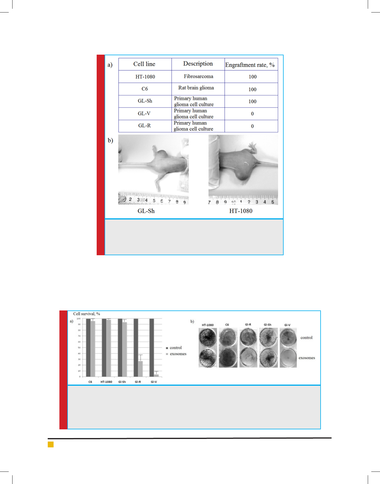
To evaluate the engraftment rate we took 5 different
tumor cell lines, which were cultivated in our laboratory.
Two of them were standard transplantable cell lines:
HT-1080, C6. The rest three lines (GL-V, GL-R, GL-Sh)
were primary lines of human gliomas, developed in our
laboratory. In the experiments with each of the cell lines
not less than 20 BALB/c Nude immune de cient athymic
mice were used. In the rst experiment engraftment
was performed through intramuscularly injections of
150,000 tumor cells per animal. As the result there were
two polar outcomes: transplantable tumor lines HT-1080
and C6 engrafted in 100% of the animals; tumor lines
GL-V, GL-R did not engraft at all ( g. 1). Engraftment
rate of the latter lines (GL-V, GL-R) did not change with
the increase in the amount of inoculated cells up to
1,000,000 cells per animal.
Analysis of survival rate of the tumor cell lines after
treatment with the exosomes from the broblasts of
BALB/c Nude mice
We assumed that negative results of engraftment of
some of the tumors in BALB/c Nude athymic mice
stemmed from the ability of exosomes to inhibit tumor
cell growth. To con rm the assumption, we decided to
compare the engraftment rate obtained in the experi-
ments with the survival rate of the same tumor cell lines
after they had been treated with the exosomes from the
broblasts of BALB/c Nude mice. To carry out the com-
parison, we used standard ultracentrifugation methods
described previously, (Lässer et al., 2012 and Shtam et al
2013) to isolate exosomes from the condensed cultural
medium after growing broblasts of BALB/c Nude mice
in it. Cells from the analyzed lines were seeded onto
24-well plates in the equal concentration. Then, equal
amounts of the isolated exosomes were added to all of
the tested tumor cell lines. In 10 days after the exosomes
had been added, the number of living cells was calcu-
lated for each of the studied tumor cell lines.
The results of the calculation are presented in g. 2.
Addition of the exosomes isolated from the broblasts
of BALB/c Nude mice did not affect the survivability
of the tumor cells HT-1080 and GL-Sh. However, exo-
some addition to the cells of the tumor lines GL-R и
GL-V resulted in signi cant decrease in their survivabil-
ity ( g. 2). Survivability of the glial cells of GL-R cell
line decreased by almost three times, and survivability
of the cells of the cell line GL-V decreased by more than
10 times. The obtained results point to the ability of the
BALB/c Nude cell exosomes to inhibit the growth of the
target cells GL-R and GL-V in in vitro experiments. It
correlates with the negative results of in vivo inocula-
tion of the cells of these tumor lines to BALB/c Nude
immune de cient mice.
Exosomes, participating in inter-cellular communica-
tion by transporting RNA and speci c proteins between
cells, are able to cause both functional and epigenetic
Roman A. Kovalev et al.
538 EXOSOMES INFLUENCE THE ENGRAFTMENT OF TUMOR CELL LINES IN ATHYMIC MICE BALB/C NUDE BIOSCIENCE BIOTECHNOLOGY RESEARCH COMMUNICATIONS
FIGURE 1. Engraftment rates of the tumor cell lines in BALB/c Nude immune
de cient athymic mice
(a) Table of engraftment rates of the tumor cell lines C6, HT-1080, GL-Sh, GL-R
и GL-V.
(b) Examples of the engrafted tumors HT-1080 and GL-Sh.
FIGURE 2. Survival rates of the tumor cell lines C6, HT-1080, GL-Sh, GL-R and GL-V after treatment with the
exosomes from the broblasts of BALB/c Nude mice
(a) Histograms of survival rates of the tumor cell lines after treatment with the exosomes from the broblasts of
BALB/c Nude mice.
(b) Survival of the cell lines C6, HT-1080, GL-Sh, GL-R and GL-V after coculturing with the exosomes from the
broblasts of BALB/c Nude mice demonstrated by means of Crystal Violet staining.
changes in the recipient cells (Grange, 2011; Kobayashi,
2014). Both discovery of p53 oncosupressor in the
exosomes of the human blood plasma and their ability
to affect proliferation of p53-negative cells con rm their
important role in control over oncogenesis (Burdakov,
2017; Jorgensen, 2015). Protein p53 is the main par-
ticipant of many signaling pathways through which cell
reactions to stress are regulated. The protein is able to stop
cell-division cycle and/or to cause apoptosis, thus pre-
venting uncontrollable cell division – reproduction of the
cells with damaged genome. In human malignant tumors
p53 gene is the one that most often is found mutated

BIOSCIENCE BIOTECHNOLOGY RESEARCH COMMUNICATIONS EXOSOMES INFLUENCE THE ENGRAFTMENT OF TUMOR CELL LINES IN ATHYMIC MICE BALB/C NUDE 539
Roman A. Kovalev et al.
(Levine, 2009; Vogelstein, 2013; Kandoth, 2013). There
are a number of mouse model studies demonstrating that
restoration of the function of wild type p53 may result in
tumor regression (Ventura, 2007; Xue, 2007).
We assume that negative effect of the exosomes con-
taining wild type p53 protein on inoculated tumor cells
may be one of the factors which prevent the engraftment
of the tumor in BALB/c Nude athymic mice.
It is a common knowledge that any experimental
model based on the usage of the athymus Nude mice
has its restrictions: immunode ciency is severe, but not
absolute (there still exists humoral adaptive immune
system and undamaged congenital immunity). Hence,
despite almost complete lack of functioning T-lympho-
cytes, both congenital immune response and high activ-
ity of NK-cells are able to restrict the speed of engraft-
ment and to decrease metastatic potency of majority of
tumors (Shultz, 2005; Shultz, 2014; Szadvari, 2016).
However, the data on the engraftment rate of differ-
ent tumor cell lines we present in this study point to a
clear division of the tumors into two groups: the tumors
engrafting in BALB/c Nude mice and the tumors which
cannot be engrafted in BALB/c Nude mice. Basing on
the vast statistics we discovered no variations in the
engraftment rate for each of the tumors used in the study
( g.1a). These facts cannot be explained only by exist-
ence of the rudimentary immunity in immune de cient
mice. We assume that one of the possible mechanisms of
the engraftment inhibition involves systemic in uence
of mouse’s own exosomes on the inoculated tumor cells.
It is necessary to remark that the cells with the wild type
of p53 protein (HT-1080) had 100% engraftment rate,
while GL-V cells lacking endogenous p53 protein (Kova-
lev, 2015) could not be engrafted at all.
Then we examined the sensitivity of all the tumor cell
lines used for inoculation to the in uence of the exosomes
isolated from the broblasts of BALB/c Nude mice. The
results we received in the experiments in vitro showed
that cells of the GL-R cell line and especially p53-nega-
tive GL-V line were sensitive to such in uence: the cells
from abovementioned tumor cell lines died as the result of
addition of the exosomes from the broblasts of BALB/c
Nude mice to the tumor cell culture.The results obtained
support the hypothesis that exosomes circulating in the
body are able to perform a defense function controlling
oncogenesis through onco suppressor p53.
Con ict of interest disclosures: We have no con ict of
interest in any part of this article.
REFERENCES
Boone JD, Dobbin ZC, Straughn JM jr., Buchsbaum DJ: (2015)
Ovarian and cervical cancer patient derived xenografts: The
past, present, and future. Gynecol Oncol 138: 486-491.
Burdakov VS, Kovalev RA, Pantina RA, Varfolomeeva EYu,
Makarov EM., Filatov MV: (2017) Exosomes transfer p53
between cells and can suppress growth and proliferation of
p53-dR. Tsitologiia 59 (9): 588-594.
Conrad SJ, El-Aswad M, Kurban E, Jeng D, Tripp BC, Nutting
C, Eversole R, Mackenzie C, Essani K: (2015) Oncolytic tana-
poxvirus expressing FliC causes regression of human colorec-
tal cancer xenografts in nude mice. J Exp Clin Cancer Res 34
(1): 19.
Frank J, Pignata C, Panteleyev AA, Prowse DM, Baden H,
Weiner L, Gaetaniello L, Ahmad W, Pozzi N, Cserhalmi-Fried-
man PB, Aita VM, Uyttendaele H, Gordon D, Ott J, Brissette JL,
Christiano AM
: (1999) Exposing the human nude phenotype.
Nature 398 (6727): 473-474.
Grange C, Tapparo M, Collino F, Vitillo L, Damasco C, Deregi-
bus MC, Tetta C, Bussolati B, Camussi G: (2011) Microvesicles
released from human renal cancer stem cells stimulate angio-
genesis and formation of lung premetastatic niche. Cancer Res
71: 5346-5356.
Haney M J, Klyachko NL, Zhao Y, Gupta R, Plotnikova EG,
He Z, PatelT, Piroyan A, Sokolsky M, Kabanov AV, Batrakova
EV: (2015) Exosomes as drug delivery vehicles for Parkinson’s
disease therapy. J Control Release 207: 18-30.
Huang SH, Li Y, Zhang J, Rong J, Ye S: (2013) Epidermal
growth factor receptor-containing exosomes induce tumor-
speci c regulatory T cells. Cancer Invest 31: 330-335.
Jørgensen MM, Bæk R, Varming K: (2015) Potentials and capa-
bilities of the extracellular vesicle (EV) array. J Extracell Vesi-
cles 4: 26048.
Kandoth C, McLellan MD, Vandin F, Ye K, Niu B, Lu C, Xie M,
Zhang Q, McMichael JF, Wyczalkowski MA, Leiserson MDM,
Miller CA, Welch JS, Walter MJ, Wendl MC, Ley TJ, Wilson
RK, Raphael BJ, Ding L: (2013) Mutational landscape and
signi cance across 12 major cancer types. Nature 502: 333–
339.
Kobayashi M, Salomon C, Tapia J, Illanes SE, Mitchell MD,
Rice GE: (2014) Ovarian cancer cell invasiveness is associated
with discordant exosomal sequestration of Let-7 miRNA and
miR-200. J Transl Med 12: 4.
Kovalev RA, Shtam TA, Karelov DV, Burdakov VS, Volnits-
kiy AV, Makarov EM, Filatov MV: (2015) Histone deacetylase
inhibitors cause the TP53-dependent induction of p21/Waf1
in tumor cells carrying mutations in TP53. Tsitologiia 57 (3):
204-211.
Lässer C, Eldh M, Lötvall J: (2012) Isolation and characterization
of RNA-containing exosomes. J Vis Exp 59: e3037.
Levine AJ, Oren M: (2009) The rst 30 years of p53: growing
ever more complex. Nat Rev Cancer 9: 749–758.
Liebman MA, Roche MI, Williams BR, Kim J, Pageau SC, Sha-
ron J: (2007) Antibody treatment of human tumor xenografts
elicits active anti-tumor immunity in nude mice. Immunol Lett
114 (1): 16–22.
Pignata C, Fiore M, Guzzetta V, Castaldo A, Sebastio G, Porta
F,
Guarino A: (1996) Congenital Alopecia and nail dystrophy

540 EXOSOMES INFLUENCE THE ENGRAFTMENT OF TUMOR CELL LINES IN ATHYMIC MICE BALB/C NUDE BIOSCIENCE BIOTECHNOLOGY RESEARCH COMMUNICATIONS
Roman A. Kovalev et al.
associated with severe functional T-cell immunode ciency in
two sibs. Am J Med Genet 65 (2): 167–170.
Shtam TA, Kovalev RA, Varfolomeeva EYU, Makarov EM,
Kil YUV, Filatov MV: (2013) Exosomes are natural carriers of
exogenous siRNA to human cells in vitro. Cell Commun Signal
11: 88.
Shultz LD, Goodwin N, Ishikawa F, Hosur V, Lyons BL, Greiner
DL: (2014) Human cancer growth and therapy in NOD/
SCID/IL2Rnull (NSG) mice. Cold Spring Harb Protoc 694-
708.
Shultz LD, Lyons BL, Burzenski LM, Gott B, Chen X, Chal-
eff S, Kotb M, Gillies SD, King M,
Mangada J, Greiner DL,
Handgretinger R: (2005) Human lymphoid and myeloid cell
devel opment in NOD/LtSz-scid IL2Rnull mice engrafted with
mobilized human hemopoietic stem cells. J Immunol 174:
6477-6489.
Srivastava A, Babu A, Filant J, Moxley KM, Ruskin R, Dha-
nasekaran D, Sood AK, McMeekin S, Ramesh R: (2016) Exploi-
tation of exosomes as nanocarriers for gene-, chemo-, and
immune-therapy of cancer. J Biomed Nanotechnol 12: 1159-
1173.
Stakleff KDS, von Gruenigen VE: (2003) Rodent models for
ovarian cancer research. Int J Gynecol Cancer 13: 405-412.
Szadvari I, Krizanova O, Babula P: (2016) Athymic nude mice
as an experimental model for cancer treatment. Physiol Res 65
(Supplementum 4): 441-453.
Ventura A, Kirsch DG, McLaughlin ME, Tuveson DA, Grimm J,
Lintault L, Newman J, Reczek EE, Weissleder R, Jacks T: (2007)
Restoration of p53 function leads to tumour regression in vivo.
Nature 445: 661–665.
Vogelstein B, Papadopoulos N, Velculescu VE, Zhou S, Diaz
LA jr, Kinzler KW: (2013) Cancer genome landscapes. Science
339: 1546–1558.
Webber J, Yeung V, Clayton A: (2015) Extracellular vesicles as
modulators of the cancer microenvironment. Semin Cell Dev
Biol 40: 27-34.
Xue W, Zender L, Miething C, Dickins RA, Hernando E, Krizh-
anovsky V, Cordon-Cardo C, Lowe SW: (2007) Senescence and
tumour clearance is triggered by p53 restoration in murine
liver carcinomas. Nature 445: 656–660.
Yu S, Cao H, Shen B, Feng J: (2015) Tumor-derived exosomes
in cancer progression and treatment failure. Oncotarget 6:
37151-37168.
Zeineldin M, Jensen D, Paranjape SR, Parelkar NK, Jokar I,
Vielhauer GA, Neufeld KL: (2014) Human cancer xenografts in
outbred Nude mice can be confounded by polymorphisms in a
modi er of tumorigenesis. Genetics 197 (4): 1365–1376.
Zuklys S, Handel A, Zhanybekova S, Govani F, Keller M, Maio
S, Mayer CE, Teh HY, Hafen K, Gallone G, Barthlott T, Pont-
ing CP, Holländer GA: (2016)
Foxn1 regulates key target genes
essential for T cell development in postnatal thymic epithelial
cells. Nat Immunol 17: 1206-1215.