
Medical
Communication
Biosci. Biotech. Res. Comm. 10(1): 213-218 (2017)
Experimental assessment of sealer bio ceramic wall
matching in rst and third apical areas by scanning
electron microscope
Torabizadeh Seyed Mohammad
1
, Shahsiah Samira
2
* and Shamohammadi Milad
3
1
Resident of Endodontic Department, School of Dentistry, Jundishapur University of Medical Sciences, Ahvaz Iran
2
Assistant Professor Department of Endodontics, School of Dentistry, Jundishapur University of Medical
Sciences, Ahvaz Iran
3
Resident of Orthodontic Department, School of Dentistry, Jundishapur University of Medical Sciences,
Ahvaz Iran
ABSTRACT
This study was aimed at investigating experimental assessment of sealer bio ceramic wall matching in 1th and third
apical area by Scanning Electron Microscope (SEM).In this experimental study, thirty single root and single channel
human teeth extracted were prepared by le rotary (FKG/Switzerland) connected to electric endodontic (Dentsply,
Maillefer, Swiss). Teeth were divided into two groups including 15 teeth: group A were obdurate with sealer bio
ceramic (BRASSELER USA Total Fill BC Sealer). Group B were lled by sealer AH-26. Root channels were led by
lateral compaction. All samples placed in 37 centigrade degrees and in 100 percent humidity. Sample was cut 3 mil-
limeters from apical end of root channel. Finally, SEM images were taken and wall matching level was measured.
Data were tested by one-way ANOVA and after LCD experiment they were analyzed. Results indicated that consider-
ing wall matching, there is no signi cant relation between similar section of two sealer section namely rst section
of AH-26 sealer with rst section of sealer bio ceramic, second section of AH-26 sealer with second section of bio
ceramic and between third sections of AH-26 sealer with third section of sealer bio ceramic while wall matching of
both sealer is similar. In sealer bio ceramic rst section has better seal and wall matching than third section. Wall
matching of sealer bio ceramic was similar to AH-26 sealer.
KEY WORDS: WALL MATCHING, SEALER BIO CERAMIC, AH-26, SCANNING ELECTRON MICROSCOPE (SEM)
213
ARTICLE INFORMATION:
*Corresponding Author:
Received 11
th
Jan, 2017
Accepted after revision 27
th
March, 2017
BBRC Print ISSN: 0974-6455
Online ISSN: 2321-4007 CODEN: USA BBRCBA
Thomson Reuters ISI ESC and Crossref Indexed Journal
NAAS Journal Score 2017: 4.31 Cosmos IF : 4.006
© A Society of Science and Nature Publication, 2017. All rights
reserved.
Online Contents Available at: http//www.bbrc.in/

214 EXPERIMENTAL ASSESSMENT OF SEALER BIO CERAMIC WALL MATCHING IN FIRST AND THIRD APICAL AREAS BIOSCIENCE BIOTECHNOLOGY RESEARCH COMMUNICATIONS
Torabizadeh Seyed Mohammad et al.
INTRODUCTION
In the past, success in endodontic treatment was identi-
ed by three factors including debridement, steriliza-
tion, and channel obturation with equal importance for
each one. In one primitive radiography study about suc-
cess and failure, Ingle indicated that 58% of treatment
failure is due to defected obturation (Ingle et al, 1994).
Forming and cleaning trend determines decontaminated
level and ability of lling root channel space; thus chan-
nel obturation is re ection of forming and cleaning.
Main reason of pulp and pre apical diseases is bacterial
(Kakehashi et al, 1965). Evidence show that root chan-
nel system cannot completely be cleaned and disinfected
and it is necessary to ll root channel space in order to
elimination of leaking (Heard and Walton 1997). Obtura-
tion of channel prevents coronary leaking and bacterial
contamination and seals apex against pre apical tissue
liquids and remained drivers in the channel (Delivanis et
al.1983 and Silva Almeida et al. 2017).
Different endo dentin materials have been recom-
mended for lling root space. Most methods use a central
material and sealer. Sealer is necessary for core material
in all methods and it provides stiff sealing against liq-
uids. Root channel sealers are necessary things for seal-
ing space of crown wall and main cone. Sealers also ll
voids and root channel irregularity, secondary, lateral
channels and space between applied gutta-percha cones
and also they act as tripper during lling. Some stand-
ards of an ideal sealer include: providing real connec-
tion between main material of obturation and crown,
not solving with tissue liquid during connection, being
bacteriostatic, and nally creates an apical and lateral
and crown seal and maintains it (Grossman and Oliet
1988 and Setia et al 2014).
Sealer can be reason of root treatment failure due to
micro leakage that happens in the space between sealer,
crown, sealer and core. Chemical connection of sealer to
channel wall on one side and on the other side obtura-
tion material of gutta-percha creates a mixture of mono
block by which there is minimum channel micro leak-
age (Pawar et al 2014 ).Most popular sealers are zinc
oxide and eugenol mixtures, hydroxide calcium sealers,
glassionomer and resins. Resin sealers are AH-26 seal-
ers (Dentsply, Maillefer and Swiss) that are used for a
long time, they have adhesion property, they don’t have
eugenol and slowly stiffens (De Moor and De Bruyne
2004; Al-Haddad and Che Ab Aziz 2016; Savadkouhi
and Fazlyab 2016; Ahuja et al. 2016).
It is stated that when this combination is in the tooth
it has ability of hydroxide apatite and chemical connec-
tion. Bioceramic sealers are the most important biocer-
mics which have best role in reducing micro leakage and
increasing prognosis of root treatment by forming mono
block and chemical connection to channel wall. One
popular bio ceramic sealer is Total Fill BC Sealer pro-
duced by BRASSELER USA factory. This sealer is acces-
sible as injectable mixed paste which is based on com-
bination of silicate calcium which is unsolvable. It does
not need water for being stiffened. It does not need to be
mixed. Working time and setting time is 4 hours in room
temperature. When channel is dry, setting time reaches
to 10 hours. Most bubble and most complexity are in
rst and third apical area of channel which has effect
on treatment prognosis (Vertucci 1984). Micro leakage
is main reason of endodentic failure which can happen
between gutta-percha and sealer, sealer and crown and
also in micro bubbles which exist in sealer. Edge match
indirectly re ects ability of sealing ller material of root
end which is very important. Assessment of edge match
of root end llers by scanning electron microscopy can
give information about ability of this material sealing
(Shokouhinejad et al. 2014; Ghorbanzadeh et al. 2014).
Bio ceramics of ceramic combinations are the best eco-
friendly mixtures which have been recently produced
and its contents include zirconium oxide, calcium sili-
cate, calcium phosphate, hydroxide and ller and other,
(Pawar et al., 2014, Utneja et al. 2015; Jitaru et al. 2016
and Silva Almeida et al. 2017).
Bioceramic sealers can increase wall matching and
can reduce microleakage specially in apical area due to
formation of mono block mixture and chemical con-
nection to channel wall thus, in this study we aimed at
assessing wall matching of bioceramic sealer in rst and
third apical areas of tooth root channel.
MATERIALS AND METHODS
PREPARATION OF SAMPLES
In this experimental laboratorial study, thirty single
root and single channel were extracted from upper jaw
and lower jaw of people due to hopeless prognise peri-
odontal. For this study double blind and teeth randomly
divided into two groups. In all groups, radiology images
were taken and they were studied considering lack of
crack, breaking, curvature, and calci cation of chan-
nel. Then teeth were oated for 5 minutes in 5/25%
sodium hypochlorite. Consequently, remained tissues
and clean mass were washed from teeth and were kept
in normal saline. Teeth crow were cut by hand piece
with high speed under water spray. Working length was
measured by entering 15 le K le (Dentsply, Maillefer,
Swiss) into root channel until le top then one millim-
eter was reduced. Channels of all roots were prepared
to 35,36% taper by rotary le connected to electric
endodential hand piece (Dentsply, Maillefer, Swiss) in all

BIOSCIENCE BIOTECHNOLOGY RESEARCH COMMUNICATIONS EXPERIMENTAL ASSESSMENT OF SEALER BIO CERAMIC WALL MATCHING IN FIRST AND THIRD APICAL AREAS 215
Torabizadeh Seyed Mohammad et al.
Table 1. Level of wall matching of sealer in lateral compaction in rst and third
apical area is shown
numbermean
Standard
deviation
MinimumMaximum
Sealer AH-26
First
section
150/0340/01090/02000/0620
Second
section
150/0370/03200/01010/0920
Third
section
150/060/01860/01100/0920
Bioceramic
sealer
First
section
150/0380/01680/02000/0750
Second
section
150/0440/02190/02000/0900
Third
section
150/0540/01850/02100/0800
teeth, number 40 race was used as widener of channel
crown area while 6% race number 40 was used for mid-
dle area of channel. Then 35,6% transferred to work-
ing length. Tooth crack must not be observed in applied
rotary les. Washing was made by 10 millimeter sodium
hypochlorite solvent, 5/25% disposable plastic syringe
with gauge 27 (BD. plastipak, India). After preparation,
root channels were washed by one-mile litter EDTA17%
and then with 5 milliliter 2/5% sodium hypochlorite for
elimination of smear layer. Finally root channels were
washed with 3 milliliters saline strilled and dried with
paper point (Dentsply, Maillefer, Swiss).
SEALING ROOT CHANNELS
Teeth are divided into two 15 teeth groups:
Group A included 15 teeth and were obturated with
35-6% gutta and sealer bioceramic ( BRASSELER USA
Total Fill BC Sealer) while their master apical gutta
was 35-6% which impregnated to bioceramic sealer
and transferred to working length. Then spreader 25
(Dentsply, Maillefer, Swiss) was transferred until it pen-
etrates. Then secondary gutta were transferred to length
of spreader in uence; this process was performed until
maximum penetration was continued to 3 to 4 mil-
limeter. Then accessible gutta were extracted by hot
chisel and remained gutta were condensed vertically in
channel span and then the hole was lled and dress-
ing. Group B including 15 teeth were lled by lateral
compaction and with gutta 35-6% and sealer AH-26.
While their master apical gutta was 35-6 percent which
impregnated by AH-26 sealer and it transferred to work-
ing length. Then spreader 25 was transferred until it
penetrated, and then secondary gutta was transferred
to 25% length of spreader penetration; this was contin-
ued until maximum penetration of spreader reached 3-4
millimeter. Next, accessible gutta was extracted by hot
chisel while remained gutta was condensed vertically in
channel span and the hole was wound dressed and lled.
All samples were kept in 37
0
degrees and 100%
humidity for one week in order to facilitate comple-
tion of sealer setting. Sample were placed in blocks and
marked from 1 to 30. these sample were taken to cut-
ting place then three 1 millimeter sections were provided
from three millimeter root apical ( rst and third apical).
Assessment by Scanning Electron Microscope (SEM) SEM
For observing by SEM, cut parts were dehydrated. Then
samples were mounted, and sputter covered with gold
then they observed under scanning electron microscope.
In order to assess wall matching following process
was performed:
Width of marginal gap was lled as maximum dis-
tance of material and root channel crown directly was
measured in thousandth scale by another researcher for
preventing bias.
Data were analyzed by SPSS software 20
th
version. In
order to compare wall matching in experimental groups
we used ANOVA test and for pairwise comparison after
experiment of LCD was applied. Level of signi cance
was p<0/05.
RESULTS
Level of wall matching of sealer in lateral compaction in
rst and third apical area is shown in table 1:
Results of ANOVA test showed that there is signi -
cant difference wall matching among groups (p=0/005).
Results of LSD test illusterated that considering wall
matching, there is no signi cant difference between
similar section of both sealer that is mean rst sec-
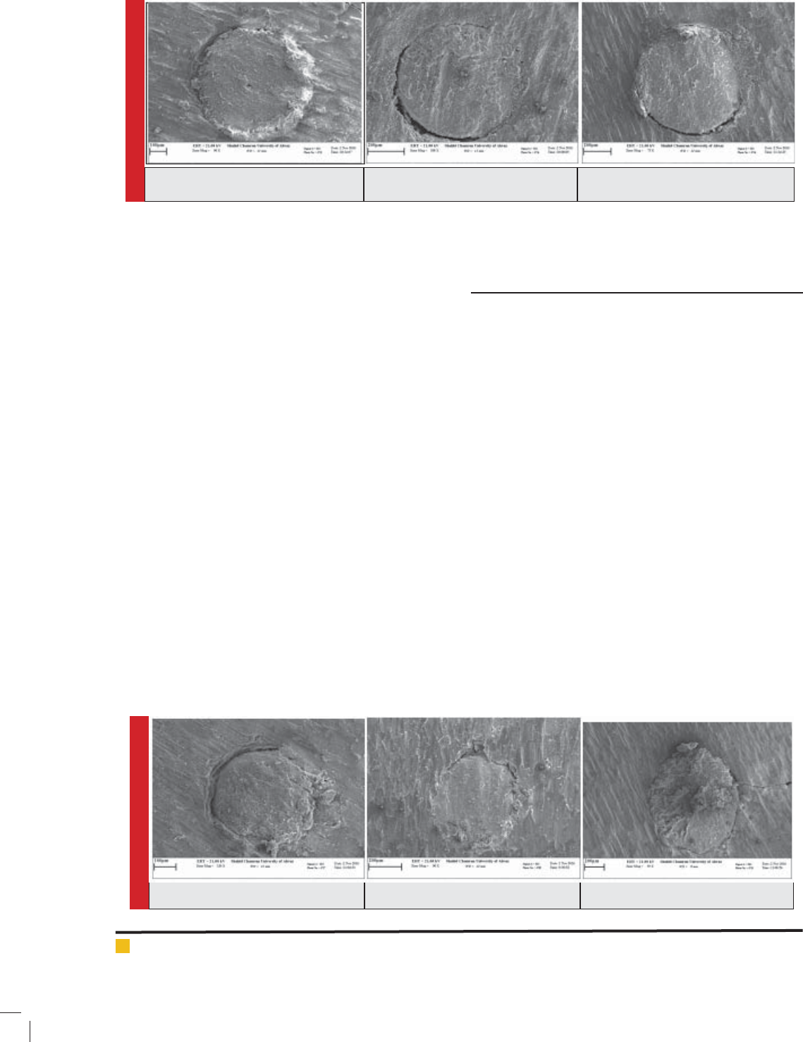
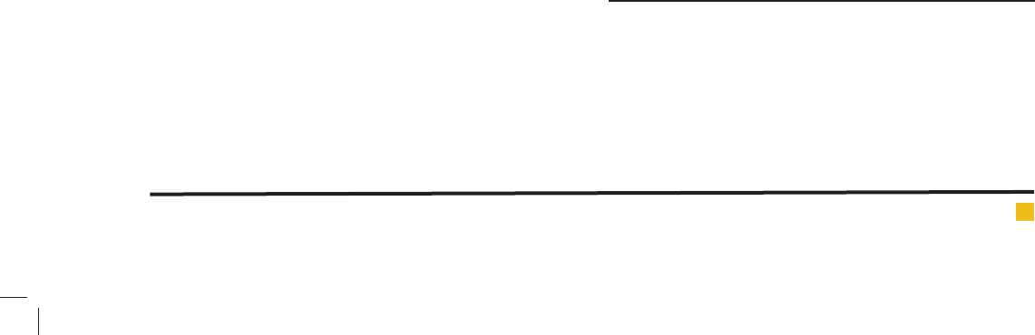
tion of AH-26 sealer( gure1) with rst section of sealer
bioceramic( gure4), second section of AH-26 ( gure2)
Torabizadeh Seyed Mohammad et al.
216 EXPERIMENTAL ASSESSMENT OF SEALER BIO CERAMIC WALL MATCHING IN FIRST AND THIRD APICAL AREAS BIOSCIENCE BIOTECHNOLOGY RESEARCH COMMUNICATIONS
with second section of bioceramic ( gure5) and third
section of AH-26 sealer ( gure3) with third section of
sealer bioceramic( gure6) (p>0/05).
In AH-26 sealer, third section signi cantly enjoys
more microscopic sapce level than rst section (p=0/001)
and second section (p=0/004) so it has less wall match-
ing. Thus rst section had signi cantly better seal and
wall matching than than third section. Second section
has signi cantly better seal and wall matching than
third section.
Cross sections: three millimeters at the end of channel
apical in AH26 sealer group from left to right includes
gure one to gure three
Figure one: rst one millimeter gure two: second
one millimeter, gure three third one millimeter
Cross sections: three millimeters at the end of chan-
nel apical in bioceramic sealer group from left to right
includes gure four to gure six
Figure four: rst one millimeter, gure ve: second
one millimeter, gure six third one millimeter
In sealer bioceramic, third section signi cantly enjoys
more microscopic space than rst section (p=0/04) so
there is less wall matching. And rst section has signi -
cantly better seal and wall matching than third section.
Third section of sealer bioceramic has signi cantly
microscopic space and less wall matching than rst sec-
tion (p=0/012) and second AH-26 sealer (P=0/29). In addi-
tion, third section of AH-26 sealer has signi cantly more
microscopic space and less wall matching than rst sec-
tion (p=0/006) and second sealer bioceramic (p=0/039).
DISCUSSION
Sealing tooth channel has main importance for preven-
tion from micro leakage and penetration bakeries into
pre apical and it is very crucial for determining prediog-
nosis of root treatment. Coronal micro leakage is consid-
ered as a vital factor in failure of root treatments. Main
factor for avoiding microleakage is applying sealers
and elimination of smear layer. For this reason, clinical
assessment of different kinds of sealers ability against
coronal penetration of bacteria’s and effect of cleaner
solvents sounds rational for puri cation of smear layer
(Farhad et al. 2007). Using sealers with appropriate
properties including connection, matchless, and tubule
penetration has two positive consequences: First, cre-
ating seal in channel due to higher interface f sealer
with crown wall; second burial of remained batteries in
crown tubules which in fact it is anti-bacterial effect of
sealers (Mohammadian et al. 2017).
Aim of this study was to investigate experimen-
tal assessment of sealer bioceramic wall matching in
1th and third apical area by Scanning Electron Micro-
scope (SEM). Gutta percha is in connection with sealers
and it is most common applied ller in root channel
(Mohammadian et al. 2017). So in this study, in order
FIGURE 1. FIGURE 2. FIGURE 3.
FIGURE 4. FIGURE 5. FIGURE 6.
Torabizadeh Seyed Mohammad et al.
BIOSCIENCE BIOTECHNOLOGY RESEARCH COMMUNICATIONS EXPERIMENTAL ASSESSMENT OF SEALER BIO CERAMIC WALL MATCHING IN FIRST AND THIRD APICAL AREAS 217
to ll root channel, gutta percha has been used.In this
study, cold lateral compaction method was used because
this method is the most common place method for obu-
ration of channel. This method is able to be used in most
clinical conditions and it provides controlling during
obturation (Cailleteau and Mullaney 1997; Anantula
and Ganta 2011; Kumar et al. 2012). In addition, cold
lateral compaction method is golden standard in endo
dentic (Anantula and Ganta 2011; Kumar et al. 2012).
Existence and nonexistence of smear layer has main
role in apical seal created by different method of obtura-
tion. Studies indicated that smear layer can act as a way
for leakage of micro-organism and it can be as a source
for growth and activities of different bacteria’s in crown
tubules (Kumar et al. 2012). Hence, in this study root
with 5/25% sodium hypoclorit solvent and 17% EDTA
were disinfected for elimination of smear layer.
In this study for assessing wall matching, SEM was
used. Because it has higher accuracy and ability of bet-
ter magni cation in interface. SEM uses electromagents
insteade of lenzes so it provides chance of more con-
trol on level of magni cation for the reseearche and at
results images are provided more transparent (Punitha P
and Shashikala K. 2011).
Results indicated that considering wall matching,
there is no signi cant relation between similar section of
two sealer section namely rst section of AH-26 sealer
with rst section of sealer bioseramic, second section
of AH-26 sealer with second section of bioceramic and
between third sections of AH-26 sealer with third sec-
tion of sealer bioceramic while wall matching of both
sealer is similar. In sealer bioceramic rst secton has bet-
ter seal and wall matching than third section. Produc-
ers claim that hydrophilic characteristic of endodentic
points of polymeric use remained humidity and form
self-sealing on setting radially and without develop-
ing in axial orientation. While alkaline nature of most
bioceramic products change crown collagen bers and
facilitates sealers penetration to crown tubules.
In this study, it was observed that penetration of sealer
bioceramic Total Fill BC is similar to resin sealer AH-26.
Mohammadian et al (2017) reported that in root apical
area ,considering sealer interface with crown, there is no
signi cant difference between two sealer bioceramic BC
and resine sealer AH-plus.
In another study, by Pawar et al (2014) have indicated
that sealer bioceramic endosequence and resin sealer
resilon epiphany enjoys better apical seal in compare
to AH-Plus resin sealer. However in this study in order
to measure seal they used color penetration method.In
study of SEM by Polineni et al (2016) considering wall
matching MM-Seal resin sealer has the least gap level byt
there was no difference between MM-Seal resin sealer
with Endosequence sealer bioceramic.Interface of sealer-
crown is a crucial area in sealed root channels. Sealers
with epoxy resin and bioceramic are not shrinked during
setting and it can be the reason of appropriate matching
in gap between crown and sealer in compare to sealers
based on eugenol,(Mohammadian et al. 2017).
In this study in AH-26 sealer third section signi -
cantly enjoyes more microscopical space than rst sec-
tion (p-0/001) and second (p-0/004) so has less wall
matching. Thus, rst section has better wall matching
than third section. Second section has signifcantly bet-
eer seal and wall matching than third section. In sealer
bioceramic, third section signi cantly has more micro-
scopic space level than rst section (p-0/04) so has less
wall matching. Hence, rst section signi cantly has bet-
ter seal and wall matching than third section.
Results ilusterated that third section of Total Fill
BC bioceramic sealer has signi cantly more microsco-
pial space and less wall matching than rst and second
sections of AH-26 sealer. In addition, third section of
AH-26 sealer has signi cantly more microscopial space
and less wall matching than rst and second sections
of Total Fill BC bioceramic sealer. These results can be
due to difference of different sections of root in apical
area. Polineni et al (2016), using SEM, coronal sections
showed that it enjoys more wall matching as compared
to apical sections.
This difference between coronal and apical level can
be due to less compaction and more diameter of crown
tubule in apical level which cause less perpetration of
sealer. Moreover, elimination of smear later in one third
apical area is problematic and it may act as physical
obstacle which is interference of sealer to root channel
crown (Polineni et al. 2016).
Generally, it must be considered that leakage totally
cannot be eliminated from treated root channels; lateral
and additional channels and other anatomic differences
along with periapical pressure have main role as pro-
gressive factor.
Considering that the areas which are commonly unaf-
fected by instrumentation and irrigation during root
canal preparation, where a more viscous material like
sealer can’t reach, can open up spaces for leakage and
decrease the chances of success.
CONCLUSION
Results indicated that there is no signi cant differ-
ence between similar sections of both sealer it means
that considering wall matching, rst section of AH-26
sealer with rst section of bioceramic sealer Total Fill
BC, second section of AH-26 sealer with second section
of sealer bioceramic sealer Total Fill BC and third section
of AH-26 sealer with third section of sealer bioceramic

Torabizadeh Seyed Mohammad et al.
218 EXPERIMENTAL ASSESSMENT OF SEALER BIO CERAMIC WALL MATCHING IN FIRST AND THIRD APICAL AREAS BIOSCIENCE BIOTECHNOLOGY RESEARCH COMMUNICATIONS
total Fill BC. In sealer bioceramic total Fill BC rst sec-
tion has signi cantly better seal and wall matching than
third section.
REFERENCES
Ahuja L, Jasuja P, Verma KG, Juneja S, Mathur A, Walia R.
(2016). A Comparative Evaluation of Sealing Ability of New
MTA Based Sealers with Conventional Resin Based Sealer: An
In-vitro Study. J Clin Diagn Res. 10 (7): Zc76-9.
Al-Haddad A, Che Ab Aziz ZA. (2016). Bioceramic-Based Root
Canal Sealers: A Review. Int J Biomater. 9753210.
Anantula K, Ganta AK. (2011). Evaluation and comparison of
sealing ability of three different obturation techniques- Lateral
compaction, Obtura II, and GuttaFlow: An in vitro study. J
Conserv Dent. 14 (1): 57-61.
Cailleteau JG, Mullaney TP. (1997). Prevalence of teaching api-
cal patency and various instrumentation and obturation tech-
niques in United States dental schools. J Endod. 23 (6): 394-6.
De Moor RJ, De Bruyne MA. (2004). The long-term sealing
ability of AH 26 and AH plus used with three gutta-percha
obturation techniques. Quintessence Int. 35 (4): 326-31.
Delivanis PD, Mattison GD, Mendel RW. (1983). The survivabil-
ity of F43 strain of Streptococcus sanguis in root canals lled
with gutta-percha and Procosol cement. J Endod. 9 (10): 407-10.
Farhad A, Havaie A, Barekateyn B, Narimani T. (2007). Com-
paring the bacterial leakage in endodontic therapy following
using EDTA as a irrigation and AH26 or tubliseal as selars.
Journal of Mashhad Dental School. 31 (1 & 2): 83-92.
Ghorbanzadeh A, Shokouhinejad N, Fathi B, Raoof M, Kho-
shkhounejad M. (2014). An In Vitro Comparison of Marginal
Adaptation of MTA and MTA-Like Materials in the Presence of
PBS at One-Week and Two-Month Intervals. J Dent (Tehran).
11 (5): 560-8.
Grossman LLI, Oliet S, Del Rio CE. (1988). Endodontic practice.
11 ed.: Lea & Febiger; p: 290-291.
Heard F, Walton RE. (1997). Scanning electron microscope
study comparing four root canal preparation techniques in
small curved canals. Int Endod J. 30 (5): 323-31.
Ingle JI, Beveridge E, Glick D, Weichmany J. (1994). The Wash-
ington study. In: Taintor G, editor. Endodontics. 280-281 ed.
Philadelphia: Lea & Febiger; p. 1-53.
Jitaru S, Hodisan I, Timis L, Lucian A, Bud M. (2016). The use
of bioceramics in endodontics - literature review. Clujul Med.
89 (4): 470-3.
Kakehashi S, Stanley H, Fitzgerald R. (1965). The effects of sur-
gical exposures of dental pulps in germ-free and conventional
laboratory rats. Oral Surg Oral Med Oral Pathol 20 (3): 340-9.
Kumar NS, Prabu PS, Prabu N, Rathinasamy S. (2012). Sealing
ability of lateral compaction, thermoplasticized gutta-percha
and owable gutta-percha obturation techniques: A compara-
tive in vitro study. J Pharm Bioallied Sci. 4 (Suppl 2): S131-5.
Mohammadian F, Farahanimastary F, Dibaji F, Kharazifard
MJ. (2017). Scanning Electron Microscopic Evaluation of the
Sealer-Dentine Interface of Three Sealers. Iran Endod J. 12 (1):
38-42.
Oltra E, Cox TC, LaCourse MR, Johnson JD, Paranjpe A. (2016).
Retreatability of two endodontic sealers, EndoSequence BC
Sealer and AH Plus: a microcomputed tomographic compari-
son. RDE. 1-8.
Pawar SS, Pujar MA, Makandar SD. (2014). Evaluation of the
apical sealing ability of bioceramic sealer, AH plus & epiph-
any: An in vitro study. J Conserv Dent. 17 (6): 579-82.
Polineni S, Bolla N, Mandava P, Vemuri S, Mallela M, Gan-
dham VM. (2016). Marginal adaptation of newer root canal
sealers to dentin: A SEM study. J Conserv Dent. 19 (4):
360-3.
Punitha P, Shashikala K. (2011). Evaluation of the Adaptation
of Resin Based Sealers Epiphany, AH plus and AH 26 to the
Root Canal Dentin by Scanning Electron Microscope. Indian
Journal of Stomatology. 2 (4).
Setia P, Sikri V, Sroa R, Sidhu B. (2014). Apical sealing ability
of two novel root canal sealers: An ex-vivo study. J Int Clin
Dent Res Organ. 5 (1): 9-13.
Shokouhinejad N, Nekoofar MH, Ashoftehyazdi K, Zahraee S,
Khoshkhounejad M. (2014). Marginal adaptation of new bioce-
ramic materials and mineral trioxide aggregate: a scanning
electron microscopy study. Iran Endod J. 9 (2): 144-8.
Silva Almeida LH, Moraes RR, Morgental RD, Pappen FG.
(2017). Are Premixed Calcium Silicate-based Endodontic Seal-
ers Comparable to Conventional Materials? A Systematic
Review of in Vitro Studies. J Endod. 23-145
Savadkouhi T. S, Fazlyab M. (2016). Discoloration Potential of
Endodontic Sealers: A Brief Review. Iran Endod J. 4 (250-254).
Utneja S, Nawal RR, Talwar S, Verma M. (2015). Current per-
spectives of bio-ceramic technology in endodontics: calcium
enriched mixture cement - review of its composition, proper-
ties and applications. Restor Dent Endod. 40 (1): 1-13.
Vertucci FJ. (1984). Root canal anatomy of the human per-
manent teeth. Oral Surg Oral Med Oral Pathol. 58 (5): 589-99.