
Medical
Communication
Biosci. Biotech. Res. Comm. 10(1): 56-62 (2017)
Low levels of ionizing radiation exposure and
cytogenetic effects in radiopharmacists
T Soltanpour Gharibdousty
1
, F Zakeri
2,3
, V Changizi
1
*, MR Rajabpour
3
and MR Farshidpour
3
1
Department of Technology of Radiology and Radiotherapy, Allied Medical Sciences School, Tehran University
of Medical Sciences, Tehran, Iran
2
Nuclear Science and Technology Research Institute, Tehran, Iran
3
Iran Nuclear Regulatory Authority, Tehran, Iran
ABSTRACT
Radiopharmaceuticals are unique medicinal formulations containing radioisotopes which are used in major clini-
cal areas for diagnosis and/or therapy. The aim of this study was to assess occupationally induced DNA damage in
workers of a radiopharmaceutical facility who are at risk of exposure to low levels of external ionizing radiation
and internal contamination. Cytokinesis-block micronucleus (CBMN) assay used as an important biological indicator
of ionizing radiation exposure in the peripheral blood lymphocytes of 35 exposed workers compared with 35 con-
trols matched for gender, age and smoking habits. Occupational dosimetry records were collected over the last year
(ranged from 1.05 to 30.73 mSv) and last 5 years exposure (ranged from 2.56 to 70.24 mSv). The results showed a
2.5 fold increase in the chromosomal damage leading to micronucleated lymphocytes in the workers of the radiop-
harmaceutical facility compared to the controls (25.82±8.67 vs. 10.52±6.83 micronuclei per 1000 binucleated cells,
p < 0.0005). The nuclear division index as a parameter of cytostasis, in the workers was signi cantly lower than
that in the controls. The mean frequency of nucleoplasmic bridges was higher in the radiation workers compared to
the controls without statistical signi cant difference (p > 0.05). Taking all the confounding factors into account, no
obvious trend of increased micronuclei as a function of either duration of employment, exposed dose, smoking or
age was observed. The present study showed that occupationally exposed individuals have higher frequencies of DNA
damage, despite the very low levels of ionizing radiation exposure.
KEY WORDS: OCCUPATIONAL EXPOSURE, RADIOPHARMACIST, MICRONUCLEI, HUMAN LYMPHOCYTE
56
ARTICLE INFORMATION:
*Corresponding Author: changizi@sina.tums.ac.ir
Received 12
th
Jan, 2017
Accepted after revision 22
nd
March, 2017
BBRC Print ISSN: 0974-6455
Online ISSN: 2321-4007 CODEN: USA BBRCBA
Thomson Reuters ISI ESC and Crossref Indexed Journal
NAAS Journal Score 2017: 4.31 Cosmos IF : 4.006
© A Society of Science and Nature Publication, 2017. All rights
reserved.
Online Contents Available at: http//www.bbrc.in/

T Soltanpour Gharibdousty et al.
INTRODUCTION
The development of nuclear energy and the growing use
of ionizing radiation in medical practice have created
deep concern regarding the long-term effects of low-dose
radiation on humans (Mozdarani and Samavat, 1996).
Positron emission tomography (PET) is an indispensable
imaging modality for the evaluation and staging of can-
cer patients by using radiotracers or radiopharmaceuti-
cals, labeled with suitable positron emitting radioisotopes
and necessitate special consideration in manufacturing
and working knowledge of radiation safety practices is
also essential. Workers in radiopharmaceutical facilities
are involved in different sections of Radionuclide produc-
tion and Radiopharmaceutical preparation and are at risk
of external radiation exposure and internal contamina-
tion during their work, (IAEA, 2012).
Generally, monitoring of individuals occupationally
exposed to ionizing radiation consists of regular lm
dosimetric control and periodic health examination
(Zakeri et al., 2003) However, personal dosimetry may
underestimate the real exposure, not only because of the
detection threshold of dosimeters but also because of
improper wearing (Zakeri and Hirobe, 2010, Sari-Min-
odier et al., 2007). Cytogenetic studies as speci c bio-
logical parameters provide additional information which
complements physical dosimetry and enables better
evaluation of radiation exposure.The cytokinesis-block
micronucleus (CBMN) assay are widely used as end
points in testing for mutagens and carcinogens (Pres-
ton, 1984, Sorsa, 1990) and micronuclei (MN) induction
re ects clastogenic and/or aneugenic events (Sari-Min-
odier et al., 2007, Zakeri and Hirobe, 2010).
MN observed in peripheral blood lymphocytes (PBL),
in the rst interphase after cell division, are small extra-
nuclear bodies resulting from chromosome breaks or
whole chromosomes lagging behind during anaphase.
These cells can be identi ed as binucleated (BN) cells by
addition of the cytoplasmic division inhibitor cytocha-
lasin B during cell culture. Ionizing radiation is a strong
clastogenic agent, and thus a potent inducer of MN (Vral
et al., 2011).
The CBMN assay has been extensively used and vali-
dated as an appropriate bio dosimetry tool to evaluate
in vivo radiation exposure of occupational, medical and
accidentally exposed individuals and to assess in vitro
radiosensitivity and cancer susceptibility (Sari-Minodier
et al., 2007, Dias et al., 2007, Thierens and Vral, 2009).
Many studies have shown that the number of radiation-
induced MN strongly correlated with dose and quality of
radiation. Most studies reported that MN in lymphocytes
was more frequent in radiation workers than in the con-
trols (Bonassi et al., 1997, Chung et al., 1996 and Vral
et al., 2011).
In this study we examined the frequency of MN in
35 radiopharmacists occupationally exposed to ioniz-
ing radiation and 35 sex- and age-matched controls.
This is important as signi cantly elevated levels of such
aberrations may be found in peripheral blood lympho-
cytes of radiation workers who are exposed within the
occupational limits recommended by the International
Commission on Radiation Protection (ICRP and 1991.).
Parameters of cell cycle progression, other biomarkers of
DNA damage such as nucleoplasmic bridges and effects
of confounding factors on MN induction have been also
investigated.
MATERIAL AND METHODS
STUDY POPULATION
All the study population was male. The exposed group
comprised 35 radiopharmacists involved in prepara-
tion and production of radiopharmaceuticals. Their age
ranged from 27 to 55 years (mean of 37.59±7.22 years)
with mean employment time of 14.35±8.71 years. The
test group was routinely monitored with lm badges
every 2 months. Their exposure ranged between 1.05
and 30.73 mSv (average of 6.59±5.83 mSv/y) during the
previous year and from 2.56 to 70.24 mSv with a mean
value of 17.89±15.04 mSv during the last 5 years.
The reference control group consisted of 35 individu-
als, who had never been occupationally exposed to ion-
izing radiation or to the known carcinogenic chemicals.
The controls matched the exposed subjects in age, sex
and smoking habits.
All subjects in this study were questioned in detail to
learn if they were systemically healthy. The question-
naire also included information about smoking habits,
medical history, drug intake and diagnostic medical irra-
diation for all individuals. For the exposed subjects, the
type, frequency and duration of occupational exposure
to ionizing radiation, radiation protection measures,
conditions of dosimeter wearing and work-related expo-
sure to other hazardous agents were questioned. All the
subjects were healthy individuals without current infec-
tions and medications in the last 6 months and no gen-
eral or dental X-ray in the last 6 months.
The study was approved by the national ethical com-
mittee. Informed consent was obtained from each person
and the study protocol conformed to the ethical guide-
lines of the World Medical Association (Declaration of
Helsinki).
Cytokinesis-block micronucleus (CBMN) assay
Peripheral blood samples (3ml) were collected into steri-
lized lithium heparin tubes by venipuncture from each
BIOSCIENCE BIOTECHNOLOGY RESEARCH COMMUNICATIONS LOW LEVELS OF IONIZING RADIATION EXPOSURE AND CYTOGENETIC EFFECTS 57
T Soltanpour Gharibdousty et al.
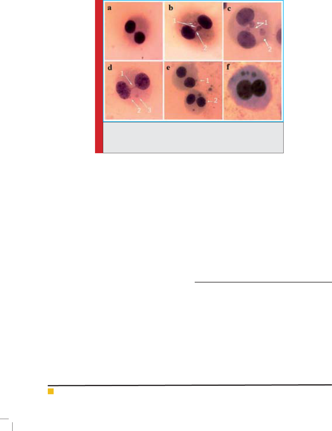
FIGURE 1. Various binucleated cells (BN) observed in the cytokinesis-block
micronucleus assay; a) a normal BN, b) a BN with a micronucleous (MN) and a
nuclear bud, c) a BN with 2 MN and one nuclear bud, d) a BN with 2 MN and
one nucleoplasmic bridge, e)Two BN with one and 2 MN, f) a BN with 3 MN.
donor and all samples were coded. Whole blood cultures
were created by mixing 0.5 ml of whole blood with 4.5
ml of RPMI 1640 medium consisting of Ham’s F10 sup-
plemented with 20% fetal calf serum (FCS), 100 U/ml
penicillin, 100 mg/ml streptomycin, 1.0% l-glutamine
and 1.0% phytohemagglutinin (PHA) for mitogenic
stimulation (all materials purchased from Gibco). The
cultures were incubated at 37 0C for 72 h. Cytochalasin
B (6 mg/ml) was added 44 h after culture initiation. The
cells were collected by centrifugation, and treated with
a mild hypotonic solution containing 0.075 M KCl for 3
min. After centrifugation and removal of the superna-
tant, the cells were xed with a fresh mixture of metha-
nol: acetic acid (3:1). Centrifugation and re-suspension
were carried out three times and the cells were then
dropped onto clean slides for detection of micronuclei
by conventional staining with 5% Giemsa (Zakeri and
Hirobe, 2010).
MN was scored in binucleated cytokinesis-blocked
cells using 400 X magni cation. The frequencies of
MN and nucleoplasmic bridges (NPBs) were determined
at the same time in binucleated cells according to the
standard criteria described by Fenech (Fenech, 1993)
were followed for identi cation of binucleated lympho-
cytes and MN. Figure 1 shows the various DNA damages
observed in the cytokinesis-block micronucleus assay.
The One thousand binucleated cells were examined for
each sample. Furthermore, Cell cycle parameters were
evaluated by classifying 1000 cells according to the
number of nuclei. The nuclear division index (NDI) was
calculated following the formula: NDI = (M1 + 2M2 +
3M3 + 4M4) /n, where M1–M4 indicate the number of
cells with 1–4 nuclei and n indicates the total number of
scored cells (Rastkhah et al., 2016)ÿÿÿÿÿþ»n4ÿÿ.
STATISTICAL EVALUATION
The frequency of MN and NDI value in the lymphocytes
of the workers and controls were compared using Stu-
dent’s t test. The in uence of age, duration of employ-
ment and physical doses was tested by linear regression
analysis and correlation test using SPSS, version 21 sta-
tistical program. The level of statistical signi cance was
set at p<0.05.
RESULTS
Demographic characteristics of control and exposed
workers (r adiopharmacists) are presented in Table
1. There was no difference in age and smoking sta-
tus distributions between control and exposed group
(Table 1).
Results of the MN, NPB and NDI frequencies have
been shown in Table 2. The mean frequency of MN/1000
binucleated cells of radiation workers were signi -
cantly higher than in the control group (25.82±8.67
vs. 10.52±6.83, p <0.0005, student’s t-test). The mean
frequency of NPBs was higher in the radiation work-
ers compared to the controls but there was no statisti-
cally signi cant difference (p > 0.05). The mean value
58 LOW LEVELS OF IONIZING RADIATION EXPOSURE AND CYTOGENETIC EFFECTS BIOSCIENCE BIOTECHNOLOGY RESEARCH COMMUNICATIONS

T Soltanpour Gharibdousty et al.
Table 1. Demographic characteristics of the radiopharmacists and the control group. Data are
expressed as mean values with standard deviation.
Groups Radiopharmacists Controls p value
Mean age, years± S.D.
(range)
(no.)
37.59±7.22
(27-55)
(35)
36.98±7.44
(25-55)
(35)
0.59
Smoking habits, no. (%)
Yes
14(40.0%) 10(28.75%) 0.32
No 21(60.0%) 25(71.25%)
Mean time exposure, years± S.D. 14.35±8.71 - -
Mean last year exposure mSv± SD
(range)
6.59±5.83
(1.05 – 30.73)
--
Mean last ve years exposure mSv± SD
(range)
17.89±15.04
(2.56 -70.24)
--
S.D: standard deviation
Table 2. Frequency of MN and NDI in lymphocytes of the radiopharmacists and the
control group
Groups Radiopharmacists Control p value
Mean MN frequency(‰) ± SD
(range)
(no.)
25.82±8.67
(11-48)
(35)
10.52±6.83
(2-23)
(35)
<0.0005a
Mean NDI frequency(‰) ± SD
(no.)
1.76 ±.02
(35)
1.84±.03
(35)
<0.0005a
Mean NPB frequency(‰) ± SD 1.02±0.47 0.85±0.37 NS
Mean MN frequency(‰) ± SD
Smokers (no.)
Non-smokers (no.)
27.46±9.01(14)
24.13±7.54(21)
p=0.35
11.51±6.89 (10)
9.54± 4.21(25)
p=0.44
MN: micronuclei, NDI: Nuclear division index, NPB: Nucleoplasmic bridge
a :obtained by Student ’s t-test
S.D: standard deviation
NS: not signi cant
of NDI; a parameter of cytostasis, for radiation workers
and controls were 1.76±.02 and 1.84±.03, respectively.
Although both are in the normal range, however, the
NDI frequency in the control group was signi cantly
higher than that in the workers (p < 0.0005, student’s
t-test).
The regression analysis showed no signi cant cor-
relation emerged between age (=0.06, p=0.7), time
exposure (=0.02, p=0.9), dose in the last year (=0.01,
p=0.9) and dose in the last 5 years (=0.10, p=0.5) with
the frequencies of the MN/1000 BN cells in the exposed
workers. There were no signi cant differences between
the mean MN/1000 BN cells in the smoker and non-
smoker exposed workers (26.12± 10.32 vs., 22.0± 6.22,
p=0.20) and between smoker and non-smoker controls
(11.31± 7.51 vs., 9.70± 4.20, p=0.48) analyzed by Stu-
dent’s t-test.
DISCUSSION
In the present study, the CBMN assay was used to evalu-
ate chromosomal damage in peripheral blood lympho-
cytes of 35 radiopharmacists working in a radiophar-
maceutical facility involving in the preparation and
production of radionuclides and radiopharmaceuti-
cals in comparison with their sex- and age-matched
controls.
Although the level of radiation dose received occu-
pationally by workers was mostly below the accepted
annual limit of 20 mSv/year, the frequencies of MN were
signi cantly higher in the workers than in the controls
(25.82±8.67 vs. 10.52±6.83 per 1000 binucleated cells,
p <0.0005). A relatively high frequency of MN forma-
tion in lymphocytes of radiation workers compared with
non-exposed individuals might be due to an accumula-
BIOSCIENCE BIOTECHNOLOGY RESEARCH COMMUNICATIONS LOW LEVELS OF IONIZING RADIATION EXPOSURE AND CYTOGENETIC EFFECTS 59

T Soltanpour Gharibdousty et al.
tion of initial DNA damage in people exposed to chronic
doses of radiation.
Many studies reported signi cantly higher MN rates
in exposed population than in controls (Sari-Minodier
et al., 2007, Zakeri and Hirobe, 2010). Maluf et al (Maluf
et al., 2001) also reported increased frequency of MN and
dicentric bridges in the lymphocytes of medical workers
exposed to X-rays. These results are also in agreement
with those who showed a signi cant higher MN val-
ues in interventional cardiologists when compared with
clinical cardiologists working outside the catheterization
laboratory (Andreassi et al., 2005).
Indeed age, gender and smoking habit are the most
important demographic variables impacting on the MN
index (Fenech, 1998). The age-related decline in the ef -
ciency of the repair processes and the accumulation of
mutations may lead to increased levels of DNA damage,
which is observed by a higher frequency of chromo-
somal aberrations (Wojda et al., 2007).
In addition to age, gender-related changes in the level
of MN frequency have been observed in many studies.
Effect of gender on MN formation has been described for
X chromosome micronucleation as being prevalent in
females (Mateuca et al., 2006). According to Fenech et al
(Fenech et al., 1994), an additional mechanism, possibly
the loss of the X chromosome, accounts for the higher
MN frequency in female. However, since the surveyed
groups in this study were all men and the mean age was
relatively the same in both groups, these effects have not
been observed.
Although cytokinetic and cytostatic effects have been
detected in heavy and moderate smokers (Calderon-
Ezquerro et al., 2007), smoking habits may or may not
affect the genotoxic effect of chronic radiation exposure
(Maffei et al., 2002, Sari-Minodier et al., 2007, Hadjide-
kova et al., 2003). Bonassi et al (Bonassi et al., 2003)
evaluated the impact of smoking on MN frequency using
pooled re-analysis of 24 databases from the Human pro-
ject. Our study indicated that smokers do not experience
an overall increase in the MN frequency compared with
non-smokers.
In this study, as reported by others, no association
between chromosome damage and absorbed dose was
found. The lack of a dose–effect relationship between
chromosome damage and chronic exposure to low lev-
els of ionizing radiation could be attributed to various
factors, which can be explained by the fact that during
chronic exposure part of the chromosomal damage can
be eliminated in vivo by the death of lymphocytes. This
can be attributed to proliferation rate of lymphocytes,
inter-individual variation, elimination of aberrant cells,
and lifespan of lymphocytes (Kubelka et al., 1992).
In this study the frequencies of NPBs were higher in
the exposed workers than in the controls without sta-
tistically signi cant difference (p > 0.05). NPBs origi-
nate from dicentric chromosomes that may be caused by
misrepair of double strand DNA breaks or telomere end
fusions. NPBs occur when centromeres of dicentric chro-
mosomes are pulled to opposite poles of the cell ana-
phase (Fenech, 2007). However, previous studies have
shown that NPB is not radiation speci c because it can
also be induced by reactive oxygen species (Umegaki
and Fenech, 2000).
NDI may be used to de ne cell cycle progression of
the lymphocytes after mitogenic stimulation and it is a
useful research tool for understanding the cell cycling
kinetics of the cultures specially after radiation expo-
sure (Rastkhah et al., 2016)ÿÿÿÿÿ-Fm4ÿÿ. Our results
showed a signi cant decrease in the NDI values of radi-
ation workers compared with non-exposed individuals
(p> 0.0005), that could be due to the chronic low dose
exposure. Signi cant decrease in the pooled NDI values
with increasing radiation doses was also observed by
other studies (Antunes et al., 2014, Pejchal et al., 2011,
IAEA, 2001.).
The increased micronuclei frequencies found in
exposed workers indicate potential genetic hazards that
may play critical role in radiation-induced carcinogene-
sis and genetic diseases in long term (Zakeri and Hirobe,
2010). It has been reported that the processes of pre-
paring and dispensing therapeutic radiopharmaceuticals
have a greater potential to expose operators to radia-
tion than do procedures for diagnosis. These operations
should therefore be performed in a controlled area with
entry restricted to essential staff only and careful con-
sideration should be given to the amount of shielding
required and to the measures to be taken to avoid radia-
tion exposure from internal contamination (AGENCY,
2011).
In radiopharmaceutical facilities, the main precau-
tions required in dealing with external irradiation will
depend on the physical characteristics of the radiation
emitted, the total activity and the physical half-life of
the radionuclide. Contamination from unsealed radioac-
tive substances may produce a further external radia-
tion hazard (Radiation and Protection, 2008).Now, it is
becoming increasingly clear that long-term radiation-
induced cancer risk can be dramatically minimized by
effective implementation of good practice of radiation
protection in radiopharmaceutical facilities and ‘respon-
sibility is on all workers and professional staff to mini-
mize the radiation exposure to themselves (Shadley
et al., 1987).
Therefore, our ndings suggest that radiation work-
ers should be aware of the necessity of applying radia-
tion protection principles (justi cation, optimization
and limitation) and con rm that the regulatory body
shall establish and enforce requirements for the protec-
60 LOW LEVELS OF IONIZING RADIATION EXPOSURE AND CYTOGENETIC EFFECTS BIOSCIENCE BIOTECHNOLOGY RESEARCH COMMUNICATIONS

T Soltanpour Gharibdousty et al.
tion of workers in existing exposure situations and sup-
port services such as education and training, technical
services, regular maintenance of equipment and quality
control program. Furthermore, evaluation and follow-up
of occupational doses should be considered as an impor-
tant part of quality assurance programs. In this regard,
cytogenetic monitoring, specially by using CBMN assay
is a valuable tool versus fIlm dosimetry following low-
dose radiation exposure and for risk assessment of per-
sonnel believed to be exposed to radiation.
ACKNOWLEDGMENT
This study has been supported by Tehran University of
Medical Sciences. Grant number:27610
REFERENCES
Agency, I. A. E. 2011. Radiation Protection And Safety Of Radi-
ation Sources: International Basic Safety Standards, Vienna,
International Atomic Energy Agency.
Andreassi, M. G., Cioppa, A., Botto, N., Joksic, G., Manfredi, S,
Federici, C, Ostojic, M., Rubino,P. & Picano, E. (2005) Somatic
DNA Damage In Interventional Cardiologists: A Case-Control
Study. Faseb J, 19, 998-9.
Antunes, A. C., Martins, V., Cardoso, J., Santos, L. & Mon-
teiro Gil, O. (2014). The Cytokinesis-Blocked Micronucleus
Assay: Dose Estimation And Inter-Individual Differences In
The Response To Gamma-Radiation. Mutat Res Genet Toxicol
Environ Mutagen, 760, 17-22.
Bonassi, S., Forni, A., Bigatti, P., Canevarollo, N., De Ferrari,
M., Lando, C., Padovani, P., Bevegni, M., Stella, M., Vecchio,
D. & Puntoni, R. (1997). Chromosome Aberrations In Hospital
Workers: Evidence From Surveillance Studies In Italy (1963-
1993). Am J Ind Med, 31, 353-60.
Bonassi, S., Neri, M., Lando, C., Ceppi, M., Lin, Y. P., Chang,
W. P., Holland, N., Kirsch-Volders, M., Zeiger, E. & Fenech, M.
(2003). Effect Of Smoking Habit On The Frequency Of Micro-
nuclei In Human Lymphocytes: Results From The Human
Micronucleus Project. Mutat Res, 543, 155-66.
Calderon-Ezquerro, C., Sanchez-Reyes, A., Sansores, R. H., Vil-
lalobos-Pietrini, R., Amador-Munoz, O., Guerrero-Guerra, C.,
Calderon-Segura, M. E., Uribe-Hernandez, R. & Gomez-Arroyo,
S. (2007). Cell Proliferation Kinetics And Genotoxicity In Lym-
phocytes Of Smokers Living In Mexico City. Hum Exp Toxicol,
26, 715-22.
Chung, H. W., Ryu, E. K., Kim, Y. J. & Ha, S. W. (1996). Chro-
mosome Aberrations In Workers Of Nuclear-Power Plants.
Mutat Res, 350, 307-14.
Dias, F. L., Antunes, L. M., Rezende, P. A., Carvalho, F. E., Silva,
C. M., Matheus, J. M., Oliveira, J. V., Jr., Lopes, G. P., Pereira,
G. A. & Balarin, M. A. (2007) Cytogenetic Analysis In Lympho-
cytes From Workers Occupationally Exposed To Low Levels Of
Ionizing Radiation. Environ Toxicol Pharmacol, 23, 228-33.
Fenech, M. (1993). The Cytokinesis-Block Micronucleus Tech-
nique And Its Application To Genotoxicity Studies In Human
Populations. Environ Health Perspect, 3, 101-7.
Fenech, M. (1998). Important Variables That In uence Base-
Line Micronucleus Frequency In Cytokinesis-Blocked Lympho-
cytes-A Biomarker For Dna Damage In Human Populations.
Mutat Res, 404, 155-65.
Fenech, M. (2007). Cytokinesis-Block Micronucleus Cytome
Assay. Nat Protoc, 2, 1084-104.
Fenech, M., Neville, S. & Rinaldi, J. (1994). Sex Is An Impor-
tant Variable Affecting Spontaneous Micronucleus Fre-
quency In Cytokinesis-Blocked Lymphocytes. Mutat Res, 313,
203-7.
Hadjidekova, V. B., Bulanova, M., Bonassi, S. & Neri, M. (2003).
Micronucleus Frequency Is Increased In Peripheral Blood Lym-
phocytes Of Nuclear Power Plant Workers. Radiat Res, 160,
684-90.
IAEA (2001). Cytogenetic Analysis For Radiation Dose Assess-
ment, Technical Report Series 405; , Vienna, .
IAEA (2012). Radioisotopes And Radiopharmaceuticals Series
No. 3, Cyclotron Produced Radionuclides: Guidance On Facility
Design And Production Of Fluorodeoxyglucose (Fdg), Vienna,.
ICRP (1991). Recommendations Of The International Commis-
sion On Radiological Protection.Publication 60., Pergamon,
Oxford.
Kubelka, D., Garaj-Vrhovac, V. & Horvat, D. (1992). Chro-
mosomal Aberrations In Persons Occupationally Exposed To
Annual X-Irradiation Doses Lower Than 25 Msv. Journal Of
Radiological Protection, 12, 33.
Maffei, F., Angelini, S., Forti, G. C., Lodi, V., Violante, F. S.,
Mattioli, S. & Hrelia, P. (2002). Micronuclei Frequencies In
Hospital Workers Occupationally Exposed To Low Levels Of
Ionizing Radiation: In uence Of Smoking Status And Other
Factors. Mutagenesis, 17, 405-9.
Maluf, S. W., Passos, D. F., Bacelar, A., Speit, G. & Erdtmann, B.
(2001). Assessment Of Dna Damage In Lymphocytes Of Work-
ers Exposed To X-Radiation Using The Micronucleus Test And
The Comet Assay. Environ Mol Mutagen, 38, 311-5.
Mateuca, R., Lombaert, N., Aka, P. V., Decordier, I. & Kirsch-
Volders, M. (2006). Chromosomal Changes: Induction, Detec-
tion Methods And Applicability In Human Biomonitoring. Bio-
chimie, 88, 1515-31.
Mozdarani, H. & Samavat, H. (1996). Cytogenetic Biomonitor-
ing Of 65 Radiology Technologists Occupationally Exposed To
Chronic Doses Of X-Irradiation In Iran. Medical Journal Of The
Islamic Republic Of Iran, 1,
43-46.
Pejchal, J., Vasilieva, V., Hristozova, M., Vilasová, Z., Vávrová,
J., Alyakov, M., Tichý, A., Zárybnická, L., Šinkorová, Z., Tam-
bor, V., Kubelková, K. & Dresler, J.( 2011). Cytokinesis-Block
Micronucleus (Cbmn) Assay/Cbmn Cytome Assay In Human
Lymphocytes After In Vitro Irradiation And Its Use In Biodo-
simetry Mil. Med. Sci. Lett. (Voj. Zdrav. Listy), 80, 28-37.
Preston, R. (1984). Cytogenetic Abnormalities As An Indica-
tor Of Mutagenic Exposure. In: Ansari Aa, De Serres Fj (Eds.),
BIOSCIENCE BIOTECHNOLOGY RESEARCH COMMUNICATIONS LOW LEVELS OF IONIZING RADIATION EXPOSURE AND CYTOGENETIC EFFECTS 61

T Soltanpour Gharibdousty et al.
Single Cell Mutation Monitoring Systems, Methodologies And
Applications. New York:Plenum Press,.
Radiation & Protection (2008). Radiation Health & Safety
Advisory Council, No14.2 Series Publication.
Rastkhah, E., Zakeri, F., Ghoranneviss, M., Rajabpour, M. R.,
Farshidpour, M. R., Mianji, F. & Bayat, M. (2016). The Cytoki-
nesis-Blocked Micronucleus Assay: Dose-Response Calibration
Curve, Background Frequency In The Population And Dose
Estimation. Radiat Environ Biophys, 55, 41-51.
Sari-Minodier, I., Orsière, T., Auquier, P., Martin, F. & Botta, A.
(2007). Cytogenetic Monitoring By Use Of The Micronucleus
Assay Among Hospital Workers Exposed To Low Doses Of Ion-
izing Radiation. Mutation Research/Genetic Toxicology And
Environmental Mutagenesis, 629, 111-121.
Shadley, J. D., Afzal, V. & Wolff, S. (1987). Characterization Of
The Adaptive Response To Ionizing Radiation Induced By Low
Doses Of X Rays To Human Lymphocytes. Radiat Res, 111, 511-7.
Sorsa, M. (1990). Use Of Cytogenetics In Detection Of Human
Exposure To Mutagens And Carcinogens In The Work Place.
In: Sherma Ak (Ed.), Advance In Cytogenetics. Calcutta : Irl.
Thierens, H. & Vral, A. (2009). The Micronucleus Assay In
Radiation Accidents. Ann Ist Super Sanita, 45, 260-4.
Umegaki, K. & Fenech, M. (2000). Cytokinesis-Block Micro-
nucleus Assay In Wil2-Ns Cells: A Sensitive System To Detect
Chromosomal Damage Induced By Reactive Oxygen Species
And Activated Human Neutrophils. Mutagenesis, 15, 261-9.
Vral, A., Fenech, M. & Thierens, H. (2011). The Micronucleus
Assay As A Biological Dosimeter Of In Vivo Ionising Radiation
Exposure. Mutagenesis, 26, 11-7.
Wojda, A., Zietkiewicz, E. & Witt, M. (2007). Effects Of Age
And Gender On Micronucleus And Chromosome Nondis-
junction Frequencies In Centenarians And Younger Subjects.
Mutagenesis, 22, 195-200.
Zakeri, F., Assaei, R. & And Varzegar, R. (2003). Chromosomal
Aberrations In Workers Occupationally Exposed To Chronic
Low-Level Ionizing Radiation. Biomonitoring By Cytogenetic
Analysis Of Radiation-Exposed Workers 9, 33-38.
Zakeri, F. & Hirobe, T. (2010). A Cytogenetic Approach To The
Effects Of Low Levels Of Ionizing Radiations On Occupation-
ally Exposed Individuals. Eur J Radiol, 73, 191-5.
62 LOW LEVELS OF IONIZING RADIATION EXPOSURE AND CYTOGENETIC EFFECTS BIOSCIENCE BIOTECHNOLOGY RESEARCH COMMUNICATIONS