
Dental
Communication
Biosci. Biotech. Res. Comm. 10(3): 580-586 (2017)
Correlation of the maxillary sinus volume with gender
and some of craniofacial indices using cone beam
computed tomography
Shahab Etemadi
1
, Golnaz Seylavi*
2
and Afshin Yadegari
3
1
Assistant Professor, Department of Oral and Maxillofacial Radiology, Isfahan (Khorasgan) Branch, Islamic
Azad University, Isfahan, Iran
2
Post Graduate Student, Department of Oral and Maxillofacial Radiology, Isfahan (Khorasgan) branch,
Islamic Azad University, Isfahan, Iran
3
Assistant Professor, Department of Oral and Maxillofacial Surgery, Isfahan (Khorasgan) Branch, Islamic
Azad University, Isfahan, Iran
ABSTRACT
Maxillary sinus volume is variable and thus, is a topic of interest for surgeons performing endoscopic surgeries. Also,
it may be helpful for sexual identi cation. This study sought to measure the maxillary sinus volume using cone beam
computed tomography (CBCT) and assess its correlation with gender and some craniofacial indices. In this descriptive,
analytical, cross-sectional study, CBCT scans of 70 patients (35 males and 35 females) older than 18 years of age were
evaluated. To measure the maxillary sinus volume, axial CBCT sections with 2mm slice thickness and Digimizer software
were used. Sinus volume was calculated using the formula: volume=sum of the thickness of each slice multiplied by
the surface area of each slice. The mean width and height of the palate in the coronal plane, anterior-posterior length
of the palate in the sagittal plane and distance between the two zygomatic buttresses were measured on the axial CBCT
sections in both males and females. The Pearson’s correlation coef cient, independent t-test and paired t-test were used
for statistical analyses. The mean maxillary sinus volume was 15.9±6.05 cm3 and 13±2.85 cm3 in males and females,
respectively. The mean volume of the maxillary sinus was larger in males, and a signi cant correlation was noted
between the mean volume of the maxillary sinus and width and height of the palate as well as the distance between the
two zygomatic buttresses (P<0.05). Despite the larger volume of the maxillary sinus in males, this parameter cannot be
used for sexual identi cation because the area under the receiver operating curve (ROC) was 62.7%.
KEY WORDS: CONE-BEAM COMPUTED TOMOGRAPHY; SEXUAL IDENTIFICATION; MAXILLARY SINUS
580
ARTICLE INFORMATION:
*Corresponding Author:
Received 19
th
July, 2017
Accepted after revision 28
th
Sep, 2017
BBRC Print ISSN: 0974-6455
Online ISSN: 2321-4007 CODEN: USA BBRCBA
Thomson Reuters ISI ESC and Crossref Indexed Journal
NAAS Journal Score 2017: 4.31 Cosmos IF: 4.006
© A Society of Science and Nature Publication, 2017. All rights
reserved.
Online Contents Available at: http//www.bbrc.in/
DOI: 10.21786/bbrc/10.3/36

Etemadi, Seylavi and Yadegari
BIOSCIENCE BIOTECHNOLOGY RESEARCH COMMUNICATIONS CORRELATION OF THE MAXILLARY SINUS VOLUME WITH GENDER 581
INTRODUCTION
Maxillary sinus is an irregular cavity within the maxil-
lary bone, which develops under the orbit. Development
of the maxillary sinus starts in the third week of preg-
nancy and continues to the pubertal period (Wang etal.
1994). Accurate measurement of the maxillary sinus
volume may be helpful prior to sinus oor augmenta-
tion for implant placement (Gray etal.2000). Maxillary
sinus volume is variable and thus, is a topic of interest
for surgeons particularly the oral and maxillofacial sur-
geons (Fernandes, 2004). Recently, demand for sexual
identi cation in forensic medicine has increased due to
an increase in rate of crime (Saccucci etal.2015). The
correlation of volumetric analyses of the maxillary sinus
and sexual identi cation has been a controversial topic
(Ariji etal. 1994; Jasim etal. 2013; Vidya etal. 2013;
Ekizoglu etal.2014; Möhlhenrich etal. 2015).
Conventional radiographs are not accurate for deter-
mination of the volume of the maxillary sinus due to the
superimposition of images of other paranasal sinuses and
craniofacial skeleton on the superior part of the maxil-
lary sinuses (Wolf etal.1993). Considering the complex
structure of the sinus, magnetic resonance imaging and
computed tomography (CT; gold standard) are used for
three-dimensional visualization of the sinus anatomy.
However, their use is limited due to high patient radi-
ation dose (CT), limited access and high cost (Schwab
etal.1998; Lenza etal.2010). Thus, this study aimed to
measure the volume of the maxillary sinus using cone
beam computed tomography (CBCT) and assess its asso-
ciation with gender and some craniofacial indices.
MATERIAL AND METHODS
This study was conducted on CBCT scans of the right
and left maxillary sinuses of 70 patients including 35
females and 35 males over 18 years of age, who pre-
sented to the Oral and Maxillofacial Radiology Depart-
ment of Islamic Azad University, Isfahan branch. Images
with optimal quality were chosen. All images had been
taken with the same CBCT unit (Galileos-Sirona, Ben-
sheim, Germany) with high resolution and 0.1 mm voxel
size and exposure settings of 85 kV and 21-35 mAs
(based on the patient’s weight) and were analyzed using
SIDEXIS software. Patients with a history of surgery or
facial asymmetry, completely or partially edentulous
patients, those with in ammatory diseases of the sinus
such as sinusitis, periodontal patients, those with tumors
such as angioma, squamous cell carcinoma and lym-
phoma, history of facial bone fracture, cysts, infection
and sinus polyps were excluded.
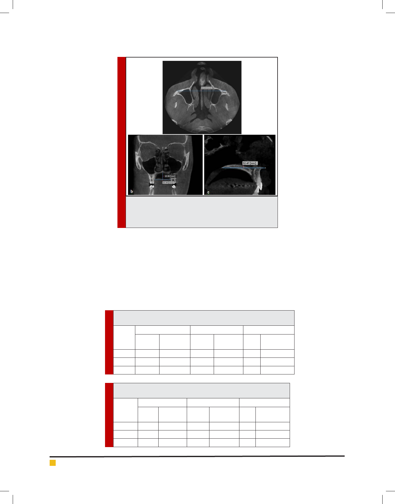
Distance between the zygomatic buttresses: Distance
between the zygomatic buttresses was calculated on
axial sections of the upper midface according to a study
by Ariji etal. [9] and Waitzman etal.(1992). To calculate
this distance, on axial sections visualizing the inferior
border of the zygomaticomaxillary suture, a horizontal
line was drawn connecting the outermost points of the
zygomatic buttresses in the two sides and its length was
measured in millimeters (Figure 1a).
Width and height of the palate: To calculate the pala-
tal width on coronal sections according to Byasaletal.
(2011), a horizontal line was drawn from the palatal
margin of the crestal bone of the rst molar tooth in
one side to the palatal margin of the crestal bone of the
other side and its length was measured. To measure the
height of the palate in this region on coronal sections,
as shown in Figure 1b, a line was drawn from the palate
perpendicular to the line connecting the alveolar crest
borders of the two sides and its length was measured.
Length of the hard palate: To measure the length of
the hard palate on sagittal sections, the distance between
the anterior nasal spine and posterior nasal spine at the
midline (where the anterior nasal spine was visible) was
measured (Figure 3C).
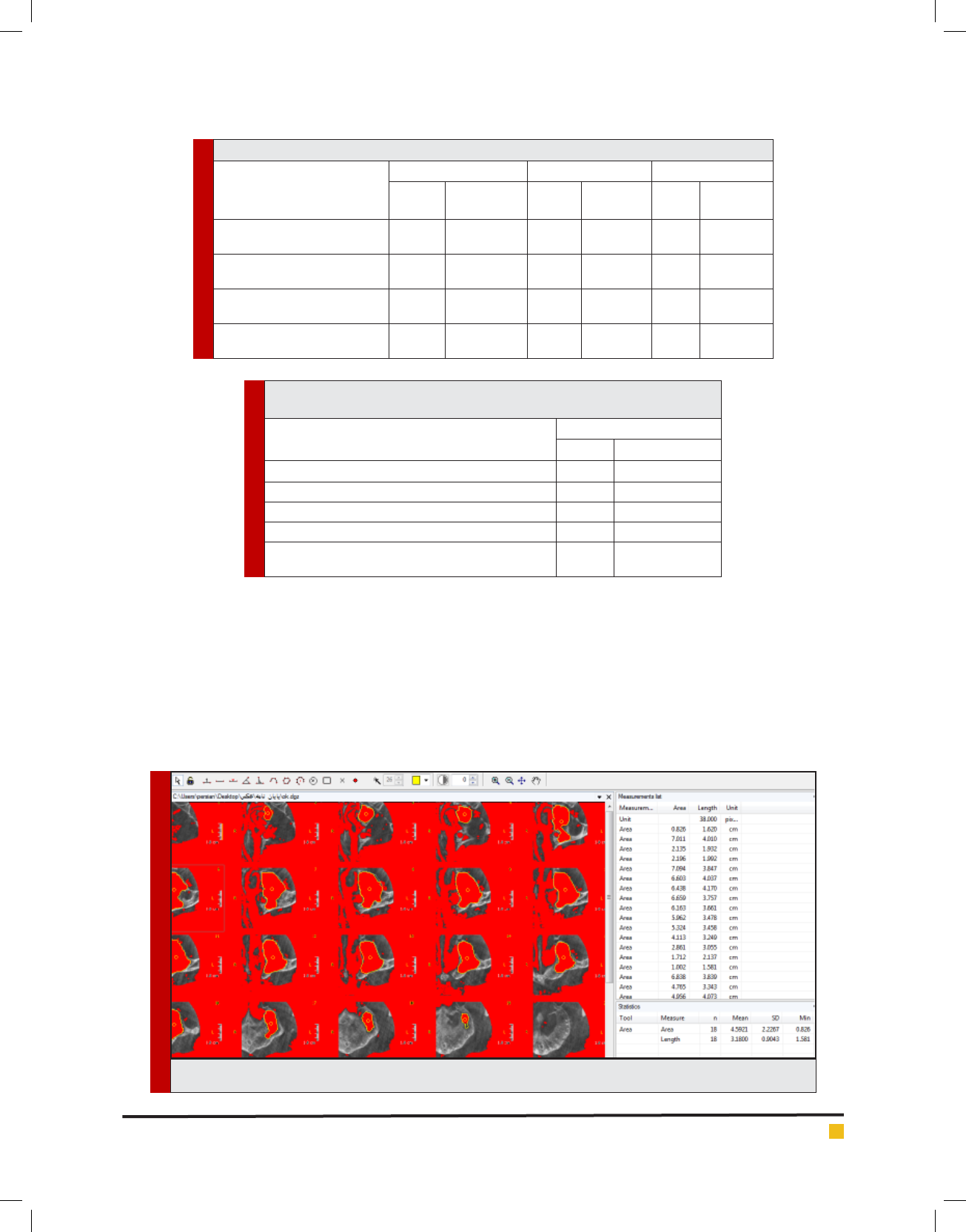
Maxillary sinus volume: To measure the maxillary
sinus volume, axial CBCT sections with 2mm slice thick-
ness and 2mm intervals were used. The CBCT sections
were subjected to Digimizer version 4.1.1 software and
the surface area of the sinus was automatically meas-
ured and stored in Excel format. The surface area of all
sections of the sinus (about 20 to 25 sections) in both
sides was calculated for each patient. Then, to calculate
the volume of the sinus in cubic centimeters, the follow-
ing formula was used: Sum of the surface area of the
sinus in each section thickness of each section (Figure
2). Data were collected and analyzed using SPSS ver-
sion 24.Independentt-test and paired t-test were used to
compare differences in the right and left sides and in the
mean size of variables.
RESULTS AND DISCUSSION
Two groups (n=35) of males and females in the age range
of 18 to 74 years were evaluated. The mean age was 37.2
years in females and 34.3 years in males. Independent
t-test showed that the mean age of males and females
was not signi cantly different (P=0.30). The mean vol-
ume of the maxillary sinus was 14.452.71 cm
3
. Inde-
pendent t-test showed that the mean volume of the sinus
in the right (P=0.02), left (P=0.006) and both (P=0.01)
sides was signi cantly greater in males than females. In
other words, a signi cant association existed between
gender and maxillary sinus volume in the right and left
sides (P<0.05, Table 1).
The area under the ROC curve was 0.627 or 62.7%
(smaller than 0.70) (Graph 1).
Etemadi, Seylavi and Yadegari
582 CORRELATION OF THE MAXILLARY SINUS VOLUME WITH GENDER BIOSCIENCE BIOTECHNOLOGY RESEARCH COMMUNICATIONS
FIGURE 1. (a) Distance between the two zygomatic buttresses.
(b) Measurement of the width and height of the palate at the
maxillary rst molar site. (C) Measurement of the anterior-pos-
terior length of the hard palate
Table 1. Comparison of the mean volume of the maxillary sinus between
males and females in the right and left sides (in cm3)
Side
Females Males Independent t-test
Mean
Standard
deviation
Mean
Standard
deviation
t P value
Righ t 13/1 2/9 15/8 6/3 2/31 0/02
Left 12/9 2/8 16/01 5/8 2/86 0/006
Total 13 2/85 15/9 6/05 2/58 0/01
Table 2. Comparison of the mean volume of the maxillary sinus in the
right and left sides based on gender (in cm3)
Gender
Right Left Independent t-test
Mean
Standard
deviation
Mean
Standard
deviation
t P value
Females 13/1 2/9 12/9 2/8 0/51 0/61
Males 15/8 6/3 16/01 5/8 0/41 0/68
Total 14/4 55/1 14/5 4/8 0/09 0/92
Paired t-test showed that the mean volume of the
maxillary sinus in the right and left sides in females
(P=0.61), males (P=0.68) and in total (P=0.92) was not
signi cantly different (Table 2).
Independent t-test showed that the mean anterior-pos-
terior length of the palate in sagittal plane was not sig-
ni cantly different between males and females (P=0.23)
but the mean distance between the two zygomatic but-
tresses in the axial plane (P<0.001), palatal width in the
rst molar region in the coronal plane (P=0.007) and
height of the palate in the rst molar region in the coro-
nal plane (P=0.002) were signi cantly greater in males
than in females. Thus, no signi cant association existed
between gender and anterior-posterior length of the pal-
ate in sagittal plane; but gender had a signi cant associa-
tion with the other three variables (Table 3).
The Pearson’s correlation coef cient showed that no
signi cant association existed between age and vol-
Etemadi, Seylavi and Yadegari
BIOSCIENCE BIOTECHNOLOGY RESEARCH COMMUNICATIONS CORRELATION OF THE MAXILLARY SINUS VOLUME WITH GENDER 583
Table 3. Comparison of the mean craniofacial parameters in males and females (in mm)
Parameter
Females Males Independent t-test
Mean
Standard
deviation
Mean
Standard
deviation
t P value
Distance between zygomatic
buttresses in axial plane
85/2 4/2 88/9 4/1 3/70 <0/001
Anterior posterior length of the
palate in sagittal plane
51/2 3/2 52/6 6/1 1/20 0/23
Width of the palate in rst
molar site in coronal plane
33/2 2/6 34/9 2/8 2/79 0/007
Height of the palate in the rst
molar site in coronal plane
11/8 1/7 13/8 3/3 3/25 0/002
Table 4. Pearson’s correlation coef cient for the correlation between the mean
size of the maxillary sinus and age and craniofacial parameters
Variable
Maxillary sinus volume
r P value
Age -0/182 0/18
Distance between zygomatic buttresses in axial plane 0/424 <0/001
Anterior posterior length of the palate in sagittal plane 0/119 0/33
Width of the palate in rst molar site in coronal plane 0/241 0/044
Height of the palate in the rst molar site in coronal
plane
0/547 <0/001
FIGURE 2. Method of measurement of maxillary sinus surface area using Digimizer software
ume of the maxillary sinus (P=0.18). The anterior-pos-
terior length of the palate in the sagittal plane had no
signi cant association with the maxillary sinus volume
(P=0.33). But an association existed between the palatal
width in the rst molar site in the coronal plane and the
volume of the maxillary sinus (P=0.044). The Pearson’s
correlation coef cient showed that a signi cant associa-
tion existed between the distance between the two zygo-
matic buttresses in the axial plane and also the height of
the palate in the rst molar region in the coronal plane
with the volume of the maxillary sinus (P<0.001, Table 4).
Maxillary sinus is the closest paranasal sinus to the
oral cavity and since implant treatments along with
open or closed sinus lift surgery (due to pneumatization
of the maxillary sinus) have greatly increased, knowl-
edge about the anatomy of the maxillary sinus is impor-

Etemadi, Seylavi and Yadegari
584 CORRELATION OF THE MAXILLARY SINUS VOLUME WITH GENDER BIOSCIENCE BIOTECHNOLOGY RESEARCH COMMUNICATIONS
tant (Gray etal. 2000). Sexual identi cation is an impor-
tant parameter in forensic medicine. Use of maxillary
sinus dimensions measured on CT scans may help in this
regard in cases where other methods fall short; however,
this method has its own drawbacks as well (Lerno, 1983).
Since a decade ago, CBCT is recommended as a modality
for maxillofacial imaging (Farman etal. 2006; Moreira
etal. 2009). This modality enables image reconstruction
without superimposition and has lower patient radiation
dose compared to CT (Moreira etal. 2009). It is believed
that the quality of CBCT images is equal to that of con-
ventional CT in visualization of maxillofacial structures
(Silva etal. 2008; Loubele etal. 2009; Carra ello etal.
2010).
In our study, the mean volume of the right maxillary
sinus was 14.45.1 cm
3
and the mean volume of the left
maxillary sinus was 14.54.8 cm
3
. Also, maxillary sinus
volume in males was signi cantly greater than that in
females (P<0.05). In 2013, Jasim etal. measured the vol-
ume and dimensions of the maxillary sinus and evalu-
ated its association with age and gender using CT. They
reported that the volume and dimensions of the maxil-
lary sinus in males were greater than that in females
and volume of the sinus decreased by an increase in age
(Jasim etal. 2013). To calculate the volume of the maxil-
lary sinus, they used a method similar to ours; however,
in our study in contrast to theirs, no signi cant associa-
tion was found between age and volume of the maxil-
lary sinus (P=0.30). This difference may be due to the
age group of patients since Jasim etal. (2013) evaluated
patients in older age group (40-69 years).
In 2016, Prabhat etal. evaluated the size and volume
of the maxillary sinus and its association with sexual
identi cation using CT. They reported that volume of
the maxillary sinus in males was signi cantly greater
than that in females. They stated that use of maxillary
sinus volume increases the accuracy of sexual identi-
cation by 83.3% (Prabhat et al. 2016). In our study,
volume of the maxillary sinus in males was greater than
that in females. However, since the area under the ROC
curve was 62.7% (<70%), maxillary sinus volume cannot
be used as a reliable indicator for sexual identi cation.
These differences in measurements may be due to ana-
tomical differences of the sinus in different geographical
areas and among different races. Another reason may be
the difference in method of calculation of sinus volume
and sample size, since our sample size was twice as big
as that of Prabhat etal.(2016).
Senturk etal. in 2015 evaluated the effect of weather
and altitude on volume of the paranasal sinuses and
found no signi cant association between the volume
of the maxillary sinus and age or between the size of
the sinus in the two sides (Sahlstrand-Johnson, 2011). In
our study, no signi cant difference was noted between
the volume of the maxillary sinus and age or between
the volume of the maxillary sinuses in the two sides
(P>0.05). Ariji etal. in 1993 evaluated the correlation
of the maxillary sinus volume and aging and found that
the mean volume of the right and left maxillary sinuses
was 14.646.32 cm
3
. They found no signi cant differ-
ence in the volume of the maxillary sinus between males
and females. Sinus volume had an ascending trend to
GRAPH 1. ROC curve for sexual identi cation using maxillary
sinus volume in the right and left sides

Etemadi, Seylavi and Yadegari
BIOSCIENCE BIOTECHNOLOGY RESEARCH COMMUNICATIONS CORRELATION OF THE MAXILLARY SINUS VOLUME WITH GENDER 585
20 years of age and then decreased. They also reported
that a signi cant association exists between the volume
of the maxillary sinuses and the distance between the
two zygomatic buttresses and the zygomatic occipital
distance (Ariji etal. 1994). The maxillary sinus volume
reported by Arihi et al. (1994) was comparable to the
value obtained in our study (14.452.71 cm
3
).
However, in our study, the volume of the maxil-
lary sinus in males was signi cantly higher than that
in females and no signi cant association was noted
between the sinus volume and age. In our study, aside
from the relationship of the maxillary sinus volume with
age and sex, its correlation with some other craniofacial
parameters such as width and height of the palate in the
rst molar area in the coronal plane, anterior-posterior
length of the palate in the sagittal plane and the distance
between the two zygomatic buttresses in the axial plane
was also eval uated and it was found that a correlation
existed between the volume of the maxillary sinus and
the distance between the two zygomatic buttresses; this
nding was comparable to that of Ariji etal. Moreover,
it was found that the greater the width and height of the
palate, the greater the size of the maxillary sinus would
be (P<0.05). But no signi cant correlation was found
between the sinus volume and the anterior-posterior
length of the hard palate (P>0.05).
CONCLUSION
The current results showed that Despite the presence of
a signi cant association between gender and volume of
the maxillary sinus (P<0.05), since the area under the
ROC curve was 0.627 or 62.7% (smaller than 0.70), vol-
ume of the maxillary sinus cannot serve as a de nite
and reliable indicator for sexual. Also, a correlation was
noted between the maxillary sinus volume and width
and height of the palate at the rst molar site and the
distance between the two zygomatic buttresses.
REFERENCES
A riji Y, Kuroki T, Moriguchi S, Ariji E, Kanda S. (1994). Age
changes in the volume of the human maxillary sinus: a study
using computed tomography. Dentomaxillofacial radiology
23(3): 163-8.
Ba ysal A, Veli I, Ucar FI, Eruz M, Ozer T, Uysal T. (2011).
Changes in mandibular transversal arch dimensions after rapid
maxillary expansion procedure assessed through cone-beam
computed tomography. Korean Journal of Orthodontics 41(3):
200-10.
Ca rra ello G, Dizonno M, Colli V, Strocchi S, Taubert SP, Leon-
ardi A, etal. (2010). Comparative study of jaws with multislice
computed tomography and cone-beam computed tomography.
La radiologia medica 115(4): 600-11.
Ek izoglu O, Inci E, Hocaoglu E, Sayin I, Kayhan FT, Can IO.
(2014). The use of maxillary sinus dimensions in gender deter-
mination: a thin-slice multidetector computed tomography
assisted morphometric study. Journal of Craniofacial Surgery
25(3): 957-60.
Fa rman AG, Scarfe WC. (2006). Development of imaging selec-
tion criteria and procedures should precede cephalometric
assessment with cone-beam computed tomography. American
Journal of Orthodontics and Dentofacial Orthopedics 130(2):
257-65.
Fe rnandes C. (2004). Volumetric analysis of maxillary sinuses
of Zulu and European crania by helical, multislice computed
tomography. The Journal of Laryngology & Otology 118(11):
877-81.
Gr ay C, Staff R, Redpath T, Needham G, Renny N. (2000).
Assessment of maxillary sinus volume for the sinus lift opera-
tion by three-dimensional magnetic resonance imaging. Den-
tomaxillofacial Radiology 29(3): 154-8.
Ja sim HH, Al-Taei JA. (2013). Computed tomographic meas-
urement of maxillary sinus volume and dimension in correla-
tion to the age and gender (comparative study among individ-
uals with dentate and edentulous maxilla). Journal of Baghdad
College of Dentistry 25(1): 87-93.
Le nza M, Lenza MdO, Dalstra M, Melsen B, Cattaneo P. (2010).
An analysis of different approaches to the assessment of upper
airway morphology: a CBCT study. Orthodontics & craniofacial
research 13(2): 96-105.
Le rno P. (1983). Identi cation par le sinus maxillaire. Odontol
leg. 216:39-40.
Lo ubele M, Bogaerts R, Van Dijck E, Pauwels R, Vanheusden S,
Suetens P, etal. (2009). Comparison between effective radia-
tion dose of CBCT and MSCT scanners for dentomaxillofacial
applications. European journal of radiology 71(3): 461-8.
Möhlhenrich SC, Heussen N, Peters F, Steiner T, Hölzle F, Mod-
abber A. (2015). Is the Maxillary Sinus Really Suitable in Sex
Determination? A Three-Dimensional Analysis of Maxillary
Sinus Volume and Surface Depending on Sex and Dentition.
Journal of Craniofacial Surgery 26(8) :723-6.
Moreira CR, Sales MA, Lopes PM, Cavalcanti MG. (2009).
Assessment of linear and angular measurements on three-
dimensional cone-beam computed tomographic images. Oral
Surgery, Oral Medicine, Oral Pathology, Oral Radiology, and
Endodontology 108(3): 430-6.
Prabhat M, Rai S, Kaur M, Pra bhat K, Bhatnagar P, Panjwani
S. (2016). Computed tomography based forensic gender deter-
mination by measuring the size and volume of the maxillary
sinuses. Journal of forensic dental sciences 8(1): 40.
Saccucci M, Cipriani F, Carderi S, Di Carlo G, D’Atti lio M,
Rodol no D, etal. (2015). Gender assessment through three-
dimensional analysis of maxillary sinuses by means of Cone
Beam Computed Tomography. European review for medical
and pharmacological sciences19(2): 185-93.
Schwab RJ . (1998). Upper airway imaging. Clinics in chest
medicine. 19(1):33-54.

Etemadi, Seylavi and Yadegari
586 CORRELATION OF THE MAXILLARY SINUS VOLUME WITH GENDER BIOSCIENCE BIOTECHNOLOGY RESEARCH COMMUNICATIONS
Sentürk M, Azgin I, Öcal R, Sakarya EU, Güler I. (2015). Volu-
metric analysis of the maxillary sinus in pediatric patients with
nasal septal deviation. ENT Updates 5(3): 107.
Silva MAG, Wolf U, Heinicke F , Bumann A, Visser H, Hirsch E.
(2008). Cone-beam computed tomography for routine orthodontic
treatment planning: a radiation dose evaluation. American Jour-
nal of Orthodontics and Dentofacial Orthopedics 133(5): 640-5.
Vidya C, Shamasundar N, Manjunatha B, Raichurkar K. (2013).
Evaluation of size and volume of maxillary sinus to determine
gender by 3D computerized tomography scan method using
dry skulls of South Indian origin. International Journal of Cur-
rent Research and Review 5(3): 97.
Waitzman AA, Posnick JC, Armstrong DC, Pron GE. (1992).
Craniofacial skeletal measurements based on computed
tomography: Part II. Normal values and growth trends. The
Cleft palate-cran iofacial journal 29(2): 118-28.
Wang R-G, Jiang S, Gu R. (1994). The cartilaginous nasal cap-
sule and embryonic development of human paranasal sinuses.
The Journal of otolaryngology 23(4): 239-43.
Wolf G, Anderhuber W, Kuhn F. (1993). Development of the
paranasal sinuses in children: implications for paranasal sinus
surgery. Annals of Otology, Rhinology & Laryngology 102(9):
705-11.