Medical
Communication
Biosci. Biotech. Res. Comm. 10(3): 475-480 (2017)
Comparison of the effects of IPS e.max heat press layered
and feldspathic porcelain on natural enamel tooth wear
Hedaiat Moradpoor
1
, Marjan Mardanpour
2
, Farnoush Golmohamadi
2
and Sahar Raissi
3
*
1
Assistant Professor, Prosthodontics Department, Faculty of Dentistry Kermanshah University of Medical
Sciences, Iran
2
Faculty of Dentistry, Kermanshah University of Medical Science, Kermanshah, Iran
3
Assistant Professor, Prosthodontics Department, Faculty of Dentistry Kermanshah University of Medical
Sciences, Iran
ABSTRACT
In an ideal situation, the tooth wear of a restorative material should be similar to dental enamel. Currently, All-ceramic
restorations are widely used; But Enamel wear is among the main disadvantages of ceramic restorations. Thus, in this
study compared the Effect of IPS e.max heat press layered and Feldspathic Porcelain on natural enamel Tooth Wear.In this
study, 20 samples were prepared from normal human teeth. The natural teeth were photographed by a stereomicroscope in
a xed position and the distance from the cusp tip to a reference point was measured. Next, 10 teeth opposed IPS e.max
heat press layered and the remaining 10 opposed Feldspathic Porcelain in a chewing simulator and subjected to 120,000
masticatory cycles. The teeth were photographed again and the greatest difference between the before and after values was
recorded. Finally data were analyzed by Independent samples T-test. The mean extent of wear on the restoration layered
IPS e.max heat press and Feldspathic Porcelain were not signi cant differences ( p-value= 0.118); although the average
wear in Feldspathic Porcelain was less than layered IPS e.max heat press. the distance from the cusp tip to a reference point
in layered IPS e.max heat Press was signi cantly difference between before and after intervention (p-value<0.001). Also,
the distance from the cusp tip to a reference point in FeldspathicPorcelain was signi cantly difference between before and
after intervention (p-value<0.001). According to the results obtained in vitro study, the mean extent of wear on the resto-
ration layered IPS e.max heat press and Feldspathic Porcelain were not signi cant differences. Also, it is recommended to
have a closer look at the factors involved in tooth enamel wear in future studies.
KEY WORDS: FELDSPATHIC PORCELAIN, IPS E.MAX HEAT PRESS LAYERED, ENAMEL TOOTH WEAR
475
ARTICLE INFORMATION:
*Corresponding Author: raissi_sahar@yahoo.com
Received 2
nd
July, 2017
Accepted after revision 29
th
Sep, 2017
BBRC Print ISSN: 0974-6455
Online ISSN: 2321-4007 CODEN: USA BBRCBA
Thomson Reuters ISI ESC and Crossref Indexed Journal
NAAS Journal Score 2017: 4.31 Cosmos IF: 4.006
© A Society of Science and Nature Publication, 2017. All rights
reserved.
Online Contents Available at:
http//www.bbrc.in/
DOI: 10.21786/bbrc/10.3/21

476 COMPARISON OF THE EFFECTS OF IPS E.MAX HEAT PRESS LAYERED AND FELDSPATHIC PORCELAIN BIOSCIENCE BIOTECHNOLOGY RESEARCH COMMUNICATIONS
Hedaiat Moradpoor et al.
INTRODUCTION
Replacement of missing teeth or repair a part of a tooth
has long been considered by dentists, Currently using a
variety of ceramic restorations has largely resolved this
problem; Ceramic restorations have advantages includ-
ing lack of fracture, lack of blanching and high strength
are also by considering the aesthetic, ease of use and
color similar to natural tooth using these restorations
have increased daily. These restorations due to the lack
of metal in their structure (metal component can cause
problems such as chemical toxicity, kerogen, change the
color of gums and creating allergic reactions to some
metals such as nickel) are preferred compared with
coated of metal - ceramic (Jung 2010) and Rosenstiel,
2015, Murillo-Gómez, 2017 and (Montazerian, 2017).
There are also disadvantages in addition to the top
bene ts of these restorations that the most important of
them is front tooth enamel erosion (Shillingburg, 2013).
Erosion is de ned as damage to the tooth surface or los-
ing volume of teeth by direct contact with the teeth or
other materials. In fact, erosion is a physiological phe-
nomenon that is happening naturally always mechani-
cally or chemically (DeGee, 1986). The sharp rise of den-
tal erosion can cause to loss of centric contacts, vertical
elevation change, Change in Functional paths during
chewing and fusion muscle fatigue (DeLong, 1989) and
(Gallegos, 1988). Dental restorations change the natu-
ral erosion rate of front teeth if have had different ero-
sion features (Sulong, 1990). So erosion between tooth
and its front restoration should be always considered
as important factor along with strength and beauty to
selecting restorative matter (Seghi, 1991).Considering
this, various methods have been tested to improve and
reduce the erosion of the front teeth in ceramicrestora-
tions; Among these methods, reinforced ceramics with
alumina oxide crystals, Lusaite, lithium DE silicate and
zirconia (Komine, 2004) and (Barath, 2003).
Given the importance of the front dental erosion by
ceramic restoration, in this study, two restorations of the
Feld spathic porcelain and layered IPS e.max heat press
ceramic were evaluated and compared in terms of the front
side tooth enamel. The reasons for choosing these two res-
torations can be reffered to improve the ceramic proper-
ties and porcelain bonding systems that has been caused
to beauty and good performance of feldspathic porcelain
(Vieira, 2004) and IPS e.max Press makers claim to improve
physical and translucency properties, lower surface hard-
ness low concentrations of crystalline phase and a smaller
sizes of crystals at different stages of cooking Empress 2
and revise in formulation of the this kind of ceramic to
obtaining high quality (Newsome 2014) and (Guess 2011).
Of course in laboratory studies that have been done in
this area, a signi cant difference has not been speci ed
in phase composition and the bending strength between
IPS e.max Press and Empress 2 (Guazzato, 2004) and
(Albakry 2003). IPS e.max Press in monolithic mode is
used for inlays, onlays, full coverage of crown... (Guess
2011). In times of short and medium term have shown in
reviewing these performances that IPS e.max Press res-
torations have good performance: Using this restoration
in onlays in the 3-year period, the ef ciency of 100%
(Guess 2009) and the crown in the 3-year period has
had the performance of 96.6% (Etman, 2008). Despite
this high performance, high erosion possibility of this
restoration on the tooth enamel can be considered as
one of the major disadvantages (Esquivel-Upshaw 2012).
Also, amount of natural tooth erosion against the lay-
ered IPS-emax heat press and feldspathic porcelain were
discussed due to lower researches and lack of consensus
on the abrasive effect of these ceramics and the need for
further studies in this area.
MATERIALS AND METHODS
This study was done as In vitro and in Reference Labo-
ratory and Research Center University of Medical Sci-
ences in Tehran. The study population was healthy teeth
recently extracted and without premolar decay of human
maxillary. Easy non-probability sampling method and
sample size were obtained from the results of previous
studies (Ahmadzadeh, 2014) and by considering =0.05,
and power of 1- = 80%, at least 20 teeth. Trend of run-
ning in this study was in this way that in each group
10 samples were prepared with dimensions of 10 × 10 ×
10 mm. in IPS-emax heat press group in order to build
cylinders of a block with cross-section dimensions 10 ×
10 × 10 is molded with incremental silicone impression
material and then wax blocks were prepared by melt-
ing the inlay10 wax. Then these wax blocks, were done
sprue, cylindering and were cast by vita ceramic Inge.
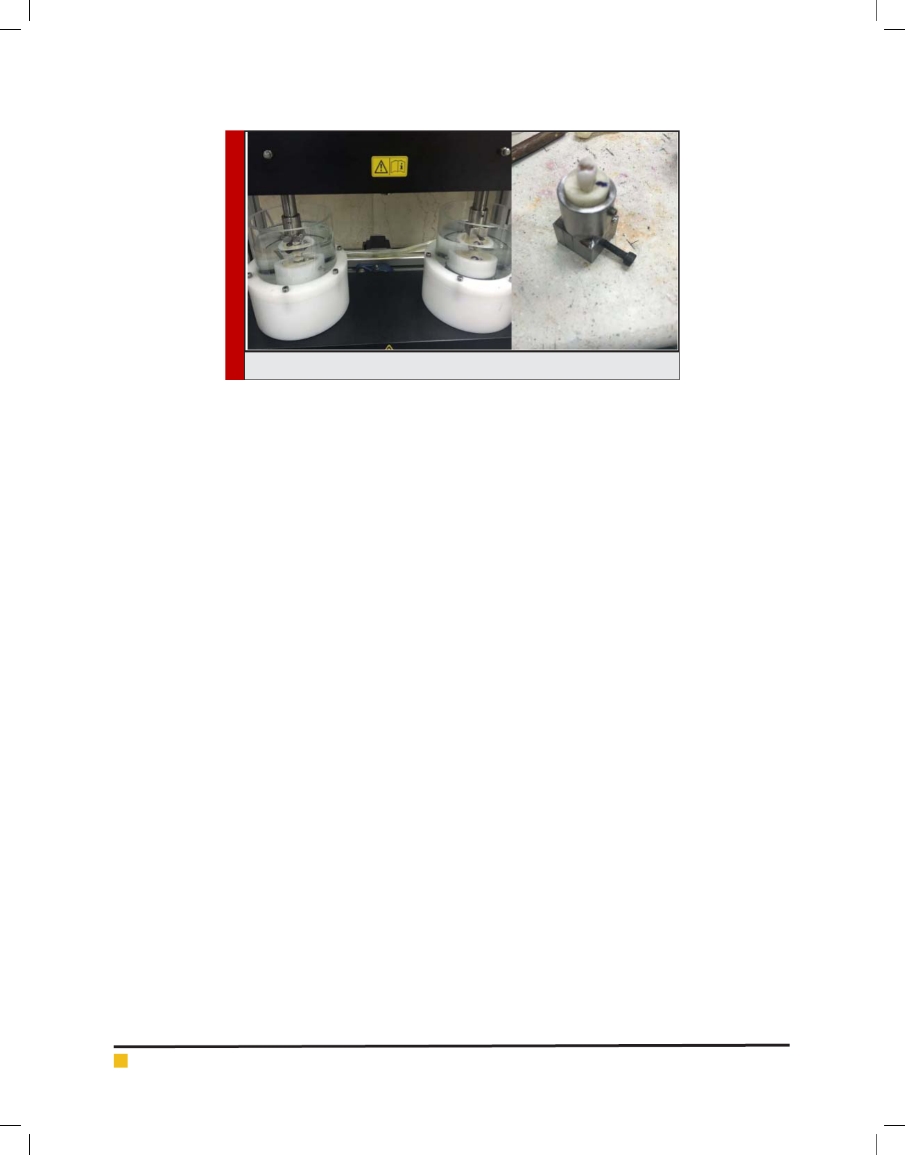
Speci c cylinder device: Chewing simulator (CS-4.2 S /
N: A100220128SM01) was used to cylindering (Figure 1).
Cylinder was heated to 800 ° C under to evaporate
and remove the wax pattern. Vita ceramic ingot was
placed within the cylinder by tongs and cylinder was
placed in speci c furnaces. After warming oven to tem-
perature of 920 ° C, ceramic melted slowly and in vac-
uum was injected into the cylinder.Sprue cut off and
the samples were sandblasted by 2-1 times of aluminum
oxide after cooling cylinder. Then the blocks were drool
with powder and liquid of VitaAkzent Plus .wax blocks
was used to make Feldspathic porcelain with dimensions
10 × 10 × 8 mm has been molded with an incremental
silicone impression material and then wax blocks were
prepared by melting the inlay10 wax. Then these blocks
were done sprue and cylindered and were cast by Vero
bond (vita mark) alloy.

BIOSCIENCE BIOTECHNOLOGY RESEARCH COMMUNICATIONS COMPARISON OF THE EFFECTS OF IPS E.MAX HEAT PRESS LAYERED AND FELDSPATHIC PORCELAIN 477
Hedaiat Moradpoor et al.
Table 1. Mean and standard deviation the amount of wear before and after treatment in two different materials
The vertical height of the buccal
cusp tip of the tooth surface
before the intervention of the
central Tafusay
The vertical height of the buccal
cusp tip of the tooth surface after
the intervention of the central
Tafusay
p-value
a
Wear rate
Average
Standard
deviation
AverageStandard deviationAverage
Standard
deviation
layered IPS
e.max heat
press
2/59900/34821/91410/3506<0/0010/68490/0673
Feldspathic
Porcelain
2/25150/47691/66560/4596<0/0010/58590/1731
p-value
b
0/0790/1910/118
a
comparison before and after the intervention (paired t test)
b
comparison between the two materials (independent samples t-test)
Porcelaining was done with VMK master (vita mark)
porcelain by thickness of 2mm and external dimensions
were measured by a gauge after cutting the sprue and
sandblasting and grinding alloy with pink and white
stone. Premolar healthy natural teeth of 4 human max-
illary as well as were selected to the total number of
samples and were stored in distilled water until erosion
time. all samples were mounted by Survivor in a plas-
tic mold in the shape of a half-cylinder in-hardening
resin (Ivoclarvivadent) before testing. Then the photos
were taken of dental teeth in Razi Metallurgical Research
Center (Razi Metallurgical Research Center) by stereomi-
croscope (VEGA II TESCAN) in xed position and a xed
place was determined for each sample. Distance from
the cusps tip to the desired location was measured by
the Motic Image Plus software. Then natural teeth were
placed in the front samples of feldspathic porcelain and
layered ips-emax heat press in chewing simulator device.
The samples were immersed in distilled water at all times
of erosion. After the erosion test, the photos were taken
of samples by stereomicroscope again in the same previ-
ous position and due to mentioned method the meas-
urement was done for each cusp again; the difference
between the two amounts was recorded and the highest
was recorded per micrometer. Data with SPSS software
version 18 and by using statistical analysis Independent
samples T test were used for statistical analysis. The sig-
ni cant level of 0.05 was considered in this study.
RESULTS AND DISCUSSION
In this study, with the help of Shapiro-Wilk test it was
determined that the data are followed from a normal
distribution (0.05 <p-value). Means of erosion rate
between the two materials of layered IPS e.max heat
press and Feldspathic Porcelain was evaluated by inde-
pendent samples t-test and erosion difference between
the two restoration was not statistically signi cant
(p-value=0.118) although the mean of erosion rate in
Feldspathic Porcelain was less than layered IPS e.max
heat press (table 1).
As it is shown in Table 1; paired t-test showed that
the difference between the mean difference of the verti-
cal height of the tip of the buccal cusps to central fossa
in tooth surface in IPS e.max heat press between before
and after the intervention (0.001> p-value) and in Feld-
spathic Porcelain between before and after the interven-
tion (0.001> p-value) was signi cant.
DISCUSSION
Ceramic materials made with respect to stiffness and
strength of them are usually resistant to erosion, but
erosion tighter than usual their front teeth is what is
made problem in this type of restorations (Heintze,
2008). In the present study, there was no statistically
signi cant difference between the mean of erosion rate
in two restorations of layered IPS e.max heat press and
Feldspathic Porcelain (0.118 = p-value), although mean
of erosion rate in Feldspathic Porcelain somewhat was
lower than layered IPS e.max the heat press.
In a study by Tian et al. (2013) that the erosion rate
of two groups of Porcelain Ceramics, and layered IPS
e.max heat press were studied, the results showed that
the erosion rate on in the layered IPS e.max heat press
ceramic has been much more than Porcelain (Tian, B.M
2013) which is inconsistent with results of this study.
In a study of Esquivel-Upshaw the rate of Front tooth
478 COMPARISON OF THE EFFECTS OF IPS E.MAX HEAT PRESS LAYERED AND FELDSPATHIC PORCELAIN BIOSCIENCE BIOTECHNOLOGY RESEARCH COMMUNICATIONS
Hedaiat Moradpoor et al.
enamel erosion has been reported by restoration of IPS
e.max heat press μm 88/3 (Esquivel-Upshaw 2012). in
study of Etman, et al that also studied the erosion of
Front tooth enamel in three types of ceramic restora-
tions, the results showed that after two years of follow-
ing the erosion mean created by Allceram Procera, μm
261, has been for IPS e.max heat press μm 215 and metal
ceramics μm156 that this difference was statistically sig-
ni cant (Etman, 2008).
In this study, Means of vertical height of the tip of
the buccal cusps to central fossa in tooth surface after
the intervention there was no signi cant difference
between the two materials (p-value=0.191); But verti-
cal height means of the tip of the buccal cusps to cen-
tral fossa tooth surface on the material of layered IPS
e.max heat press there was a statistically signi cant
difference between before and after the intervention
(0.001> p-value) and in Feldspathic Porcelain between
before and after the intervention (0.001> p -value). It
is clear that the signi cant erosion has occurred with
regard to these results. But the erosion differences have
not shown obvious difference between two restora-
tions. Heintze et al. by examining erosion rate of front
ceramic teeth and factors affecting on it, have recog-
nized that factors such as the shape of the samples,
surface treatment, enamel thickness, type of milling
device and its geometry shape, grains size and crystal
size are effective in erosion (Heintze, 2008).
In fact, the erosion is related to the friction created
between the two levels, so it can be concluded that
surface properties and conducted interaction will have
important impact by contact in the two levels. Smooth
surfaces have less resistance and therefore less erosion
compared to uneven surfaces; the erosion rate of them
on front teeth will be also more whatever the surface
roughness of restorative materials to be more. For this
reason, it has always tried to maintain the strength of
the restoration; its surface roughness is reduced as much
as possible. According to studies conducted, the surface
roughness is for feldspathic porcelains of MPa 6560 and
for IPS e.max Press, MPa 5800 that due to the proximity
of value of this variable in two restorations examined,
lake of difference in Front tooth erosion is also justi ed
(Al-Shehri 2002).
Another important factor in front tooth erosion frac-
ture toughness. All ceramic materials that have been used
in dentistry so far, has been with much lower fracture
toughness than metals. According to studies for fracture
toughness of IPS e.max Press MPam 1/22/ 1- 4/3 (14)
and for feldspathic porcelain on range of MPam 1/290/0
up to be 1.56 (23), as well as natural tooth enamel frac-
ture toughness has been reported in the range of MPam
1/237/1-7/0 (Heintze, S.D 2008) and natural teeth dentin
has been reported in MPam 1/208/3 (24), that compared
with metals have less fracture toughness between 20 and
100 times. Lee et al in New Zealand during a study have
shown that gold brigade 3 compared to IPS e.max Press
has less abrasive and friction on the tooth enamel and
can be seen signi cant cracks in front enamel surface
of samples of IPSe.max Press examples in comparison
with gold brigade 3 (Lee 2012). Lee and et al differences
in fracture toughness in different materials know as one
of the Justi er factor; so that during the action and the
inclusion of occlusal forces on the surface of the resto-
ration are with micro fracture and lead to bumps and
abuses such as inclusion Crystalline that are going off
the surface of the material; as result leads to accumula-
tion of a lot of pressure on enamel and troughs Gaug-
ing. In addition dug particles can act like an Abrasive
and to create three-dimensional erosion; So it can be
expected that less erosion is created in front teeth in the
restorations with higher fracture toughness (Anusavice,
2013). Given that in terms of fracture toughness as well
as there was no signi cant differences between two
FIGURE 1. A prepared sample and chewing simulator

BIOSCIENCE BIOTECHNOLOGY RESEARCH COMMUNICATIONS COMPARISON OF THE EFFECTS OF IPS E.MAX HEAT PRESS LAYERED AND FELDSPATHIC PORCELAIN 479
Hedaiat Moradpoor et al.
restorations evaluated in this study, so the same Front
tooth erosion is justi able.
CONCLUSION
According to the results obtained in present experimen-
tal study, the means of erosion rate in two restorations
of layered IPS e.max heat press and Feldspathic Porce-
lain has no statistically signi cant difference with each
other, although mean of erosion rate in the Feldspathic
Porcelain has been somewhat lower than layered IPS
e.max heat the press; The results showed that the ero-
sion on both the restorations after intervention has been
signi cant compared to before intervention; As a result,
it can be concluded that these two restorations causes to
signi cant erosion of front tooth enamel, but have no
signi cant difference with each other.
SUGGESTIONS
It is suggested that in future research erosion caused be
evaluated by natural tooth enamel and other high appli-
cable restorations and also be compared by two restora-
tions of layered IPS e.max heat press and Feldspathic
Porcelain. It is also possible to design and carry out
long-term laboratory studies of this kind, to provide the
information necessary to assess the relationship between
laboratory and clinical erosion data.
REFERENCES
Ahmadzadeh, A., Ashtiani, A.H., Epakchi, S., Pormehdi, M.,
Neshati, A., Golmohamadi, F. and Mousavi, N., 2014. Com-
parison of the Effect of Feldspathic Porcelain and Zirconia on
Natural Tooth Wear. Journal of Islamic Dental Association of
IRAN (JIDAI), 26(3), p.3.
Albakry, M., Guazzato, M. and Swain, M.V., 2003. Fracture
toughness and hardness evaluation of three pressable all-
ceramic dental materials. Journal of dentistry, 31(3), pp.181-
188.
Al-Shehri, S., 2002. Relative fracture toughness and hardness
of dental ceramics. Saudi Dent J, 14(2), p.67.
Anusavice, K.J., Shen, C. and Rawls, H.R., 2013. Phillips’ sci-
ence of dental materials. Elsevier Health Sciences.
Barath, V.S., Faber, F.J., Westland, S. and Niedermeier, W.,
2003. Spectrophotometric analysis of all-ceramic materials
and their interaction with luting agents and different back-
grounds. Advances in Dental Research, 17(1), pp.55-60.
DeGee, A.J., Pallav, P. and Davidson, C.L., 1986. Effect of abra-
sion medium on wear of stress-bearing composites and amal-
gam in vitro. Journal of Dental Research, 65(5), pp.654-658.
DeLong, R., Sasik, C., Pintado, M.R. and Douglas, W.H., 1989.
The wear of enamel when opposed by ceramic systems. Dental
Materials, 5(4), pp.266-271.
El Mowafy, O.M. and Watts, D.C., 1986. Fracture toughness of
human dentin. Journal of dental research, 65(5), pp.677-681.
Esquivel-Upshaw, J.F., Rose, W.F., Barrett, A.A., Oliveira, E.R.,
Yang, M.C., Clark, A.E. and Anusavice, K.J., 2012. Three years
in vivo wear: core-ceramic, veneers, and enamel antagonists.
Dental Materials, 28(6), pp.615-621.
Etman, M.K., Woolford, M. and Dunne, S., 2008. Quantitative
measurement of tooth and ceramic wear: in vivo study. Inter-
national Journal of Prosthodontics, 21(3).
Gallegos, L.I. and Nicholls, J.I., 1988. In vitro two-body wear
of three veneering resins. The Journal of prosthetic dentistry,
60(2), pp.172-178.
Guazzato, M., Proos, K., Quach, L. and Swain, M.V., 2004.
Strength, reliability and mode of fracture of bilayered por-
celain/zirconia (Y-TZP) dental ceramics. Biomaterials, 25(20),
pp.5045-5052.
Guess, P.C., Schultheis, S., Bonfante, E.A., Coelho, P.G., Fer-
encz, J.L. and Silva, N.R., 2011. All-ceramic systems: labora-
tory and clinical performance. Dental Clinics of North Amer-
ica, 55(2), pp.333-352.
Guess, P.C., Strub, J.R., Steinhart, N., Wolkewitz, M. and Stap-
pert, C.F., 2009. All-ceramic partial coverage restorations—
midterm results of a 5-year prospective clinical splitmouth
study. journal of dentistry, 37(8), pp.627-637.
Heintze, S.D., Cavalleri, A., Forjanic, M., Zellweger, G. and
Rousson, V., 2008. Wear of ceramic and antagonist—a system-
atic evaluation of in uencing factors in vitro. dental materials,
24(4), pp.433-449.
Jung, Y.S., Lee, J.W., Choi, Y.J., Ahn, J.S., Shin, S.W. and Huh,
J.B., 2010. A study on the in-vitro wear of the natural tooth
structure by opposing zirconia or dental porcelain. The journal
of advanced prosthodontics, 2(3), pp.111-115.
Komine, F., Tomic, M., Gerds, T. and Strub, J.R., 2004. In uence
of different adhesive resin cements on the fracture strength
of aluminum oxide ceramic posterior crowns. The Journal of
prosthetic dentistry, 92(4), pp.359-364.
Lee, A., 2012. Wear behaviour of human enamel opposing
lithium disilicate glass ceramic and type III gold (Doctoral dis-
sertation, University of Otago).
Montazerian, M. and Zanotto, E.D., 2017. Restorative Dental
Glass-Ceramics: Current Status and Trends. In Clinical Appli-
cations of Biomaterials (pp. 313-336). Springer International
Publishing.
Morena, R., Lockwood, P.E. and Fairhurst, C.W., 1986. Fracture
toughness of commercial dental porcelains. Dental Materials,
2(2), pp.58-62.
Murillo-Gómez, F., Rueggeberg, F.A. and De Goes, M.F., 2017.
Short-and Long-Term Bond Strength Between Resin Cement
and Glass-Ceramic Using a Silane-Containing Universal Adhe-
sive. Operative Dentistry.
Newsome, P., 2014. Use of ceramic materials in the restoration
of posterior teeth. Primary dental journal, 3(2), p.42.
Rosenstiel, S.F., Land, M.F. and Fujimoto, J., 2015. Contem-
porary Fixed Prosthodontics-E-Book. Elsevier Health Sciences.

Hedaiat Moradpoor et al.
480 COMPARISON OF THE EFFECTS OF IPS E.MAX HEAT PRESS LAYERED AND FELDSPATHIC PORCELAIN BIOSCIENCE BIOTECHNOLOGY RESEARCH COMMUNICATIONS
Seghi, R.R., Rosenstiel, S.F. and Bauer, P., 1991. Abrasion of
human enamel by different dental ceramics in vitro. Journal of
dental research, 70(3), pp.221-225.
Shillingburg, H.T., Sather, D.A., Wilson, E.L., Cain, J.R., Mitch-
ell, D.L., Blanco, L.J. and Kessler, J.C., 2013. Fundamentals of
xed prosthodontics. Acta stomatol Croat, 47(2), p.177.
Sulong, M.Z.A.M. and Aziz, R.A., 1990. Wear of materials used
in dentistry: a review of the literature. The Journal of pros-
thetic dentistry, 63(3), pp.342-349.
Tian, B.M., Zhang, S.F., He, L., Guo, J.W., Yu, J.T. and Wu, X.H.,
2013. An experimental study of the wear behavior of dental
feldspathic glass-ceramic and lithium disilicate glass-ceramic.
Zhonghua kou qiang yi xue za zhi= Zhonghua kouqiang
yixue zazhi= Chinese journal of stomatology, 48(11), pp.683-
688.
Vieira, L.C.C., Araújo, É. and Monteiro Júnior, S., 2004. Effect
of different ceramic surface treatments on resin microtensile
bond strength. Journal of Prosthodontics, 13(1), pp.28-35.