
Sports Science
Communication
Biosci. Biotech. Res. Comm. 10(2): 133-137 (2017)
The effect of micro needling therapy on the quality of
life in women suffering from skin-deep varicose legs
Vahideh Riyahi
1
and Bijan Goodarzi
2
1
Master in Sports Pathology and Corrective Exercise, Young Researchers and Elite Club, Borujerd Branch,
Islamic Azad University, Borujerd, Iran
2
Assisstant Professor, Department of Physical Education and Sports Sciences, Borujerd Branch, Islamic Azad
University, Borujerd, Iran
ABSTRACT
The aim of the present research was to examine the effect of micro needling therapy on quality of life in women suf-
fering from skin- deep varicose leg. For this purpose 20 patients with varicose veins of the lower limb with the height
of 6/1 ± 163/1 cm, weight 4/6 ± 70/1 kg and fat content 6/5 ± 32/4 were selected purposefully, as the subjects. After
randomization of subjects into two groups of 10 subjects for 20 weeks, every 4 weeks for a total of 5 sessions a meet-
ing with patients was conducted using Micro needling, the control group did not have any therapeutic intervention.
To analyze the data, independent t-test at a signi cance level of 05/0> P were used. The ndings show that between
the measured values of quality of life index before and after the test respectively, there is signi cant increase and a
signi cant reduction of 05/0> P. The results showed that the use of micro-needling can be considered as a safe and
effective treatment in enhancing the quality of life in patients with varicose veins of lower limbs.
KEY WORDS: VARICOSE VEINS, MICRO NEEDLING, QUALITY OF LIFE
133
ARTICLE INFORMATION:
*Corresponding Author: bijan35454@yahoo.com
Received 11
th
March, 2017
Accepted after revision 11
th
June, 2017
BBRC Print ISSN: 0974-6455
Online ISSN: 2321-4007 CODEN: USA BBRCBA
Thomson Reuters ISI ESC and Crossref Indexed Journal
NAAS Journal Score 2017: 4.31 Cosmos IF : 4.006
© A Society of Science and Nature Publication, 2017. All rights
reserved.
Online Contents Available at: http//www.bbrc.in/
INTRODUCTION
The term Varicose veins which stems from its Latin ori-
gin Varix meaning “twisted” refers to dilated veins or
swelling of the veins. Varicose veins, is the most com-
mon human vascular disease that affects about 20-10%
of the population. Varicose veins are veins long, dilated
and twisted and which often seen on the interior sur-
faces of the lower limbs. The highest prevalence of
varicose veins is estimated in women aged 49-40 years.
Varicose veins are large, fully swollen and sometimes
palpable, dilated and elongated, and sometimes higher
than 4 mm in diameter. Swollen and web-like varicose
veins are very common (Harrie and Kendall 2014).

134 THE EFFECT OF MICRO NEEDLING THERAPY ON THE QUALITY OF LIFE IN WOMEN BIOSCIENCE BIOTECHNOLOGY RESEARCH COMMUNICATIONS
Vahideh Riyahi and Bijan Goodarzi
More than 40 percent of women over 50 have the dis-
ease. Varicose veins are skin-deep dilated and zigzagged
veins due to structural defects and performance of the
saphenous veins or inherent weakness of the vein wall or
rarely due to the Artery - IV stulas. More than 20 mil-
lion adult Americans suffer from varicose vein disease
and the disease causes a sickly condition which results
in the reduction of a patient’s performance (Chung,
McCray et al. 2000). These veins are related to leg swell-
ing, pain, dermatitis, phlebitis and ulcers, Patients with
venous insuf ciency often complain of pain in their legs
that worsen with prolonged standing and relieved by
elevating the feet. On examination of the shank diam-
eter, edema, varicose veins and super cial veins with
erythematic, dermatitis, and hyper pigmentation can
be seen in the lower leg and the skin near the external
ankle might become ulcerous. Mostly, varicose veins are
asymptomatic and only regarding beauty aspects require
medical attention themselves. If symptomatic, the pain,
diffuse and non-speci c fullness and heaviness in the
legs, especially after standing a long time are probable.
Sometimes swelling in the ankles and feet can be seen
in the prominent vessels (Chung, McCray et al. 2000).
So far, various methods for reducing the symptoms of
varicose veins including pain in the legs after prolonged
standing or sitting, night pain and feeling of heaviness
in the legs are provided. Avoiding long time standing
or sitting position, avoiding long-time exposure of legs
in the sun and heat or prolonged contact with garter or
belt, avoidance of continuous wearing of strip (band)
or stockings, avoiding simple sugars and lipids and
the use of medications such as Flavonoid derivatives,
sodium morrhuate are among the methods of reducing
the symptoms of varicose veins (Stiegmann, Sun et al.
1988). Micro needling is a method recently used for skin
diseases. Micro needling is a device for general skin nee-
dling which has a comparable impact with laser treat-
ments and chemical peelings. This device can be used
exclusively or as a complement to other treatment meth-
ods. The disposable needles’ points are short and tiny,
it includes 12 micro needles that should be discarded
after each treatment. For treatment, needle penetration
depth must be between 25% to 2/5 mm set with a speed
of 25 beats per second and beat for 90 beats per seconds
which nally between 300 and 1,000 tiny holes in sec-
onds are created in the surface. Micro needling is used
to stimulate the skin to make natural collagen. Further-
more, treatment with this device helps penetration and
absorption of drugs to the skin, skin rejuvenation and
wrinkles, acne scars healing, improvement of body craze
and the overall appearance of the skin and shrinkage of
the size of the pores. Backlog and increasing the elastic-
ity of the skin, new collagen production, surface and
deep wrinkles reduction, improving crazes, and improv-
ing decrements of pigmented lesions’ penetration, mate-
rials, creams and vitamins to the skin after treatment
with micro needling can be named as the advantages of
micro needling (Van Stiegmann and Goff 1988).
The aim of this study is to examine the effect of micro
needling on the quality of life in the women suffering
from skin-deep varicose foot.
Hypotheses: A 20-week period of micro needling on
the surface varicose leg in women has no signi cant
effect on quality of life. A 20-week period micro nee-
dling on the surface varicose leg in women has no sig-
ni cant effect on quality of life.
RESEARCH METHODS
The study population included all women with varicose
veins in the age range between 40 to 60 years who have
gone to health centers for treatment. The sample con-
sisted of a total of 20 diagnosed women, of those, 10
patients were randomly divided into a control group and
10 other were selected as the experimental group.
After introducing the participants about the purpose
of the study, in the rst step, measuring pain intensity
was performed using questionnaires. After the initial
evaluation of samples, micro needling was conducted
for 20 weeks and every four weeks for a total of 5 ses-
sion meetings under the supervision of an expert. It
should be noted that the research subjects before the
study must not have experience in any use of micro nee-
dling. Moreover, the use of any antidepressant medica-
tions during the study will be banned and any treatment
with these drugs should be avoided for at least 7 days
before the study begins. The study regarding the lack of
any physical and psychological risks to the subjects will
be approved by sports medicine and orthopedic special-
ists.
Subjects who were 20 patients were divided randomly
into two equal groups. Group A: control group, includ-
ing subjects who during the 20-week study did not
have any therapeutic intervention. The second group,
the experimental group who used micro needling for 20
weeks and a session for each 4 weeks and a total of 5
sessions. In the present study to determine effects on
quality of life of micro needling in two stages: one day
before the beginning and one day after the end of the
second phase of the 20-week period micro needling was
measured using indicators mentioned.
Using deep micro needling (adjustable needle tip 2
mm) by a dermatologist every 4 weeks for 20 weeks and
a total of 5 sessions was performed on all the surface
varicose veins of the lower limb (Dhurat, et al. 2013) . To
evaluate the quality of life, questionnaire FAMAS, Ver-
sion 2 is used which includes 59 questions under the title

BIOSCIENCE BIOTECHNOLOGY RESEARCH COMMUNICATIONS THE EFFECT OF MICRO NEEDLING THERAPY ON THE QUALITY OF LIFE IN WOMEN 135
Vahideh Riyahi and Bijan Goodarzi
of 7 subscales as follows. Motility 7 cases, Symptoms 7
cases, emotional well-being 7 cases, general satisfaction
7 cases, social or family Welfare 7, thinking, and fatigue
9, other concerns 15, and each questionnaire ranges
from number zero to the number 236. The questions
were in line with the subject and assumptions which
after being translated into Persian has been review by
several professors and experts and according to them
the necessary corrections have been made. It should be
noted that in previous studies of content validity of the
questionnaire have been made. Regarding the reliabil-
ity, internal consistency method and Cronbach’s alpha
coef cient is studied in a prototype and questions which
decrease the reliability (Stability Coef cient) have been
modi ed (Cella, Dineen et al. 1996)
RESULTS AND DISCUSSION
In order to classify and regulate the data, descriptive
statistics and to analyze the data and raw information
inferential statistics are used. In statistical data analysis
of the differences of quality of life between the groups,
independent t test will be utilized, Paired t-test were
used for the detection of pre-test and post-test scores
and their signi cant differences in each group. For sta-
tistical analysis, SPSS software was used. At the begin-
ning of the study, after grouping there is no signi cant
differences in age, weight and height of the two groups
(0/05> P).
SURVEYING NORMAL DATA DISTRIBUTION
To study the normal distribution of data Kolmogorov-
Smirnov test was used. According to this test, distribu-
tion is normal when the amount is signi cantly higher
than 0/05. The results are provided in table (1-4).
Based on the results of Kolmogorov-Smirnov test,
distribution data for all studied variables were normal.
Therefore, parametric statistical tests were used for data
analysis and hypothesis testing.
A 20-week period micro needling has no signi cant
effect on quality of life in women with skin-deep
varicose of legs
To check this hypothesis rst the statistical paired t-test
(paired) were used to compare pre-test and post-test of
each group (Table 2-4) and then t-test was used for com-
parison of independent groups (Table 4-4) at the signi -
cant level P ≤.0 / 05.
Table (1-4). Independent t-test for pretest variables
The signi cance
level
Average and
Standard deviation
Variable
0/62 5/2±44/5 Age (years)
0/71 6/1±163/1 Height (Cm)
0/49 4/6±70/1 Weight (kg)
0/81 5/6±4/32 Fat percentage
Table (2-4). Kolmogorov-Smirnov test results for the variables before and after the test
The signi cance
level
Average and
Standard deviation
Group Variables
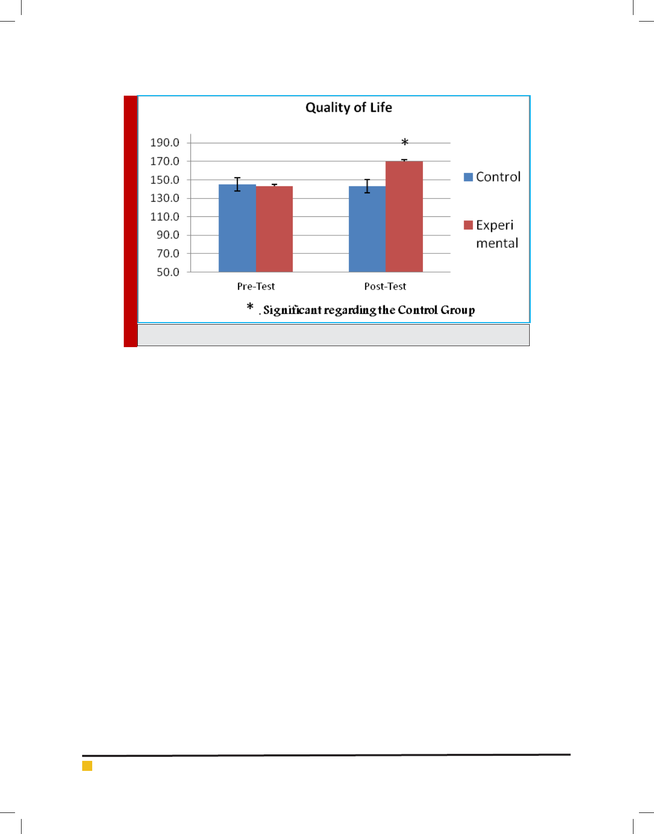
491/0 2/25±5/145 pre-Test
Control
Quality of Life
178/0 4/22±7/143 Post-Test
491/0 6/30±5/143 Pre-Test
Experimental
318/0 8/28±31/170 Post-Test
Table 5-4. paired t-test results regarding pretest and posttest of the quality of life
The signi cance
level
t
Degrees of
freedom
Within-group
mean difference
Post-Test Pre-Test Group Variable
090/0 82/0 9 2/2 4/22±7/143 2/25±5/145 Control
Quality of Life
001/0 1/6 9 37/27 8/28±31/170 6/30±5/143 Experimental
Table 6-4. results of independent t-test of quality of life in post-test
Two-tailed
signi cance level
t
Degrees of
freedom
The mean difference
between groups
Variable
001/0 1/4 19 17/25 Quality of life
136 THE EFFECT OF MICRO NEEDLING THERAPY ON THE QUALITY OF LIFE IN WOMEN BIOSCIENCE BIOTECHNOLOGY RESEARCH COMMUNICATIONS
Vahideh Riyahi and Bijan Goodarzi
Results showed that the experimental group in post-
test after the exercise protocol in comparison to the pre-
test show signi cant difference in the quality of life and
because of that the experimental group was signi cantly
increased in the quality of life. But in the control group
no signi cant difference was observed in the quality of
life in the pretest and post-test. Also according to the
results from the table, in comparing the change in the
groups in post-test, signi cant difference was observed
between the changes. Therefore, the null hypothesis of
no effect of 20 weeks of micro-needling on the quality
of life in the women suffering from skin-deep varicose
leg is rejected and it can be stated that this type of inter-
vention is to improve quality of life in these individuals.
The results of micro needling to enhance quality of life
in people with varicose veins might be because micro-
needle contains 12 short and ne needle with penetra-
tion depth of 2 mm and a speed of 25 beats per second
to 90 beats per second which ultimately creates between
300 to 1,000 tiny holes in seconds in the surface and it
stimulates skin’s natural production of collagen (Pahwa,
Pahwa et al. 2012) and leads to ulcer wrinkles removal,
improvement of the body crazes, betterment of the over-
all appearance of the skin and shrinkage of the size of
the pores (Majid 2009). In addition, accumulation and
increasing elasticity of the skin, new collagen produc-
tion, surface and deep wrinkles reduction, improvement
of body craze and healing of pigmented lesions, happens
through micro needling (Aust, Fernandes et al. 2008).
Tiny pores made on the skin causes the production of
new blood vessels and collagen bers. Micro needling
by stimulating stem cells and broblasts in the skin does
restorative work in the best way possible. Given that in
the skin-deep varicose, faulty and defective veins are at
the surface, 20 weeks use of micro needling stimulates
vascular and skin tissues, and complications of varicose
veins is reduced, thereby enhancing quality of life.
The results showed that the use of micro-needling can be
used as a safe and effective treatment in enhancing quality
of life in patients with varicose veins in lower limbs.
REFERENCES
Aust, M. C., D. Fernandes, P. Kolokythas, H. M. Kaplan and P.
M. Vogt (2008). Percutaneous collagen induction therapy: an
alternative treatment for scars, wrinkles, and skin laxity.Plastic
and reconstructive surgery 121(4): 1421-1429.
Bartsch, T. and P. Goadsby (2005). Anatomy and physiology
of pain referral patterns in primary and cervicogenic headache
disorders. Headache Currents 2(2): 42-48.
Cella, D., K. Dineen, B. Arnason, A. Reder, K. Webster, G. Kara-
batsos, C. Chang, S. Lloyd, F. Mo and J. Stewart (1996). Valida-
tion of the functional assessment of multiple sclerosis quality
of life instrument. Neurology 47(1): 129-139.
Chung, C. Y., W. H. McCray, S. Dhaliwal, T. Haywood, M. Black,
J.-B. Liu and L. S. Miller (2000). Three-dimensional esophageal
varix model quanti cation of variceal volume by high-resolu-
tion endoluminal US. Gastrointestinal endoscopy 52(1): 87-91.
Cooper, A. and F. Plum (1987). Biochemistry and physiology of
brain ammonia. Physiological reviews 67(2): 440-519.
Crick, S. J., M. N. Sheppard, S. Y. HO, L. Gebstein and R. H.
Anderson (1998). Anatomy of the pig heart: comparisons with
FIGURE 2-4. Comparison of quality of life in both experimental and control groups.

BIOSCIENCE BIOTECHNOLOGY RESEARCH COMMUNICATIONS THE EFFECT OF MICRO NEEDLING THERAPY ON THE QUALITY OF LIFE IN WOMEN 137
Vahideh Riyahi and Bijan Goodarzi
normal human cardiac structure. Journal of anatomy 193(01):
105-119.
Dhurat, R., M. Sukesh, G. Avhad, A. Dandale, A. Pal and P.
Pund (2013). A randomized evaluator blinded study of effect of
microneedling in androgenetic alopecia: a pilot study.” Inter-
national journal of trichology 5(1): 6.
Donovan, J. and H. Win eld (1992). Laparoscopic varix liga-
tion. The Journal of urology 147(1): 77-81.
Gray, H. (2009). Gray’s Anatomy: With original illustrations by
Henry Carter, Arcturus Publishing.
Harrie, R. P. and C. J. Kendall (2014). Case Study 137 Orbital
Varix. Clinical Ophthalmic Echography, Springer: 315-315.
Henwood, T. R., S. Riek and D. R. Taaffe (2008). “Strength ver-
sus muscle power-speci c resistance training in community-
dwelling older adults.” The Journals of Gerontology Series A:
Biological Sciences and Medical Sciences 63(1): 83-91.
Jensen, D., R. Jutabha, T. Kovacs, G. Randall, S. Cheng, M.
Freeman, M. Jensen and J. Gornbein (1995). “Endoscopic
hemostasis of severe gastric varix hemorrhage in a randomized
prospective blinded study of rubber band ligation vS. sclero-
therapy.” Gastrointestinal Endoscopy 41(4): 365.
Katz, A. M. (2010). Physiology of the Heart, Lippincott Wil-
liams & Wilkins.
Lamont, L. A., W. J. Tranquilli and K. A. Grimm (2000). Physi-
ology of pain. Veterinary clinics of North America: Small ani-
mal practice 30(4): 703-728.
Majid, I. (2009). Microneedling therapy in atrophic facial scars:
an objective assessment. Journal of cutaneous and aesthetic
surgery 2(1): 26.
Mann, K. G. (1999). Biochemistry and physiology of blood
coagulation. Thrombosis and haemostasis 82(2): 165-174.
Marchand, S. (2008). The physiology of pain mechanisms:
from the periphery to the brain.m Rheumatic Disease Clinics of
North America 34(2): 285-309.
Melzack, R. (1975). The McGill Pain Questionnaire: major
properties and scoring methods. Pain 1(3): 277-299.
Melzack, R. (1987). The short-form McGill pain questionnaire.
Pain 30(2): 191-197.
Melzack, R., P. Wall, A. Steptoe and J. Wardle (1994). Pain
mechanisms: a new theory. Psychosocial processes and health:
A reader: 112-131.
Pahwa, M., P. Pahwa and A. Zaheer (2012). Tram track effect
after treatment of acne scars using a microneedling device.
Dermatologic Surgery 38(7pt1): 1107-1108.
Patel, N. B. (2010). Physiology of pain. Guide to pain manage-
ment in low-resource settings: 13.
Rowlands, S. and M. Permezel (1998). 1 Physiology of pain
in labour. Baillière’s clinical obstetrics and gynaecology 12(3):
347-362.
Salavati, M., M. Mazaheri, H. Negahban, S. Sohani, M. Ebra-
himian, I. Ebrahimi and A. Kazemnejad (2008). Validation of
a Persian-version of Knee injury and Osteoarthritis Outcome
Score (KOOS) in Iranians with knee injuries. Osteoarthritis and
Cartilage 16(10): 1178-1182.
Scott, J. and E. Huskisson (1976). Graphic representation of
pain.Pain 2(2): 175-184.
Stiegmann, G., J. Sun and W. Hammond (1988). Results of
experimental endoscopic esophageal varix ligation.The Ameri-
can surgeon 54(2): 105-108.
Van Stiegmann, G. and J. S. Goff (1988). Endoscopic esopha-
geal varix ligation: preliminary clinical experience. Gastroin-
testinal endoscopy 34(2): 113-117.