
Toxicological
Communication
Biosci. Biotech. Res. Comm. 10(2): 97-101 (2017)
Effect of cadmium chloride on nucleus preopticus
in
Heteropneustes fossilis
and its recovery by herbal
compound, Ashawagandha
K. Mukati*, B. Nagar and L. Bhattacharya
Endocrinology and Physiology unit, School of Studies in Zoology and Biotechnology, Vikram University,
Ujjain (M.P.)- 456010, India
ABSTRACT
The effect of cadmium chloride on nucleus preopticus in Heteropneustes fossilis was analysed histologicalyl. Fish
when treated with cadmium chloride of 0.5 ppm for 7,14, and 21 days exhibited degeneration, hypertrophy, reduced
neurosecretory material and vacuolization in NPO. Their size was signi cantly (p<0.001) increased after cadmium
chloride exposure. In ashawagandha recovery group, these nuclei displayed a gradual reorganization in structural
detail and size. Recovery by a herbal compaund ashawagandha exhibited reduced hypertrophy and vacuolization in
NPO.
KEY WORDS: ASHAWAGANDHA, CADMIUM CHLORIDE,
HETEROPNEUSTUS FOSSILIS
, NUCLEUS PREOPTICUS
97
ARTICLE INFORMATION:
*Corresponding Author: k1.kavitamukati@gmail.com
Received 11
th
March, 2017
Accepted after revision 28
th
June, 2017
BBRC Print ISSN: 0974-6455
Online ISSN: 2321-4007 CODEN: USA BBRCBA
Thomson Reuters ISI ESC and Crossref Indexed Journal
NAAS Journal Score 2017: 4.31 Cosmos IF : 4.006
© A Society of Science and Nature Publication, 2017. All rights
reserved.
Online Contents Available at: http//www.bbrc.in/
INTRODUCTION
Heavy metals occur naturally in the environment and
are found in varying levels in ground and surface water.
Heavy metals are reported as pollutants which caused
the metabolic, physiological and structural alterations in
sh (Jiraungkoorakul et al., 2007; 2008; 2009). Among
heavy metal cadmium has been shown to be responsi-
ble for a number of reproductive abnormalities in sh
(Sharma et al.,2013 ). The pituitary gland is one of the
most important endocrine organs of sh. The histol-
ogy of the pituitary of teleost sh has been described
by a number of authors (Balcı et al., 2006; Ozen and
Timur, 1993; Hibiya, 1982). The heavy metals (lead,
cadmium, mercury etc.) are known to interfere with the
endocrine system of model organisms such as mam-
mals, sh etc. and lead to a disturbance in hormonal
metabolism, hormone-regulated cellular of physiologi-
cal processes(Colborn and Clement,1992 ; Kavlock et
al.,1996). However, metal deterioration of hypothalamic

98 EFFECT OF CADMIUM CHLORIDE ON NUCLEUS PREOPTICUS IN
HETEROPNEUSTES FOSSILIS
BIOSCIENCE BIOTECHNOLOGY RESEARCH COMMUNICATIONS
Mukati, Nagar and Bhattacharya
nuclei in sh are not well illustrated. Since only in Het-
eropneustus fossilis (Shukla and Pandey,1984; Pandit
and Bhattacharya, 2013)
MATERIAL AND METHODS
Heteropneustus fossilis measuring about length 12±5
cm and weight 25±5 gm were used in the present study.
Cadmium was used for present study in the form of cad-
mium chloride (CdCl
2
). The dose of cadmium chloride
was decided after determination of LC 50 value. It was
found to be 0.5 ppm. The herbal compound Ashawagan-
dha ( Withania somnifera) is used as recovery agent of
damaged tissue. Fishes were acclimatized to laboratory
condition for 7 days before the commencement of the
experiment and were treated with 0.01 KMno4 solution to
remove dermal infection . Fishes were fed with chopped
prawn twice a day. The 72 hrs LC50 value of cadmium
chloride was found to be 0.5 ppm in H. fossilis.
The shes were divided in three groups having36
shes in each one.
neurohypophysis.Nucleus preopticus is well developed
in H. fossilis. These cells were spherical in shape with
evenly distributed cytoplasm and their nuclear mate-
rial. The nucleus usually contains a single nucleolus but
sometime more than two nuclei were also observed. The
perikarya of NPO cells were loaden with neurosecretory
material. The neurosecretory cells of NPO were posi-
tive to AF and CAHP. Cellular differentiation was more
marked after using CAHP technique. The hypothalamus
of control sh exhibited the NPO neurons presented in
their active secreting stage. The hypothalamic nuclei
(NPO) exhibited strong af nity to CAHP and AF stain.
(Fig. 1, 3 and 5)
TREATED GROUPS
The hypothalamus of cadmium chloride treated sh after
7 days duration exhibited the NPO neurons were present
in necrotic condition. The NPO neurons exhibited thick
cell boundaries and clumping of cytoplasm. The cell as
well as nuclei appeared turgid. The neurosecretory mate-
rial in the perikarya of the cells was coarse. The cad-
mium chloride treatment induced deformities in these
neuronal cells (Fig.2).
In sh exposed to cadmium chloride for 14 days
duration, the hypothalamic nuclei (NPO and NLT) exhib-
ited cytoplasmic and nuclear abnormalities and deple-
tion of Neurosecretory material. The NPO cells showed
degeneration in their cytoplasmic contents. Their cell
boundaries were disappeared (Fig. 4).
After the treatment of cadmium chloride of 21 days
duration these neurons presented exhausted condition
due to cadmium stress. Some of the NPO cells showed
hypertrophy. Degenerated cytoplasm and disappeared
cell boundaries were observed in these cells. They had
vacuolated cytoplasm (Fig. 6). Similar results were found
by (Shukla and Pandey, 1984) in hypothalamic nuclei
of Sarotherodon mossambicus exposed to DDT. While
Katti and Sathaynesan (1986) reported degeneration of
the NPO neurons in Clarias batrachus exposed to lead
nitrate. Our ndings corroborates with study of Ram and
Joy (1988). The observed that the neurons containing
less quantity of neuroscretory material and exhibited
various degrees of degeneration and nuclear necrosis in
Channa punctatus exposed to HgCl2 and methyl mercu-
ric chloride of the dose.
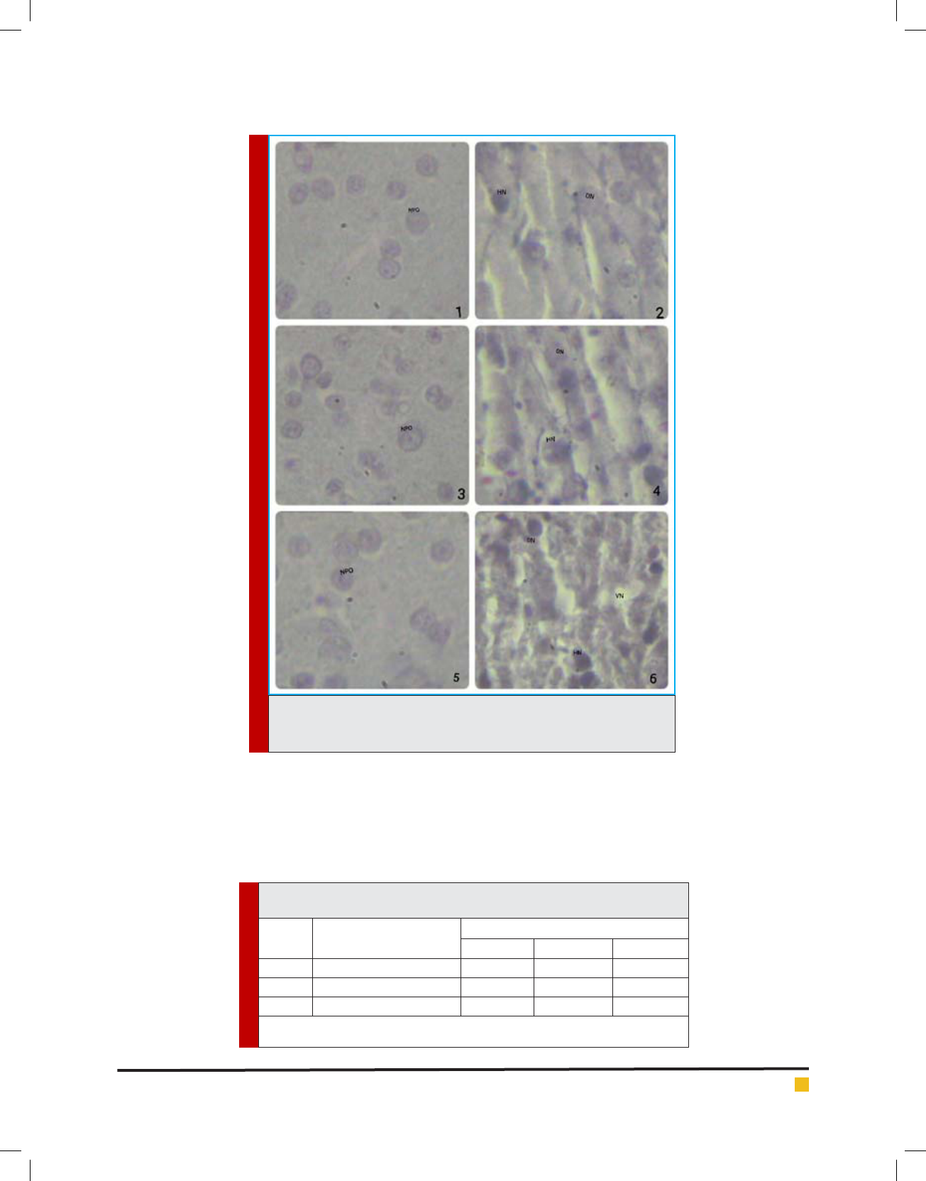
Fig-1. Control group (7 days): Showing NPO cells
stained brightly and neurosecretory material on the
periphery of NPO cells visible.
Fig-2. Treated group (7 days): Showing fusion of NPO
cells and atrophie condition, among neurons visible.
Fig-3. Control group (14 days): Showing NPO cell
bodies with evenly distributed cytoplasm and clearly
visible neurosecretory material.
S. No. Group Treatment
1 Control Water without CdCl2 + plain
food
2 Treated Exposed to 0.5ppm CdCl2 +
plain food
3 Recovery by
Ashawagandha
Exposed to 0.5ppm CdCl2 +
Ashawgandha with food
Fishes of all experiment groups (control, treated and
recovery)were sacri ced each after 7,14, and 21 days.
Pituitary with brain were xed in aqueous Bouin’s solu-
tion for 24 hours. The material was washed with water,
dehydrated and cleared through graded alcohol and
xyline respectively after ltration in paraf n blocks
were prepared and section of 5 μ thickness were cut and
stretched on albumenized slides. The slides were stained
with Chrome Alum Haematoxyline Phloxine (CAHP)
(Gomori, 1941) stains and mount in DPX for histological
observation. All the data and results for nal observa-
tion were processed in the form of microphotographs
and table. The diameter of NPO were recorded and dif-
ference if any were compared by statistical analysis
using student ‘ t’ test (Bancroft, 1966).
RESULTS AND DISCUSSION
The hypothalamo-hypophysial neurosecretory system is
a unique endocrine apparatus consisting of the cells of
nucleus preopticus (NPO) in neurohypophysis. The nerve
bers of these hypothalamic nuclei terminate in the
BIOSCIENCE BIOTECHNOLOGY RESEARCH COMMUNICATIONS EFFECT OF CADMIUM CHLORIDE ON NUCLEUS PREOPTICUS IN
HETEROPNEUSTES FOSSILIS
99
Mukati, Nagar and Bhattacharya
PLATE 1. Microphotographs of sections through hypothalamus showing
NPO of Heteropneustes fossilis in control and treated group (7, 14 21 days,
CAHP X1000)
Fig- 4. Treated group (14 days) : Showing deformed
NPO cell bodies and their hypertrophied nature.
Fig-5. Control group (21 days): Showing NPO cells
with their normal structural con guration.
Fig-6. Treated group (21days): Showing hypertro-
phied nature and vacuolization of cytoplasm in cell.
Recovery group: After 21 days treatment of cadmium
chloride the shes were administered with Ashawagandha.
In 7 days: After 7 days duration in Ashawagndha
group the NPO exhibited reformed condition. Vacuoliza-
tion was still present in their cytoplasm. Some of them
were appeared with regenerated cytoplasm and their
Table 1. Diameter of Nucleus preopticus of Heteropneustes fossilis in control
and experimental group.
S. No Experimental group
Days of exposure
7 days 14 days 21 days
1 Control 10.4±0.06 10.9±0.05 11.2±0.03
2 Treated 13.6±0.07** 15.2±0.06*** 17.9±0.03***
3 Recovery Ashawagandha 11.0±0.02** 12.2±0.04** 12.3±0.01**
All values are expressed in Mean ± SEM, Total no. of samples for each observation: 10, Signi cant
level (**p < 0.05, ***p < 0.001).
100 EFFECT OF CADMIUM CHLORIDE ON NUCLEUS PREOPTICUS IN
HETEROPNEUSTES FOSSILIS
BIOSCIENCE BIOTECHNOLOGY RESEARCH COMMUNICATIONS
Mukati, Nagar and Bhattacharya
Hgcl2 exposure NPO size were increased in the spirulina
and chorella were effective to recover the histological
changes in the NPO of H.fossilis.
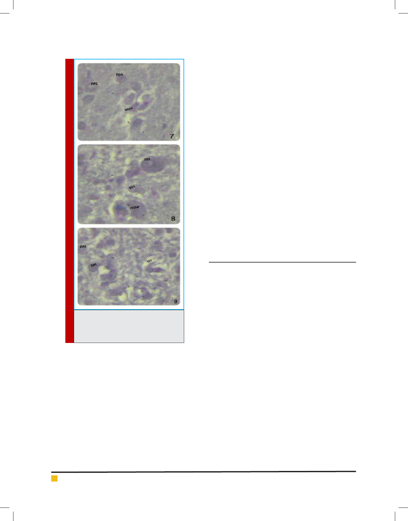
Fig-7. Ashawagandha Recovery Group (7 days):
Showing accumulation of neurosecretory material in
NPO cells and vacuolization in their cytoplasm still
persist.
Fig-8. Ashawagndha Recovery Group (14 days):
Showing still exhibited hypertrophied condition in a few
NPO cells, most of them in normal appearance.
Fig-9. Ashawagndha Recovery Group (21 days):
Showing regenerated cytoplasm and nuclear contents of
cell body of NPO.
ABBERVIATIONS
C-Cytoplasm, NPO-Nucleus preopticus
NSM-Neurosecretory material DN-Degenerated
neurons
HN-Hypertrophied neurons, VN-Vacuolized neu-
rons
RH-Reduced hypertrophy, RN-Regenerated neu-
rons
RV-Reduced vacuolization
ACKNOWLEDGEMENTS
The authors are grateful to Dr. M.S. Parihar, Professor
and Head, School of Studies in Zoology and Biotechnol-
ogy Vikram University, Ujjain(M.P.) for providing neces-
sary facilities to complete this work.
REFERENCES
Balcı, B., I
˙
kiz, B.R., Mutaf, B.F. (2006), Histomorphological
comparison of pituitary gland of dusky grouper (Epinephelus
guaza L., 1758) and blacktip grouper (Epinephelus alexandri-
nus V., 1828). JFAS, 23: 183–186
Bancroft, J.D. and Stevens, A. (1986), Theory and practice of
histological techniques, 2nd edn. Churchill Livingstone, New
York , 570.
Colborn, T. and C. Clement. (1992), Chemically induced altera-
tions in sexual and functional development: the wildlife/
human connection Princeton Scienti c Publishing. Princeton,
N.J.
Gomori G. (1941), Chromalum-haematoxylin phloxin method.
Am. J. Pathol. 17, 395
Hibiya, T. (1982): An Atlas of Fish Histology. Gustow Fischer
Verlag, Stuttgart.
Jiraungkoorakul, W., Sahaphong, S., Kangwanrangsan, N. and
Kim. M. H., Histopathologcal study. (2006), The effect of ascor-
bic acid on cadmium exposure in sh (Puntius altus). J.Fish.
Aquat. Sci., 1: 191-199
PLATE 2. Microphotographs of sections
through hypothalamus showing NPO of
Heteropneustes fossilis in recovery group
(7, 14, 21 days, CAHP X1000)
nuclear contents (Fig. 7 ) In 14 days In Ashawagndha
recovery group the NPO cells showed reduced hypertro-
phy and increased population with some extent of neu-
rosecretory material ( Fig. 8 ) In 21 days After 21 days
duration the shes of Ashawangndha recovery group
showed signs of reconstitution in the neurons. In recov-
ery group cellular hypertrophy still persisted in a few of
NPO neurons. Most of them were in normal appearance.
They had regenerated cytoplasm and nuclear contents
(Fig. 9)
These observations were in good agreement with
the study of Shukla and Pandey (1984). They postu-
lated decreased size of hypothalamic nuclei in S. mos-
sambicus exposed to DDT and BHC when kept in plain
water. Pandit and Bhattacharya (2013) observed after

Mukati, Nagar and Bhattacharya
BIOSCIENCE BIOTECHNOLOGY RESEARCH COMMUNICATIONS EFFECT OF CADMIUM CHLORIDE ON NUCLEUS PREOPTICUS IN
HETEROPNEUSTES FOSSILIS
101
Jiraungkoorakul, W., Sahaphong, S., Kangwanrangsan, N. and
Kim. M. H., Histopathologcal study. (2007), The effect of ascor-
bic acid on cadmium exposure in gill of Puntius altus.int. J.
Zool. Res., 3: 77-85
Jiraungkoorakul,W., Sahaphong, S., Kangwanrangsan, N.
and Zakaria. S. (2008), The protective in uence of ascorbic
acid against the genotoxicity of waterborne lead exposure in
Nile tilapia oreochromis niloticus (L.). J.Fish Biol.,73 : 355-
366
Katti S.R. and Sathyanesan A.G. (1986), Changes in the
hypothalamo-neurohypophysial complex to lead treated sh,
Clarias batrachus (L), Z Mikrosk-anat. Forsch. Leipzig. 100,
347
Kavlock, R.J., G.P. Daston, C. DeRosa, P. Fenner- Crisp, L.E.
Gray, S. Kaatari, G. Lucier, M. Hecker, M.J. Mac, C. Mazka, R.
Miller, J. Moore, R. Rolland, G. Scott, D. Sheehan, T. Sinks and
H.A. Tilson. (1996), Research needs for the risk assessment of
endocrine disruptors: a report of the US-EPA sponsored work-
shop. Environ. Health. Perspect., 102: 715-740
Ozen, M.R., Timur, G. (1993), Lighted microscopic identi ca-
tion of cell types in carp, pike and vimba pituitary gland with
histochemical methods. Dog
˘
a Turk. J. Biol.,17: 311–320
Pandit, N. and Bhattacharya, L. (2013), Effect of mercuric chlo-
ride on nucleus preopticus in Heteropneustes fossilis and their
recovery by spirulina and chlorella, International Journal of
Research in BioSciences. 2 (4), 26-31,
Ram R.N. and Joy K.P. (1988), Mercurial induced changes in
hypothalamo-neurohypophysial complex in relation to repro-
duction in teleostean sh, Channa punctatus (Bloch), Bull.
Environ. Contam. Toxicol. 41, 329
Sharma, S., Vyas, V., Tamot, S., and Manhor, S. (2013), His-
tological changes in the testis of air–breathing sh, Hetero-
pnuestes fossilis( Bloch) following Cadmium chloride exposure
vol. 3, No.2,1216-1221
Shukla L. and Pandey A.K. (1984), Histopathological responses
of hypothalamic nuclei and their recovery in DDT stressed sh,
Sarotherodon mossambicus, Nat. Acad. Sci. Lett. 7(10), 317-321