
Medical
Communication
Biosci. Biotech. Res. Comm. 9(4): 725-728 (2016)
Ex vivo
comparative analysis of root-canal dentin
removal by MTwo and K3 rotary les using spiral
computed tomography
Hengameh Akhavan
1
, Mehrdad Panjnoush
2
, Neda Montaser Kuhsari
3
, Azin Sadighnia
4
and Sohrab Tour Savadkouhi
1
*
1
Assistant Professor, Department of Endodontics, Dental Branch, Islamic Azad University, Tehran, Iran
2
Assistant Professor, Department of Oral and Maxillofacial Radiology, Dental Branch, Tehran University of
Medical Sciences, Tehran, Iran
3
Student of Dentistry, Dental Branch, Islamic Azad University, Tehran, Iran
4
Dentist
ABSTRACT
The main objective in root canal preparation is to develop an enlarged and shaped taper from apical to coronal,
maintaining the original canal shape. The aim of this study is analysis of root canals dentin removal shaped by MTwo
(Group M) and K3 (Group K) rotary les using spiral-computed tomography. A total of 40 mesiobuccal root canals of
mesial roots with curvatures ranging 20-35 degree, and working length ranging 15-17 mm included the study. The
initial images were reconstructed and cross- sections corresponding to distance 2, 4.5 and 7mm from the anatomic
apex. Group M was prepared with Mtwo les with master apical le size 40 (single length technique) and group K
was prepared with K3 les (VT technique, %4), with master apical le size 40. Post instrument images was recorded
as same as the initial ones. Dentinal thickness measured in buccal, mesial, lingual and distal sections of each root
canal. Statistical analysis was performed with one sample test Kolmogorov- Smirnov and T-test for comparing sam-
ples between groups. The mean total root dentine removal between group M (0.17+-0.04) and group K (0.20+_0.07)
was statistically different (P<0.001). Group K showed 15% more dentin removal compared group M. Recording to our
ndings K3 had showed more dentin removal than Mtwo especially in apical and coronal third of curved root canals,
so Mtwo acted better than K3.
KEY WORDS: MTWO, K3, RESIDUAL DENTIN THICKNESS, ROOT CANAL CBCT
725
ARTICLE INFORMATION:
*Corresponding Author: s_savadkouhi@yahoo.com
Received 25
th
Oct, 2016
Accepted after revision 12
th
Dec, 2016
BBRC Print ISSN: 0974-6455
Online ISSN: 2321-4007
Thomson Reuters ISI ESC and Crossref Indexed Journal
NAAS Journal Score 2015: 3.48 Cosmos IF : 4.006
© A Society of Science and Nature Publication, 2016. All rights
reserved.
Online Contents Available at: http//www.bbrc.in/
726
EX VIVO
COMPARATIVE ANALYSIS OF ROOT-CANAL DENTIN REMOVAL BIOSCIENCE BIOTECHNOLOGY RESEARCH COMMUNICATIONS
Hengameh Akhavan et al.
INTRODUCTION
The main objective in root canal preparation is to
develop an enlarged and shaped taper from apical to
coronal, maintaining the original canal shape. Shap-
ing procedure directly related not only to the length but
also to the position and curvature of each individual
root canal (Schilder, 1974). When a curvature is present,
endodontic preparations become challenging. Devia-
tion from the original curvature can lead to excessive
or inappropriate dentine removal, which weakens the
tooth, resulting in stripping perforations or fracture of
the root. Several instrument systems have been recom-
mended in the past with different features. Today, the
use of rotary Ni-Ti les has signi cantly reduced devia-
tions from the original root canal shape compared with
stainless steel hand les, (Kandaswamy et al 2009 Talati
et al 2013 and Stavileci et al., 2015).
Several methods have been introduced for assess-
ment of post canal instrumentation shaping such as
scanning electron microscope, radiographs, photo-
graphic assessment and computer manipulation (Hüls-
mann, Peters, & Dummer, 2005). The methods are inva-
sive in nature; hence accurate repositioning of pre- and
post-instrumented specimens is dif cult (Cumbo et al
2015).
Spiral computed tomography; a non-invasive tech-
nology has been advocated for pre-and post-instrumen-
tation evaluation of canal. It can render cross-sectional
(cut plane) and 3D images that are highly accurate and
quanti able. At any level, the amount of post instru-
mentation dentinal removal and canal transportation
can be analyzed without loss of specimen (Narayan
et al., 2012, Maitin et al., 2013 and Deepak et al., 2015).
The aim of this study is analysis of root canals dentin
removal shaped by M Two and K3 rotary les using spi-
ral computed tomography.
MATERIAL AND METHODS
In this experimental study human extracted mandibular
rst molars, which had no restorations and had been
extracted due to extensive destruction of coronal struc-
tures or periodontal problems subjected the study. The
teeth were disinfected in 1% hypochlorite solution for
one hour and kept in sterile normal saline solution until
further processing. Assessment radiographic images
were taken using E-speed lms (AGFA, Heraeus Kulzer
GmbH; Hanau, Germany) with 70 kVp and 8 mA.
A total of 40 mesiobuccal root canals of mesial roots
with curvatures ranging 20-35 degree, and working
length ranging 15-17 mm were included in the study.
They were randomly divided in two experimental groups
of 18 canals each, and the remaining two for control
group. The teeth were imbedded in a self-cure acrylic
material (Acropars OP, Marlic medical industries, Tehran,
Iran) and submitted to CBCT analysis (Somatom sensa-
tion 16 CT scanner, Siemens LTD Berlin, Germany). The
initial images were reconstructed and cross- sections
corresponding to distance 2, 4.5 and 7mm from the ana-
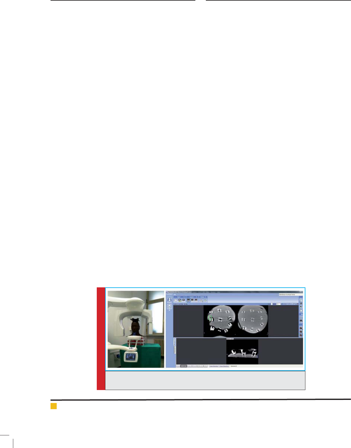
tomic apex (Syngo CT software VB20, Siemens) (Fig.1).
The access cavities of all samples were prepared and
Initial ling was done from 08 to 15 size k le and both
experimental groups were instrumented with X-Smart
(Dentsply Maillefer, Ballaigues, Switzerland). Between
each instrument, the root canals were irrigated with 5
ml of 5.25% sodium hypochlorite and nally ushed
with normal saline. Single length technique was used in
a gentle in and outward motion according to the manu-
facturers’ instruction. Group M was prepared with Mtwo
les with master apical le size 40 and group K was
prepared with K3 les (VT technique, %4), with master
apical le size 40 according to manufactures protocol.
Post instrument images was recorded as same as the ini-
tial ones. Dentinal thickness measured in buccal, mesial,
FIGURE 1. CBCT analysis (Somatom sensation 16 CT scanner, Siemens LTD Berlin,
Germany and Syngo CT software VB20, Siemens)

BIOSCIENCE BIOTECHNOLOGY RESEARCH COMMUNICATIONS
EX VIVO
COMPARATIVE ANALYSIS OF ROOT-CANAL DENTIN REMOVAL 727
Hengameh Akhavan et al.
lingual and distal sections of each root canal. Statistical
analysis was performed with one sample test Kolmogo-
rov- Smirnov and T-test for comparing samples between
groups.
RESULTS
No specimens excluded because of le fracture or other
mishaps. The two specimens in control group showed
the xing method was accurate and there was no dif-
ference between rst and second scans. In 2mm sections
from the apices, root canal dentin removal for group
M (0.12 +- 0.20) and group K (0.14 +- 0.04) was statis-
tically different (P<0.001). Group K showed 16% more
dentin removal compared group M.In 4.5mm sections
from the apices, root canal dentin removal for group M
(0.18 +- 0.04) and group K (0.18 +- 0.04) was the same
(P<0.09).
In 7mm sections from the apices, root canal dentin
removal for group M (0.21 +- 0.01) and group K (0.27
+- 0.06) was statistically different (P<0.001). Group K
showed 27% more dentin removal compared group M
(table-1). The mean total root dentine removal between
group M (0.17+-0.04) and group K (0.20+_0.07) was sta-
tistically different (P<0.001). Group K showed 15% more
dentin removal compared group M (table-2).
DISCUSSION
NiTi rotary les have superior shaping ability to main-
tain canal centered during instrumentation compared to
stainless steel hand les (Gergi, Rjeily, Sader, & Naaman,
2010). But this advantage is differ among different
brands of NiTi rotary les, depends on their speci cs.In
this study, we analyzed the effect of canal instrumenta-
tion on dentin removal using MTwo and K3 rotary les.
The method used for dentin removal in the current study
(spiral tomography) allowed us to evaluate morphologi-
cal changes after root canal preparation in a conserva-
tive manner (Özer, 2011).
Within the limits of an “in vitro” study, the cone
beam computed tomography offers a method that is
relatively simple and economical and provides useful
information about the action of instruments in the canal
space (Gergi et al., 2010; Maitin et al., 2013; Özer, 2011;
Tasdemir, Aydemir, Inan, & Ünal, 2005). An alternative
conservative method of assessing root canal instrumen-
tation techniques is the microcomputer tomography that
is more expensive and requires well-trained operators in
order to obtain valid results (Peters et al 2001; You et al.,
2011; Zhao et al 2013).
Cumbo et al reported that M Two and BioRace
rotary les had comparable results in amount of den-
tine removal in different levels of root canals (Cumbo et
al., 2015). For this comparison they had used specimens
with single root canals with a curvature lower than 20
degrees that is lower than our specimens (20-35 degrees).
They had used resin blocks and digital photographs for
shaping ability assessment (the Bramante technique) but
the technique they had used is not conservative com-
pared the technique had used in this study (CBCT).
In an ex vivo study by Maitin et al, they reported
the MTwo rotary les had better well centered prepa-
ration in coronal and mid root compared to ProTaper,
K3 and Race (Maitin et al., 2013). They had used sin-
gle rooted specimens with a curvature ranging 20 to 40
degrees comparable to our specimens (20-35 degrees).
The severity of curvature makes the preparation proce-
dure more challenging. The result of our study con rms
their results and shows MTwo has little dentine removal
and better centering ability compared to K3 rotary les.
In contrast of our results Narayan et al concluded that
MTwo rotary les shaped root canals enlarger in volume
and with compared K3 and Race. This difference can be
explained by difference in shaping procedures (Narayan
et al., 2012).
Recording to our ndings K3 had showed more dentin
removal than Mtwo especially in apical and coronal third
of curved root canals, so M two acted better than K3.
The authors deny any con icts of interest related to
this study.
REFERENCES
Cumb o, E., Russo, R., & Gallina, G. (2015). Assessment of Root
Canal Enlargement Using Mtwo and BioRace Rotary Files. The
Scienti c World Journal, 2015.
Table 1: Root dentine removal in different sections
Rotary
systems
Root
sections
Dentin removal
(mm)
Coef cient
Variations
MTwo 2mm 0.12+-0.02 17
4.5mm 0.18+-0.04 22
7mm 0.21+-0.01 5
K3 2mm 0.14+-0.04 28
4.5mm 0.18+-0.04 22
7mm 0.27+-0.06 22
Table 2: The mean total root dentine removal
Rotary
systems
Mean dentin
removal (mm)
Coef cient
Variations
MTwo 0.17+-0.04 23
K3 0.20+-0.07 35

728
EX VIVO
COMPARATIVE ANALYSIS OF ROOT-CANAL DENTIN REMOVAL BIOSCIENCE BIOTECHNOLOGY RESEARCH COMMUNICATIONS
Hengameh Akhavan et al.
Deep ak, J., Ashish, M., Patil, N., Kadam, N., Yadav, V., & Jag-
dale, H. (2015). Shaping Ability of 5th Generation Ni-Ti Rotary
Systems for Root Canal Preparation in Curved Root Canals
using CBCT: An In Vitro Study. Journal of international oral
health: JIOH, 7(Suppl 1), 57.
Gerg i, R., Rjeily, J. A., Sader, J., & Naaman, A. (2010). Com-
parison of canal transportation and centering ability of twisted
les, Path le-ProTaper system, and stainless steel hand K- les
by using computed tomography. Journal of Endodontics, 36(5),
904-907.
Hüls mann, M., Peters, O. A., & Dummer, P. M. (2005). Mechani-
cal preparation of root canals: shaping goals, techniques and
means. Endodontic topics, 10(1), 30-76.
Kanda swamy, D., Venkateshbabu, N., Porkodi, I., & Pradeep,
G. (2009). Canal-centering ability: An endodontic challenge.
Journal of conservative dentistry, 12(1), 3.
Maiti n, N., Arunagiri, D., Brave, D., Maitin, S. N., Kaushik, S.,
& Roy, S. (2013). An ex vivo comparative analysis on shaping
ability of four NiTi rotary endodontic instruments using spi-
ral computed tomography. Journal of conservative dentistry,
16(3), 219.
Naray an, G. S., Venkatesan, S. M., Karumaran, C., Indira, R.,
Ramachandran, S., & Srinivasan, M. (2012). A comparative
evaluation on the cleaning and shaping ability of three nickel
titanium rotary instruments using computerized tomography-
An ex vivo study. Contemporary clinical dentistry, 3(6), 151.
Özer, S. Y. (2011). Comparison of root canal transportation
induced by three rotary systems with noncutting tips using
computed tomography. Oral Surgery, Oral Medicine, Oral
Pathology, Oral Radiology, and Endodontology, 111(2), 244-
250.
Peters , O., Schönenberger, K., & Laib, A. (2001). Effects of four
Ni–Ti preparation techniques on root canal geometry assessed
by micro computed tomography. International endodontic
journal, 34(3), 221-230.
Schilde r, H. (1974). Cleaning and shaping the root canal. Dent
Clin North Am, 18, 269-296.
Stavile ci, M., Hoxha, V., Görduysus, Ö., Tatar, I., Laperre, K.,
Hostens, J. Muhaxheri, E. (2015). Evaluation of root canal prep-
aration using rotary system and hand instruments assessed by
micro-computed tomography. Medical science monitor basic
research, 21, 123.
Talati, A ., Moradi, S., Forgani, M., & Monajemzadeh, A. (2013).
Shaping ability of nickel-titanium rotary instruments in curved
root canals. Iranian endodontic journal, 8(2), 55-58.
Tasdemir, T., Aydemir, H., Inan, U., & Ünal, O. (2005). Canal
preparation with Hero 642 rotary Ni–Ti instruments com-
pared with stainless steel hand K‐ le assessed using computed
tomography. International endodontic journal, 38(6), 402-408.
You, S.-Y., K im, H.C., Bae, K.S., Baek, S.-H., Kum, K.Y. & Lee,
W. (2011). Shaping ability of reciprocating motion in curved
root canals: a comparative study with micro–computed tomog-
raphy. Journal of Endodontics, 37(9), 1296-1300.
Zhao, D., She n, Y., Peng, B., & Haapasalo, M. (2013). Micro–
computed tomography evaluation of the preparation of
mesiobuccal root canals in maxillary rst molars with Hy ex
CM, Twisted Files, and K3 instruments. Journal of Endodon-
tics, 39(3), 385-388.